Click image to see more details
-

-
-
-
-
+3
Product Info Summary
| SKU: | PB9213 |
|---|---|
| Size: | 100 μg/vial |
| Reactive Species: | Human, Mouse, Rat |
| Host: | Rabbit |
| Application: | IHC, WB |
Customers Who Bought This Also Bought

Product info
Product Name
Anti-Heme oxygenase 2/HMOX2 Antibody Picoband®
SKU/Catalog Number
PB9213
Size
100 μg/vial
Form
Lyophilized
Description
Boster Bio Anti-Heme oxygenase 2/HMOX2 Antibody Picoband® catalog # PB9213. Tested in IHC, WB applications. This antibody reacts with Human, Mouse, Rat. The brand Picoband indicates this is a premium antibody that guarantees superior quality, high affinity, and strong signals with minimal background in Western blot applications. Only our best-performing antibodies are designated as Picoband, ensuring unmatched performance.
Storage & Handling
Store at -20˚C for one year from date of receipt. After reconstitution, at 4˚C for one month. It can also be aliquotted and stored frozen at -20˚C for six months. Avoid repeated freeze-thaw cycles.
Cite This Product
Anti-Heme oxygenase 2/HMOX2 Antibody Picoband® (Boster Biological Technology, Pleasanton CA, USA, Catalog # PB9213)
Host
Rabbit
Contents
Each vial contains 4 mg Trehalose, 0.9 mg NaCl and 0.2 mg Na2HPO4.
Clonality
Polyclonal
Isotype
Rabbit IgG
Immunogen
E.coli-derived human HMOX2 recombinant protein (Position: S2-M316). Human HMOX2 shares 89% and 90% amino acid (aa) sequences identity with mouse and rat HMOX2, respectively.
Cross-reactivity
No cross-reactivity with other proteins
Reactive Species
PB9213 is reactive to HMOX2 in Human, Mouse, Rat
Observed Molecular Weight
36 kDa
Calculated molecular weight
36.0 kDa
Background of HMOX2
Heme oxygenase 2 (HMOX2), also known as HO-2, is an enzyme that in humans is encoded by the HMOX2 gene. It is mapped to 16p13.3. HMOX2 belongs to the heme oxygenase family. Heme oxygenase cleaves the heme ring at the alpha methene bridge to form biliverdin. Biliverdin is subsequently converted to bilirubin by biliverdin reductase. Under physiological conditions, the activity of heme oxygenase is highest in the spleen, where senescent erythrocytes are sequestrated and destroyed. Heme oxygenase 2 could be implicated in the production of carbon monoxide in brain where it could act as a neurotransmitter.
Antibody Validation
Boster validates all antibodies on WB, IHC, ICC, Immunofluorescence, and ELISA with known positive control and negative samples to ensure specificity and high affinity, including thorough antibody incubations.
Application & Images
Applications
PB9213 is guaranteed for IHC, WB Boster Guarantee
Recommend Dilution
| Application | Dilution | Species |
|---|---|---|
| Western blot | 0.1-0.5μg/ml | Human, Mouse, Rat |
| Immunohistochemistry (Paraffin-embedded Section) | 2-5μg/ml | Human |
Tested application
Suggested blocking solution with 5% non-fat milk or BSA; (*)Recommended protein loading: 20-40 µg per lane
Use TE buffer pH 9.0 for antigen retrieval; (*) citrate buffer pH 6.0 is an alternative.
Validation Images & Assay Conditions
Click image to see more details
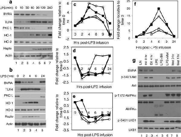
TLR4 is a negative regulator of biliverdin/BVRA signaling. ( a ) Healthy donor’s blood was untreated (lane 1) or treated with LPS (10 ng/ml) for the indicated times (lanes 2–7). Leukocytes were isolated and analyzed. Abbreviations are: Heme oxygenase 1, HO-1; Heme oxygenase 2, HO-2; Haptoglobin, Hapto. ( b ) In an earlier study , subjects were administered LPS (1 ng/kg) in vivo and blood was drawn at the indicated times post LPS infusion. Leukocyte lysates available from that study were normalized for protein content and analyzed by western blotting. ( c – f ) In another prior study , leukocytes from four subjects administered LPS in vivo were analyzed for changes in gene expression over a period of 24 hours post LPS infusion. Data from that study , available through GEO dataset GSE3284, revealed a temporal ( c ) increase in TLR4 mRNA, ( d ) decline in BVRA mRNA, ( e ) decrease in PKCζ mRNA ( f ) increase in haptoglobin mRNA expression. In ( c – f ) each symbol represents a subject. ( g ) At time 0 (0 hr) healthy donor’s blood was untreated (UN; lane 1), treated for 1 hour with biliverdin (Bili 0 hr; 50 μM; lane 2), treated for 4 hours with LPS (10 ng/ml; lanes 4–6) to trigger a decline in BVRA expression, or for 1 hour with metformin (Met; 10 μM; lane 7). Four hours later (time 4 hr) blood samples were treated for 1 hour with biliverdin (Bili 4 hr; 50 μM; lane 3) or metformin (Met 4 hr; 10 μM; lane 8). Samples pretreated with LPS for 4 hours (lanes 4–6), were then treated for 1 hr with biliverdin (Bili; 50 μM; lane 5) or metformin (Met; 10 μM; lane 6). Leukocytes were isolated and analyzed by western blotting.
Index in PubMed under a CC BY license. PMID: 31065010

Click image to see more details
Western blot analysis of PHMOX2 using anti-HMOX2 antibody (PB9213).
Electrophoresis was performed on a 10% SDS-PAGE gel at 80V (Stacking gel) / 120V (Resolving gel) for 2 hours. The sample well of each lane was loaded with 30 ug of sample under reducing conditions.
Lane 1: huamn Jurkat whole cell lysates,
Lane 2: human Hela whole cell lysates,
Lane 3: human K562 whole cell lysates,
Lane 4: human PANC-1 whole cell lysates,
Lane 5: rat PC-12 whole cell lysates,
Lane 6: mouse RAW264.7 whole cell lysates.
After electrophoresis, proteins were transferred to a nitrocellulose membrane at 150 mA for 50-90 minutes. Blocked the membrane with 5% non-fat milk/TBS for 1.5 hour at RT. The membrane was incubated with rabbit anti-HMOX2 antigen affinity purified polyclonal antibody (PB9213) at 0.5 μg/mL overnight at 4°C, then washed with TBS-0.1%Tween 3 times with 5 minutes each and probed with a goat anti-rabbit IgG-HRP secondary antibody (Catalog # BA1054) at a dilution of 1:5000 for 1.5 hour at RT. The signal is developed using an ECL Plus Western Blotting Substrate (Catalog # AR1196-200) with Tanon 5200 system. A specific band was detected for HMOX2 at approximately 36 kDa. The expected band size for HMOX2 is at 36 kDa.

Click image to see more details
IHC analysis of HMOX2 using anti-HMOX2 antibody (PB9213).
HMOX2 was detected in a paraffin-embedded section of human appendix mucinous adenocarcinoma tissue. Heat mediated antigen retrieval was performed in EDTA buffer (pH 8.0, epitope retrieval solution). The tissue section was blocked with 10% goat serum. The tissue section was then incubated with 2 μg/ml rabbit anti-HMOX2 Antibody (PB9213) overnight at 4°C. Peroxidase Conjugated Goat Anti-rabbit IgG was used as secondary antibody and incubated for 30 minutes at 37°C. The tissue section was developed using HRP Conjugated Rabbit IgG Super Vision Assay Kit (Catalog # SV0002) with DAB as the chromogen.

Click image to see more details
IHC analysis of HMOX2 using anti-HMOX2 antibody (PB9213).
HMOX2 was detected in a paraffin-embedded section of human colorectal adenocarcinoma tissue. Heat mediated antigen retrieval was performed in EDTA buffer (pH 8.0, epitope retrieval solution). The tissue section was blocked with 10% goat serum. The tissue section was then incubated with 2 μg/ml rabbit anti-HMOX2 Antibody (PB9213) overnight at 4°C. Peroxidase Conjugated Goat Anti-rabbit IgG was used as secondary antibody and incubated for 30 minutes at 37°C. The tissue section was developed using HRP Conjugated Rabbit IgG Super Vision Assay Kit (Catalog # SV0002) with DAB as the chromogen.

Click image to see more details
IHC analysis of HMOX2 using anti-HMOX2 antibody (PB9213).
HMOX2 was detected in a paraffin-embedded section of human lung adenocarcinoma tissue. Heat mediated antigen retrieval was performed in EDTA buffer (pH 8.0, epitope retrieval solution). The tissue section was blocked with 10% goat serum. The tissue section was then incubated with 2 μg/ml rabbit anti-HMOX2 Antibody (PB9213) overnight at 4°C. Peroxidase Conjugated Goat Anti-rabbit IgG was used as secondary antibody and incubated for 30 minutes at 37°C. The tissue section was developed using HRP Conjugated Rabbit IgG Super Vision Assay Kit (Catalog # SV0002) with DAB as the chromogen.

Click image to see more details
IHC analysis of HMOX2 using anti-HMOX2 antibody (PB9213).
HMOX2 was detected in a paraffin-embedded section of human ovarian serous adenocarcinoma tissue. Heat mediated antigen retrieval was performed in EDTA buffer (pH 8.0, epitope retrieval solution). The tissue section was blocked with 10% goat serum. The tissue section was then incubated with 2 μg/ml rabbit anti-HMOX2 Antibody (PB9213) overnight at 4°C. Peroxidase Conjugated Goat Anti-rabbit IgG was used as secondary antibody and incubated for 30 minutes at 37°C. The tissue section was developed using HRP Conjugated Rabbit IgG Super Vision Assay Kit (Catalog # SV0002) with DAB as the chromogen.

Click image to see more details
IHC analysis of HMOX2 using anti-HMOX2 antibody (PB9213).
HMOX2 was detected in a paraffin-embedded section of human thyroid cancer tissue. Heat mediated antigen retrieval was performed in EDTA buffer (pH 8.0, epitope retrieval solution). The tissue section was blocked with 10% goat serum. The tissue section was then incubated with 2 μg/ml rabbit anti-HMOX2 Antibody (PB9213) overnight at 4°C. Peroxidase Conjugated Goat Anti-rabbit IgG was used as secondary antibody and incubated for 30 minutes at 37°C. The tissue section was developed using HRP Conjugated Rabbit IgG Super Vision Assay Kit (Catalog # SV0002) with DAB as the chromogen.
Specific Publications For Anti-Heme oxygenase 2/HMOX2 Antibody Picoband® (PB9213)
Loading publications
Recommended Resources
Here are featured tools and databases that you might find useful.
- Boster's Pathways Library
- Protein Databases
- Bioscience Research Protocol Resources
- Data Processing & Analysis Software
- Photo Editing Software
- Scientific Literature Resources
- Research Paper Management Tools
- Molecular Biology Software
- Primer Design Tools
- Bioinformatics Tools
- Phylogenetic Tree Analysis
Customer Reviews
Have you used Anti-Heme oxygenase 2/HMOX2 Antibody Picoband®?
Share your experimental results or join a short interview to earn up to $1,000 in product credits or other rewards.
0 Reviews For Anti-Heme oxygenase 2/HMOX2 Antibody Picoband®
Customer Q&As
Have a question?
Find answers in Q&As, reviews.
Can't find your answer?
Submit your question
2 Customer Q&As for Anti-Heme oxygenase 2/HMOX2 Antibody Picoband®
Question
We are currently using anti-Heme oxygenase 2/HMOX2 antibody PB9213 for human tissue, and we are well pleased with the WB results. The species of reactivity given in the datasheet says human. Is it true that the antibody can work on dog tissues as well?
Verified Customer
Verified customer
Asked: 2020-01-16
Answer
The anti-Heme oxygenase 2/HMOX2 antibody (PB9213) has not been validated for cross reactivity specifically with dog tissues, but there is a good chance of cross reactivity. We have an innovator award program that if you test this antibody and show it works in dog you can get your next antibody for free. Please contact me if I can help you with anything.
Boster Scientific Support
Answered: 2020-01-16
Question
Has PB9213 been tested with rat samples, specifically with WB and IHC-P?
Verified customer
Asked: 2019-06-26
Answer
The Anti-Heme Oxygenase 2/HMOX2 Antibody Picoband™ (PB9213) didn't work well on rat samples.
Boster Scientific Support
Answered: 2019-07-02
