Click image to see more details
Product Info Summary
| SKU: | DZ41310 |
|---|---|
| Size: | 200 μl/vial |
| Reactive Species: | Human |
| Host: | Rabbit |
| Application: | IF, ICC, WB |
Customers Who Bought This Also Bought

Product info
Product Name
Anti-Human CCNE1 Antibody
SKU/Catalog Number
DZ41310
Size
200 μl/vial
Form
Liquid
Description
Boster Bio Anti-CCNE1 Antibody catalog # DZ41310. This antibody reacts with Human.
Storage & Handling
At -20°C for one year, at 4°C for one month. Avoid repeated freezing and thawing.
Cite This Product
Anti-Human CCNE1 Antibody (Boster Biological Technology, Pleasanton CA, USA, Catalog # DZ41310)
Host
Rabbit
Contents
Each vial contains 20mM PBS, 50% glycerol, 0.02% NaN3.
Clonality
Polyclonal
Isotype
Rabbit IgG
Reactive Species
DZ41310 is reactive to CCNE1 in Human
Calculated molecular weight
47.1 kDa
Application & Images
Applications
DZ41310 is guaranteed for IF, ICC, WB Boster Guarantee
Recommend Dilution
WB: 1:1000
ICC/IF: 1:100-1:200
Validation Images & Assay Conditions
Click image to see more details
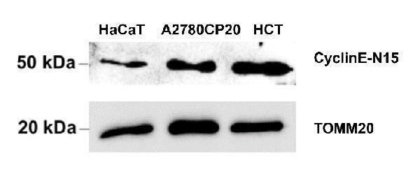
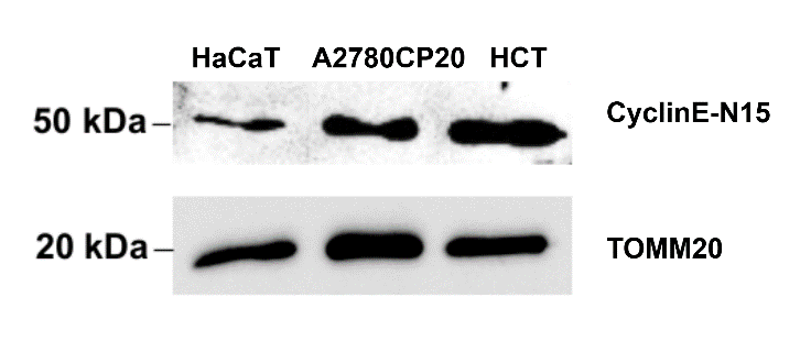
Western blot analysis of CCNE1 using anti-CCNE1 antibody (DZ41310).
Electrophoresis was performed on a 5-20% SDS-PAGE gel at 80V (Stacking gel) / 120V (Resolving gel) for 2 hours. The sample well of each lane was loaded with 30 ug of sample under reducing conditions.
Lane 1: human HaCaT whole cell lysates,
Lane 2: human A2780cp20 whole cell lysates,
Lane 3: human HCT whole cell lysates.
After electrophoresis, proteins were transferred to a nitrocellulose membrane at 150 mA for 50-90 minutes. Blocked the membrane with 5% non-fat milk/TBS for 1.5 hour at RT. The membrane was incubated with rabbit anti-CCNE1 antigen affinity purified polyclonal antibody (DZ41310) at 1:1000 overnight at 4°C, then washed with TBS-0.1%Tween 3 times with 5 minutes each and probed with a Anti-rabbit/mouse IgG horseradish peroxidase-conjugated at a dilution of 1:10000 for 1.5 hour at RT. The signal is developed using an Biorad Chemidoc system. A specific band was detected for CCNE1 at approximately 50 kDa. The expected band size for CCNE1 is at 47 kDa.

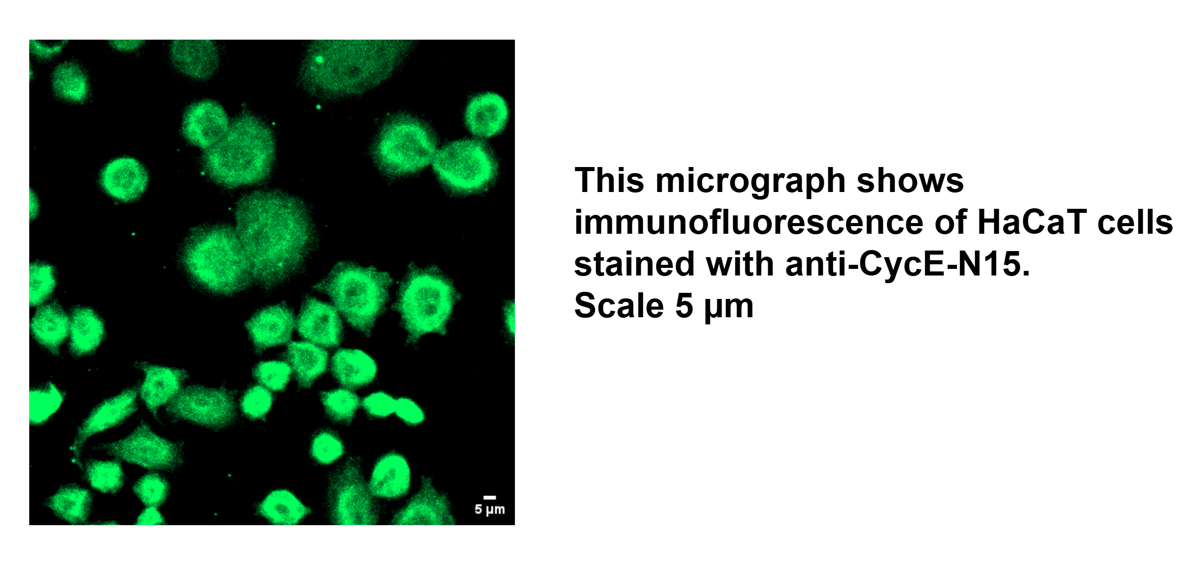
Click image to see more details
IF analysis of CCNE1 using anti-CCNE1 antibody (DZ41310).
CCNE1 was detected in an immunocytochemical section of HaCaT cells. The cells were fixed in 4% paraformaldehyde for 15 minutes at room temperature, permeabilized with 0.1% Triton X-100 for 10 minutes, and blocked with 1% BSA for 1 hour. And then incubated with 1:100-1:200 rabbit anti-CCNE1 Antibody (DZ41310) overnight at 4°C. Goat anti-rabbit IgG conjugated to Alexa Fluor 488 was used as secondary antibody at 1:1000 dilution and incubated for 1-2 hours at RT. The section was counterstained with DAPI. Visualize using Zeiss LSM 900 Confocal Microscope with Airyscan 2 and filter sets appropriate for the label used.
Specific Publications For Anti-Human CCNE1 Antibody (DZ41310)
Loading publications
Recommended Resources
Here are featured tools and databases that you might find useful.
- Boster's Pathways Library
- Protein Databases
- Bioscience Research Protocol Resources
- Data Processing & Analysis Software
- Photo Editing Software
- Scientific Literature Resources
- Research Paper Management Tools
- Molecular Biology Software
- Primer Design Tools
- Bioinformatics Tools
- Phylogenetic Tree Analysis
Customer Reviews
Have you used Anti-Human CCNE1 Antibody?
Share your experimental results or join a short interview to earn up to $1,000 in product credits or other rewards.
2 Reviews For Anti-Human CCNE1 Antibody
A strong nuclear signal for Cyclin E was observed in proliferating cells by IF. Results were reproducible and matched the expected patterns.
Excellent
| SKU | DZ41310 |
|---|---|
| Application | Immunofluorescence |
| Sample | Human HaCaT cell |
| Sample Processing Description | Cells were fixed in 4% paraformaldehyde for 15 minutes at room temperature, permeabilized with 0.1% Triton X-100 for 10 minutes and blocked in 1% BSA for 1 hour before incubation with the primary antibody. |
| Primary Antibody | Human CCNE1 Antibody |
| Primary Incubation | 1:100-1:200, overnight at 4 ℃ |
| Secondary Antibody | Goat anti-rabbit IgG conjugated to Alexa Fluor 488 |
| Secondary Incubation | 1:1000, 1-2 hours in room temperature |
| Other Reagents used | PBS, 0.1% Triton X-100, 1% BSA, DAPI for nuclear counterstaining |
| Detection | Fluorescence microscopy using AlexaFluor488 (excitation 488 nm, emission 519 nm) using Zeiss LSM 900 Confocal Microscope with Airyscan 2 |
| Results Summary | The CyclinE-N15 antibody (DZ41310) worked fine in IF application using human cell lines. In IF, the nuclear localization of Cyclin E matched the known expression pattern. A fluorescent secondary antibody (Alexa Fluor 488) was used for detection and gave clear signal. Overall, a reliable antibody for D15 transcript Cyclin E detection in human cell systems. |
Kasturi Mitra
Verified customer
Submitted 2025-11-05
Using freshly prepared blocking buffer helped reduce background. Results were reproducible and matched the expected patterns.
Excellent
| SKU | DZ41310 |
|---|---|
| Application | Western Blot |
| Sample | Human HaCaT cell, Human A2780 cell, Human HCT cell |
| Sample Processing Description | Dissection, Homogenization, Sample boiling in 1X Lamelli, SDS-PAGE, Standard Western Blotting |
| Primary Antibody | Human CCNE1 Antibody |
| Primary Incubation | 1:1000, overnight at 4 ℃ |
| Secondary Antibody | Anti-rabbit/mouse IgG horseradish peroxidase-conjugated |
| Secondary Incubation | 1:10000, 1-2 hours in room temperature |
| Detection | Biorad Chemidoc |
| Results Summary | Using freshly prepared blocking buffer helped reduce background. Results were reproducible and matched the expected patterns. |
Kasturi Mitra
Verified customer
Submitted 2025-11-05
Customer Q&As
Have a question?
Find answers in Q&As, reviews.
Can't find your answer?
Submit your question
