Click image to see more details
-

-
-
-
-
+9
Product Info Summary
| SKU: | RP1002 |
|---|---|
| Size: | 100 μg/vial |
| Reactive Species: | Human |
| Host: | Rabbit |
| Application: | IHC, WB |
Customers Who Bought This Also Bought

Product info
Product Name
Anti-IFN gamma Antibody Picoband®
SKU/Catalog Number
RP1002
BA14299 is an alternative SKU for this antibody, used in previous lots.
Size
100 μg/vial
Form
Lyophilized
Description
Boster Bio Anti-IFN gamma Antibody catalog # RP1002. Tested in IHC, WB applications. This antibody reacts with Human. The brand Picoband indicates this is a premium antibody that guarantees superior quality, high affinity, and strong signals with minimal background in Western blot applications. Only our best-performing antibodies are designated as Picoband, ensuring unmatched performance.
Storage & Handling
Store at -20˚C for one year from date of receipt. After reconstitution, at 4˚C for one month. It can also be aliquotted and stored frozen at -20˚C for six months. Avoid repeated freeze-thaw cycles.
Cite This Product
Anti-IFN gamma Antibody Picoband® (Boster Biological Technology, Pleasanton CA, USA, Catalog # RP1002)
Host
Rabbit
Contents
Each vial contains 0.9mg NaCl, 0.2mg Na2HPO4, 0.05mg NaN3. Carrier free (No BSA) form available in stock. If you want this antibody carrier free please specify "Carrier Free" or "No BSA" in your order note.
Clonality
Polyclonal
Isotype
Rabbit IgG
Immunogen
E. coli-derived human IFN gamma recombinant protein (Position: Q24-Q166).
Cross-reactivity
No cross-reactivity with other proteins
Reactive Species
RP1002 is reactive to IFNG in Human
Observed Molecular Weight
17 kDa
Calculated molecular weight
19.3 kDa
Background of IFNG
Interferon-gamma (IFN-gamma) is an inflammatory cytokine that has been implicated in the development of fibrosis in inflamed tissues. The production of IFN-gamma, which is under genetic control, can influence the development of fibrosis in lung allografts. IFN-gamma is also produced by natural killer (NK) cells and most prominently by CD8 cytotoxic T cells, and is vital for the control of microbial pathogens. Interferon gamma is believed to be crucial for host defence against many infections. Genetically determined variability in IFN-gamma and expression might be important for the development of tuberculosis. IFN-gamma activates human macrophage oxidative metabolism and antimicrobial activity. In addition to having antiviral activity, IFN-gamma has important immunoregulatory functions. IFN-gamma plays an important role in the control of neointima proliferation.
Antibody Validation
Boster validates all antibodies on WB, IHC, ICC, Immunofluorescence, and ELISA with known positive control and negative samples to ensure specificity and high affinity, including thorough antibody incubations.
Application & Images
Applications
RP1002 is guaranteed for IHC, WB Boster Guarantee
Recommend Dilution
| Application | Dilution | Species |
|---|---|---|
| Immunohistochemistry (Paraffin-embedded Section) | 0.5-1μg/ml | Human |
| ELISA | 0.1-0.5μg/ml | Human, - |
| Western blot | 0.1-0.5μg/ml | Human |
Tested application
Use TE buffer pH 9.0 for antigen retrieval; (*) citrate buffer pH 6.0 is an alternative.
Validation Images & Assay Conditions
Click image to see more details
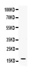
Figure . Western blot analysis of IFN gamma using anti-IFN gamma antibody (RP1002).
Electrophoresis was performed on a 5-20% SDS-PAGE gel at 70V (Stacking gel) / 90V (Resolving gel) for 2-3 hours. The sample well of each lane was loaded with 50ug of sample under reducing conditions.
Lane: Recombinant Human IFN gamma Protein 0.5ng
After Electrophoresis, proteins were transferred to a Nitrocellulose membrane at 150mA for 50-90 minutes. Blocked the membrane with 5% Non-fat Milk/ TBS for 1.5 hour at RT. The membrane was incubated with rabbit anti-IFN gamma antigen affinity purified polyclonal antibody (Catalog # RP1002) at 0.5 μg/mL overnight at 4°C, then washed with TBS-0.1%Tween 3 times with 5 minutes each and probed with a goat anti-rabbit IgG-HRP secondary antibody at a dilution of 1:10000 for 1.5 hour at RT. The signal is developed using an Enhanced Chemiluminescent detection (ECL) kit (Catalog # EK1002) with Tanon 5200 system. A specific band was detected for IFN gamma at approximately 17KD. The expected band size for IFN gamma is at 17KD.

Click image to see more details
IHC analysis of IFN gamma using anti-IFN gamma antibody (RP1002).
IFN gamma was detected in a paraffin-embedded section of human lung cancer tissue. Heat mediated antigen retrieval was performed in EDTA buffer (pH 8.0, epitope retrieval solution). The tissue section was blocked with 10% goat serum. The tissue section was then incubated with 1 μg/ml rabbit anti-IFN gamma Antibody (RP1002) overnight at 4°C. Peroxidase Conjugated Goat Anti-rabbit IgG was used as secondary antibody and incubated for 30 minutes at 37°C. The tissue section was developed using HRP Conjugated Rabbit IgG Super Vision Assay Kit (Catalog # SV0002) with DAB as the chromogen.

Click image to see more details
ROC curve analysis for evaluating the sensitivity and specificity of the IP-10 level, IFN-γ/IL-4 ratio, or their combination to predict significant fibrosis among CHB patients. ( a ) ROC curve analysis for serum IP-10 (with the cut-of f value of 300 pg/mL), the serum IFN-γ/IL-4 ratio (with the cut off value of 1.8), and the combination of IP-10 and the IFN-γ/IL-4 ratio; ( b ) Specificity and sensitivity for IP-10, the IFN-γ/IL-4 ratio, and their combination to predict significant liver fibrosis among patients with CHB.
Index in PubMed under a CC BY license. PMID: 28067328

Click image to see more details
Intrahepatic mRNA levels of IP-10, IFN-γ, and IL-4 in chronic hepatitis B patients with or without fibrosis. Real-time qRT-PCR was conducted to quantify the mRNA levels of intrahepatic IP-10, IFN-γ, and IL-4 in the CHB patients without or with fibrosis as described in the Materials and Methods section. The relative mRNA levels of intrahepatic IP-10, IFN-γ, and IL-4 were calculated by comparative Ct analysis after normalization for the quantity of GAPDH in the same samples and were represented as 2 - △ △ Ct values for controls (the F0 group), which were set equal 1. ♦♦♦ Differs from controls (the F0 group), P < 0.05; ★ ★ ★ differs from mild or minimal fibrosis (the F1–2 group), P < 0.05; ▴ ▴ ▴ differs from moderate fibrosis (the F3–4 group), P < 0.05. (a) IP-10; (b) IFN-γ; (c) IL-4.
Index in PubMed under a CC BY license. PMID: 28067328

Click image to see more details
Statistical analysis of the correlation between the serum IP-10 level or the IFN-γ/IL-4 ratio with liver fibrosis among chronic hepatitis B patients. Spearman’s correlation analysis of the association between ( a ) IP-10; ( b ) IFN-γ; ( c ) the IFN-γ/IL-4 ratio and TGF-β1. Spearman’s correlation analysis of the association between serum ( d ) IFN-γ; ( e ) IL-4; ( f ) the IFN-γ/IL-4 ratio and IP-10.
Index in PubMed under a CC BY license. PMID: 28067328

Click image to see more details
Serum Levels of IFN-γ, TNF-α, perforin and granzyme B in different clinical stages of chronic HBV-infected patients. HCs, healthy controls; IT, chronic HBV carriers; IA, CHB patients; IH, HBV-ACLF. Nemenyi test following Kruskal-Wallis H test were used for comparing IFN-γ, TNF-α, perforin and granzyme B levels between two compared groups. Compared with HCs group, ◆ ◆ ◆ P < 0.01, ◆ ◆ P < 0.05; Compared with IT group, ★ ★ ★ P < 0.01, ★ ★ P < 0.05; Compared with IA group, ▲▲▲ P < 0.01.
Index in PubMed under a CC BY license. PMID: 28273905

Click image to see more details
Levels of TNF-α ( a ) and IFN-γ ( b ) in the supernatants with or without co-cultured NK cells. Student-Newman-Keuls q test following one-way ANOVA were used for comparing IFN-γ, TNF-α, perforin and granzyme B levels between two compared groups. Compare with HepG2 cells group, ◆ ◆ ◆ P < 0.01; Compare with HBV-HepG2 cells group, ◇ ◇ ◇ P < 0.01; Compare with NK cells group, ★ ★ ★ P < 0.01. Levels of perforin ( c ) and granzyme B ( d ) in the supernatants in the presence IFN-α with or without NKG2D blocking. Compare with control group, ▲▲▲ P < 0.01; Compare with + IFN-α group, ■■■ P < 0.01.
Index in PubMed under a CC BY license. PMID: 28273905

Click image to see more details
Analysis of NKG2D and IFN-γ mRNA levels in co-cultured cells (NK + HepG2/HBV-HepG2) of Group A ( a , NK + HepG2) and Group B ( b , NK + HBV-HepG2). Nemenyi test following Kruskal-Wallis H test were used for comparing mRNA expressions of NKG2D and IFN-γ between two compared groups. Compared with Control group (NK + HepG2 or NK + HBV-HepG2), ◆ ◆ ◆ P < 0.01; Compared with Control + NKG2D mAb group, ★ ★ ★ P < 0.01; Compared with Control + IFN-α group, ▲▲▲ P < 0.01; Analysis of the levels of NKG2D and IFN-γ protein in different groups ( c ). The density of NKG2D and IFN-γ protein was the highest in group of NK + HBV-HepG2 + IFN-α, followed by group of NK + HBV-HepG2 + IFN-α + NKG2DmAb and group of NK + HBV-HepG2 + NKG2DmAb.
Index in PubMed under a CC BY license. PMID: 28273905

Click image to see more details
Representative graphs of intrahepatic IFN-γ + cells (A, 200×) and NKG2D + cells (B, 200×) expressions. ( a ) HCs, healthy controls, ( b ) IT, chronic HBV carriers, ( c ) IA, CHB patients, ( d ) IH, HBV-ACLF patients. ( e ) Collective analysis of results from all 4 groups. IFN-γ + cells were distributed mainly in the inflammatory sites and periportal areas that were infiltrated with lymphocytes. NKG2D + cells were mainly distributed in Disse’s space of hepatic lobule in HCs and chronic HBV carriers, and mainly in periportal areas in CHB and HBV-ACLF group. Nemenyi test following Kruskal-Wallis H test were used for comparing intrahepatic IFN-γ + and NKG2D + cells expressions between two groups. Compared with HCs group, ◆ ◆ ◆ P < 0.01; Compared with IT group, ★ ★ ★ P < 0.01; Compared with IA group, ▲▲▲ P < 0.01.
Index in PubMed under a CC BY license. PMID: 28273905

Click image to see more details
Intrahepatic expression of NKG2D mRNA ( a ) and IFN-γ mRNA ( b ). HCs, healthy controls (the relative expression were defined as 1.00); IT, chronic HBV carriers; IA, CHB patients; IH, HBV-ACLF. Nemenyi test following Kruskal-Wallis H test were used for comparing mRNA expressions of NKG2D and IFN-γ between two groups. Compared with HCs group, ◆ ◆ ◆ P < 0.01; Compared with IT group, ★ ★ ★ P < 0.01; Compared with IA group, ▲▲▲ P < 0.01.
Index in PubMed under a CC BY license. PMID: 28273905

Click image to see more details
Percentages of NK (CD3 − CD56 + ) cells ( a – d , m ) in PBMC, NK cell group 2D receptor (NKG2D) + ( e – h , n ) and IFN-γ + ( i – l , o ) NK cells within total NK cells. HCs, healthy controls; IT, chronic HBV carriers; IA, CHB patients; IH, HBV-ACLF patients. Student-Newman-Keuls q test following one-way ANOVA were used for comparing percentages of NK cells, and Nemenyi test following Kruskal-Wallis H test were used for comparing percentages of NKG2D + and IFN-γ + NK cells between two groups. Compared with HCs group, ◆ ◆ ◆ P < 0.01; Compared with IT group, ★ ★ ★ P < 0.01, ★ ★ P < 0.05; Compared with IA group, ▲▲▲ P < 0.01.
Index in PubMed under a CC BY license. PMID: 28273905

Click image to see more details
Intrahepatic protein expression of IP-10, IFN-γ, IL-4, TGF-β1, and α-SMA as well as the IFN-γ/IL-4 ratio in chronic hepatitis B patients with or without fibrosis. The protein expression of intrahepatic (a, b, c, and d) IP-10, (e, f, g, and h) IFN-γ, and (i, g, k, and l) IL-4. In addition, the protein levels of intrahepatic IP-10, IFN-γ, IL-4, TGF-β1, and α-SMA were quantified based on the value of integrated optical density (IOD) and represented as histograms, from which the IFN-γ/IL-4 ratio was calculated. ♦♦♦ Differs from controls (the F0 group), P < 0.05; ★ ★ ★ differs from mild or minimal fibrosis (the F1–2 group), P < 0.05; ▴ ▴ ▴ differs from moderate fibrosis (the F3–4 group), P < 0.05.
Index in PubMed under a CC BY license. PMID: 28067328

Click image to see more details
Serum levels of IP-10, IFN-γ, IL-4, and TGF-β1 as well as the IFN-γ/IL-4 ratio in chronic hepatitis B patients with or without fibrosis. The levels of serum IP-10, IFN-γ, IL-4, and TGF-β1 in CHB patients with or without liver fibrosis were determined by ELISA, and the IFN-γ/IL-4 ratio was calculated. ♦♦♦ Differs from controls (the F0 group), P < 0.05; ★ ★ ★ differs from mild or minimal fibrosis (the F1–2 group), P < 0.05; ▴ ▴ ▴ differs from moderate fibrosis (the F3–4 group), P < 0.05. ( a ) IP-10; ( b ) IFN-γ; ( c ) IL-4; ( d ) TGF-β1; ( e ) the IFN-γ/IL-4 ratio.
Index in PubMed under a CC BY license. PMID: 28067328
Specific Publications For Anti-IFN gamma Antibody Picoband® (RP1002)
Loading publications
Recommended Resources
Here are featured tools and databases that you might find useful.
- Boster's Pathways Library
- Protein Databases
- Bioscience Research Protocol Resources
- Data Processing & Analysis Software
- Photo Editing Software
- Scientific Literature Resources
- Research Paper Management Tools
- Molecular Biology Software
- Primer Design Tools
- Bioinformatics Tools
- Phylogenetic Tree Analysis
Customer Reviews
Have you used Anti-IFN gamma Antibody Picoband®?
Share your experimental results or join a short interview to earn up to $1,000 in product credits or other rewards.
0 Reviews For Anti-IFN gamma Antibody Picoband®
Customer Q&As
Have a question?
Find answers in Q&As, reviews.
Can't find your answer?
Submit your question
4 Customer Q&As for Anti-IFN gamma Antibody Picoband®
Question
My team were well pleased with the WB result of your anti-IFN gamma antibody. However we have seen positive staining in leukocyte secreted. using this antibody. Is that expected? Could you tell me where is IFNG supposed to be expressed?
Verified Customer
Verified customer
Asked: 2019-06-11
Answer
According to literature, leukocyte does express IFNG. Generally IFNG expresses in secreted. Regarding which tissues have IFNG expression, here are a few articles citing expression in various tissues:
Blood, Pubmed ID: 15489334
Boster Scientific Support
Answered: 2019-06-11
Question
We have seen staining in human blood. What should we do? Is anti-IFN gamma antibody supposed to stain blood positively?
R. Jha
Verified customer
Asked: 2017-03-08
Answer
From what I have seen in literature blood does express IFNG. From what I have seen in Uniprot.org, IFNG is expressed in leukocyte, blood, among other tissues. Regarding which tissues have IFNG expression, here are a few articles citing expression in various tissues:
Blood, Pubmed ID: 15489334
Boster Scientific Support
Answered: 2017-03-08
Question
We are currently using anti-IFN gamma antibody RP1002 for human tissue, and we are happy with the IHC results. The species of reactivity given in the datasheet says human. Is it possible that the antibody can work on pig tissues as well?
S. Krishna
Verified customer
Asked: 2016-06-17
Answer
The anti-IFN gamma antibody (RP1002) has not been validated for cross reactivity specifically with pig tissues, though there is a good chance of cross reactivity. We have an innovator award program that if you test this antibody and show it works in pig you can get your next antibody for free. Please contact me if I can help you with anything.
Boster Scientific Support
Answered: 2016-06-17
Question
We purchased anti-IFN gamma antibody for WB on leukocyte in a previous experiment. I am using human, and We want to use the antibody for IHC next. I would like examining leukocyte as well as blood in our next experiment. Do you have any suggestion on which antibody would work the best for IHC?
S. Jones
Verified customer
Asked: 2013-07-15
Answer
I looked at the website and datasheets of our anti-IFN gamma antibody and it appears that RP1002 has been validated on human in both WB and IHC. Thus RP1002 should work for your application. Our Boster satisfaction guarantee will cover this product for IHC in human even if the specific tissue type has not been validated. We do have a comprehensive range of products for IHC detection and you can check out our website bosterbio.com to find out more information about them.
Boster Scientific Support
Answered: 2013-07-15
