
Click image to see more details
Product Info Summary
| SKU: | RP1012 |
|---|---|
| Size: | 100 μg/vial |
| Reactive Species: | Human |
| Host: | Rabbit |
| Application: | WB |
Customers Who Bought This Also Bought

Product info
Product Name
Anti-Interleukin-6 IL6 Antibody Picoband®
SKU/Catalog Number
RP1012
Size
100 μg/vial
Form
Lyophilized
Description
Boster Bio Anti-Interleukin-6 IL6 Antibody catalog # RP1012. Tested in WB applications. This antibody reacts with Human. The brand Picoband indicates this is a premium antibody that guarantees superior quality, high affinity, and strong signals with minimal background in Western blot applications. Only our best-performing antibodies are designated as Picoband, ensuring unmatched performance.
Storage & Handling
Store at -20˚C for one year from date of receipt. After reconstitution, at 4˚C for one month. It can also be aliquotted and stored frozen at -20˚C for six months. Avoid repeated freeze-thaw cycles.
Cite This Product
Anti-Interleukin-6 IL6 Antibody Picoband® (Boster Biological Technology, Pleasanton CA, USA, Catalog # RP1012)
Host
Rabbit
Contents
Each vial contains 0.9mg NaCl, 0.2mg Na2HPO4, 0.05mg NaN3. Carrier free (No BSA) form available in stock. If you want this antibody carrier free please specify "Carrier Free" or "No BSA" in your order note.
Clonality
Polyclonal
Isotype
Rabbit IgG
Immunogen
E. coli-derived human IL-6 recombinant protein (Position: P29-M212).
Cross-reactivity
No cross-reactivity with other proteins
Reactive Species
RP1012 is reactive to IL6 in Human
Observed Molecular Weight
20 kDa
Calculated molecular weight
23.7 kDa
Background of IL6
Interleukin-6 (IL-6) is a protein that in humans is encoded by the IL6 gene. IL-6 is an interleukin that acts as both a pro-inflammatory and anti-inflammatory cytokine. It is secreted by T cells and macrophages to stimulate immune response to trauma, especially burns or other tissue damage leading to inflammation. IL-6 is one of the most important mediators of fever and of the acute phase response. IL-6 is also essential for hybridoma growth and is found in many supplemental cloning media such as briclone. Bowcock et al. (1988) assigned the IL6 gene to chromosome 7p21. By in situ hybridization and Southern blot analysis of mouse-human hybrid cell lines, Sutherland et al. (1988) mapped the IL-6 gene to chromosome 7p15.
Antibody Validation
Boster validates all antibodies on WB, IHC, ICC, Immunofluorescence, and ELISA with known positive control and negative samples to ensure specificity and high affinity, including thorough antibody incubations.
Application & Images
Applications
RP1012 is guaranteed for WB Boster Guarantee
Recommend Dilution
| Application | Dilution | Species |
|---|---|---|
| Western blot | 0.1-0.5μg/ml | Human |
Validation Images & Assay Conditions
Click image to see more details
Western blot analysis of IL-6 using anti-IL-6 antibody (RP1012).
Electrophoresis was performed on a 5-20% SDS-PAGE gel at 70V (Stacking gel) / 90V (Resolving gel) for 2-3 hours.
Lane 1: recombinant human IL6 protein 10 ng,
Lane 2: recombinant human IL6 protein 5 ng.
After electrophoresis, proteins were transferred to a nitrocellulose membrane at 150 mA for 50-90 minutes. Blocked the membrane with 5% non-fat milk/TBS for 1.5 hour at RT. The membrane was incubated with rabbit anti-IL-6 antigen affinity purified polyclonal antibody (Catalog # RP1012) at 0.5 μg/mL overnight at 4°C, then washed with TBS-0.1%Tween 3 times with 5 minutes each and probed with a goat anti-rabbit IgG-HRP secondary antibody at a dilution of 1:5000 for 1.5 hour at RT. The signal is developed using an Enhanced Chemiluminescent detection (ECL) kit (Catalog # EK1002) with Tanon 5200 system.

Click image to see more details
Real-time PCR analysis of upregulated or downregulated gene expression in response to HIF-1alpha (A) Aliquots of the same RNA preparations used for microarray hybridization were analyzed by quantitative real-time PCR . In three pairwise comparisons, the upregulation-folds of IGFBP5, IRS4, TNFAIP6, SOCS1, IL-6, VEGF-A mRNA expression were calculated. The mean and standard error are shown (p < 0.05). (B) Aliquots of the same RNA preparations used for microarray hybridization were analyzed by quantitative real-time PCR. In three pairwise comparisons, the downregulation-folds of IGFBP3, ZNF569, SOCS2, SIRPa and XRCC4 mRNA were calculated. The mean and standard error are shown (p < 0.05).
Index in PubMed under a CC BY license. PMID: 20003295

Click image to see more details
Western blot analysis of regulation of protein expression by HIF-1alpha in NCI-H446 cells . According to different treatments, all the cells were divided into four groups: control group (the cells cultured under normoxic conditions of 20% O2), Ad5-HIF-1alpha transfection group, hypoxia group (the cells cultured under normoxic conditions of 1% O2) and Ad5-siHIF-1alpha transfection group (after transfection, the cells were cultured under normoxic conditions of 1% O2). (A) Western blot analysis for IGFBP5 protein expressed by the cells of four groups. (B) Western blot analysis for SOCS1 protein expressed by the cells of four groups. (C) Densitometric analysis of the IGFBP5 and SOCS1 bands compared to the corresponding β-actin bands (*p < 0.05 expression of IGFBP5 or SOCS1 protein in Ad5-HIF-1alpha group vs. control group; ** p < 0.05 expression of IGFBP5 or SOCS1 protein in hypoxia group vs. control group; *** p < 0.05 expression of IGFBP5 or SOCS1 protein in Ad5-siHIF-1alpha group vs. control group). (D) Western blot analysis for IL-6 protein expressed by the cells of four groups. (E) Western blot analysis for STAT3 protein expressed by the cells of four groups. (F) Densitometric analysis of the IL-6 and STAT3 bands compared to the corresponding β-actin bands (*p < 0.05 expression of IL-6 or STAT3 protein in Ad5-HIF-1alpha group vs. Ad5-siHIF-1alpha group group.)
Index in PubMed under a CC BY license. PMID: 20003295

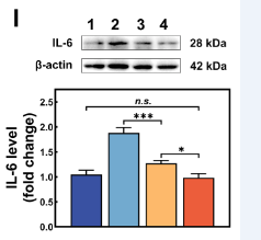
Click image to see more details
Western blot analysis of IL-6 using anti-IL-6 antibody (RP1012).
Electrophoresis was performed on a 5-20% SDS-PAGE gel at 70V (Stacking gel) / 90V (Resolving gel) for 2-3 hours.
Lane 1-4: human HK-2 whole cell lysates.
After electrophoresis, proteins were transferred to a nitrocellulose membrane at 150 mA for 50-90 minutes. Blocked the membrane with 5% non-fat milk/TBS for 1.5 hour at RT. The membrane was incubated with rabbit anti-IL-6 antigen affinity purified polyclonal antibody (Catalog # RP1012) at 1:1000 overnight at 4°C, then washed with TBS-0.1%Tween 3 times with 5 minutes each and probed with a goat anti-rabbit IgG-HRP secondary antibody for 1 hour at RT. The signal is developed using an Enhanced Chemiluminescent detection (ECL) kit (Catalog # EK1002) with Tanon 5200 system.
Specific Publications For Anti-Interleukin-6 IL6 Antibody Picoband® (RP1012)
Loading publications
Recommended Resources
Here are featured tools and databases that you might find useful.
- Boster's Pathways Library
- Protein Databases
- Bioscience Research Protocol Resources
- Data Processing & Analysis Software
- Photo Editing Software
- Scientific Literature Resources
- Research Paper Management Tools
- Molecular Biology Software
- Primer Design Tools
- Bioinformatics Tools
- Phylogenetic Tree Analysis
Customer Reviews
Have you used Anti-Interleukin-6 IL6 Antibody Picoband®?
Share your experimental results or join a short interview to earn up to $1,000 in product credits or other rewards.
1 Reviews For Anti-Interleukin-6 IL6 Antibody Picoband®
Using Boster’s IL-6 antibody (RP1012) in WB on HK-2 cells produced clear, specific bands with high sensitivity and cost-effectiveness, outperforming previously tested antibodies.
Excellent
| SKU | RP1012 |
|---|---|
| Application | Western Blot |
| Sample | human HK-2 cells |
| Sample Processing Description | Cell samples were directly lysed in RIPA buffer, mixed with loading buffer at the appropriate ratio, and denatured at 98 °C. Twenty microliters of each protein sample were loaded per lane onto SDS-PAGE. |
| Other Reagents | 5% non-fat milk |
| Primary Antibody | Interleukin-6 IL6 Antibody Picoband® |
| Primary Incubation | 1:1000, overnight at 4 ℃ |
| Secondary Antibody | HRP Conjugated AffiniPure Goat Anti-rabbit IgG (H+L) |
| Secondary Incubation | 1 h in RT |
| Detection | Substrate: ECL substrate |
| Results Summary | Previously, WB experiments were performed using antibodies from two domestic and international suppliers, which showed poor specificity and were expensive. Subsequently, Boster’s IL-6 antibody (Cat. RP1012) was used, demonstrating high specificity, strong titer, cost-effectiveness, and yielding clear bands. |
Jingwen Zhang, Ocean University of China
Verified customer
Submitted 2026-02-27
Customer Q&As
Have a question?
Find answers in Q&As, reviews.
Can't find your answer?
Submit your question
3 Customer Q&As for Anti-Interleukin-6 IL6 Antibody Picoband®
Question
We are currently using anti-IL6 antibody RP1012 for human tissue, and we are content with the WB results. The species of reactivity given in the datasheet says human. Is it true that the antibody can work on horse tissues as well?
Verified Customer
Verified customer
Asked: 2019-10-28
Answer
The anti-IL6 antibody (RP1012) has not been validated for cross reactivity specifically with horse tissues, but there is a good chance of cross reactivity. We have an innovator award program that if you test this antibody and show it works in horse you can get your next antibody for free. Please contact me if I can help you with anything.
Boster Scientific Support
Answered: 2019-10-28
Question
We have been able to see staining in human lung. Any tips? Is anti-IL6 antibody supposed to stain lung positively?
Verified Customer
Verified customer
Asked: 2018-04-12
Answer
From literature lung does express IL6. From Uniprot.org, IL6 is expressed in left coronary artery, fibroblast, lung, among other tissues. Regarding which tissues have IL6 expression, here are a few articles citing expression in various tissues:
Fibroblast, Pubmed ID: 3758081
Lung, Pubmed ID: 15489334
Boster Scientific Support
Answered: 2018-04-12
Question
My team were happy with the WB result of your anti-IL6 antibody. However we have seen positive staining in fibroblast secreted. using this antibody. Is that expected? Could you tell me where is IL6 supposed to be expressed?
A. Krishna
Verified customer
Asked: 2013-06-25
Answer
Based on literature, fibroblast does express IL6. Generally IL6 expresses in secreted. Regarding which tissues have IL6 expression, here are a few articles citing expression in various tissues:
Fibroblast, Pubmed ID: 3758081
Lung, Pubmed ID: 15489334
Boster Scientific Support
Answered: 2013-06-25
