Click image to see more details
-

-
-
-
-
+6
Product Info Summary
| SKU: | A00736-1 |
|---|---|
| Size: | 100 μg/vial |
| Reactive Species: | Human |
| Host: | Rabbit |
| Application: | ELISA, Flow Cytometry, IF, ICC, WB |
Customers Who Bought This Also Bought

Product info
Product Name
Anti-HuR/ELAVL1 Antibody Picoband®
SKU/Catalog Number
A00736-1
Size
100 μg/vial
Form
Lyophilized
Description
Boster Bio Anti-HuR/ELAVL1 Antibody Picoband® catalog # A00736-1. Tested in WB, ICC, IF, Flow Cytometry, ELISA applications. This antibody reacts with Human. The brand Picoband indicates this is a premium antibody that guarantees superior quality, high affinity, and strong signals with minimal background in Western blot applications. Only our best-performing antibodies are designated as Picoband, ensuring unmatched performance.
Storage & Handling
At -20°C for one year from date of receipt. After reconstitution, at 4°C for one month. It can also be aliquotted and stored frozen at -20°C for six months. Avoid repeated freezing and thawing.
Cite This Product
Anti-HuR/ELAVL1 Antibody Picoband® (Boster Biological Technology, Pleasanton CA, USA, Catalog # A00736-1)
Host
Rabbit
Contents
Each vial contains 4 mg Trehalose, 0.9 mg NaCl, 0.2 mg Na2HPO4.
Clonality
Polyclonal
Immunogen
E.coli-derived human HuR/ELAVL1 recombinant protein (Position: M1-S232).
Reactive Species
A00736-1 is reactive to ELAVL1 in Human
Observed Molecular Weight
36 kDa
Calculated molecular weight
36.1 kDa
Background of ELAVL1
The protein encoded by this gene is a member of the ELAVL family of RNA-binding proteins that contain several RNA recognition motifs, and selectively bind AU-rich elements (AREs) found in the 3' untranslated regions of mRNAs. AREs signal degradation of mRNAs as a means to regulate gene expression, thus by binding AREs, the ELAVL family of proteins play a role in stabilizing ARE-containing mRNAs. This gene has been implicated in a variety of biological processes and has been linked to a number of diseases, including cancer. It is highly expressed in many cancers, and could be potentially useful in cancer diagnosis, prognosis, and therapy.
Antibody Validation
Boster validates all antibodies on WB, IHC, ICC, Immunofluorescence, and ELISA with known positive control and negative samples to ensure specificity and high affinity, including thorough antibody incubations.
Application & Images
Applications
A00736-1 is guaranteed for ELISA, Flow Cytometry, IF, ICC, WB Boster Guarantee
Recommend Dilution
| Application | Dilution | Species |
|---|---|---|
| Western blot | 0.25-0.5 μg/ml | Human |
| Immunocytochemistry/Immunofluorescence | 5 μg/ml | Human |
| Flow Cytometry (Fixed) | 1-3 μg/1x106 cells | Human |
| ELISA | 0.1-0.5 μg/ml |
Validation Images & Assay Conditions
Click image to see more details
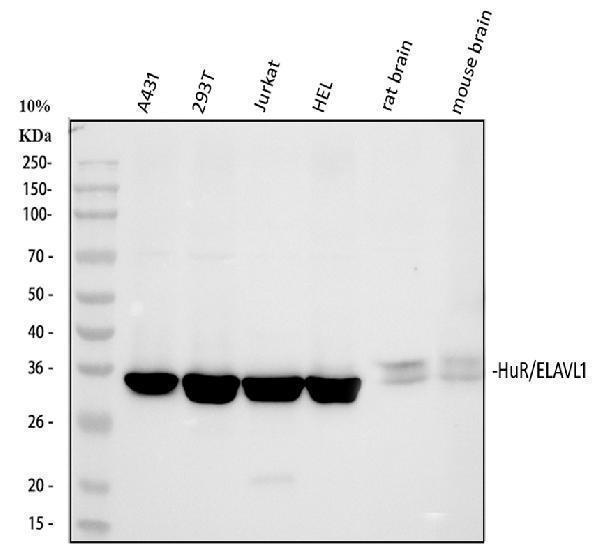
Western blot analysis of HuR/ELAVL1 using anti-HuR/ELAVL1 antibody (A00736-1).
Electrophoresis was performed on a 8% SDS-PAGE gel at 80V (Stacking gel) / 120V (Resolving gel) for 2 hours. The sample well of each lane was loaded with 30 ug of sample under reducing conditions.
Lane 1: human A431 whole cell lysates,
Lane 2: human 293T whole cell lysates,
Lane 3: human Jurkat whole cell lysates,
Lane 4: human HEL whole cell lysates,
Lane 5: rat brain tissue lysates,
Lane 6: mouse brain tissue lysates.
After electrophoresis, proteins were transferred to a nitrocellulose membrane at 150 mA for 50-90 minutes. Blocked the membrane with 5% non-fat milk/TBS for 1.5 hour at RT. The membrane was incubated with rabbit anti-HuR/ELAVL1 antigen affinity purified polyclonal antibody (A00736-1) at 1:1000 overnight at 4°C, then washed with TBS-0.1%Tween 3 times with 5 minutes each and probed with a goat anti-rabbit IgG-HRP secondary antibody at a dilution of 1:5000 for 1.5 hour at RT. The signal is developed using an ECL Plus Western Blotting Substrate (Catalog # AR1196-200) with Tanon 5200 system. A specific band was detected for HuR/ELAVL1 at approximately 36 kDa. The expected band size for HuR/ELAVL1 is at 36 kDa.

Click image to see more details
IHC analysis of HuR/ELAVL1 using anti-HuR/ELAVL1 antibody (A00736-1).
HuR/ELAVL1 was detected in a paraffin-embedded section of human breast cancer tissue. Heat mediated antigen retrieval was performed in EDTA buffer (pH 8.0, epitope retrieval solution). The tissue section was blocked with 10% goat serum. The tissue section was then incubated with 1:100 rabbit anti-HuR/ELAVL1 Antibody (A00736-1) overnight at 4°C. Peroxidase Conjugated Goat Anti-rabbit IgG was used as secondary antibody and incubated for 30 minutes at 37°C. The tissue section was developed using HRP Conjugated Rabbit IgG Super Vision Assay Kit (Catalog # SV0002) with DAB as the chromogen.

Click image to see more details
IHC analysis of HuR/ELAVL1 using anti-HuR/ELAVL1 antibody (A00736-1).
HuR/ELAVL1 was detected in a paraffin-embedded section of human ovarian cancer tissue. Heat mediated antigen retrieval was performed in EDTA buffer (pH 8.0, epitope retrieval solution). The tissue section was blocked with 10% goat serum. The tissue section was then incubated with 1:100 rabbit anti-HuR/ELAVL1 Antibody (A00736-1) overnight at 4°C. Peroxidase Conjugated Goat Anti-rabbit IgG was used as secondary antibody and incubated for 30 minutes at 37°C. The tissue section was developed using HRP Conjugated Rabbit IgG Super Vision Assay Kit (Catalog # SV0002) with DAB as the chromogen.

Click image to see more details
IHC analysis of HuR/ELAVL1 using anti-HuR/ELAVL1 antibody (A00736-1).
HuR/ELAVL1 was detected in a paraffin-embedded section of mouse brain tissue. Heat mediated antigen retrieval was performed in EDTA buffer (pH 8.0, epitope retrieval solution). The tissue section was blocked with 10% goat serum. The tissue section was then incubated with 1:100 rabbit anti-HuR/ELAVL1 Antibody (A00736-1) overnight at 4°C. Peroxidase Conjugated Goat Anti-rabbit IgG was used as secondary antibody and incubated for 30 minutes at 37°C. The tissue section was developed using HRP Conjugated Rabbit IgG Super Vision Assay Kit (Catalog # SV0002) with DAB as the chromogen.

Click image to see more details
IHC analysis of HuR/ELAVL1 using anti-HuR/ELAVL1 antibody (A00736-1).
HuR/ELAVL1 was detected in a paraffin-embedded section of rat brain tissue. Heat mediated antigen retrieval was performed in EDTA buffer (pH 8.0, epitope retrieval solution). The tissue section was blocked with 10% goat serum. The tissue section was then incubated with 1:100 rabbit anti-HuR/ELAVL1 Antibody (A00736-1) overnight at 4°C. Peroxidase Conjugated Goat Anti-rabbit IgG was used as secondary antibody and incubated for 30 minutes at 37°C. The tissue section was developed using HRP Conjugated Rabbit IgG Super Vision Assay Kit (Catalog # SV0002) with DAB as the chromogen.

Click image to see more details
Western blot analysis of HuR/ELAVL1 using anti-HuR/ELAVL1 antibody (A00736-1).
Electrophoresis was performed on a 10% SDS-PAGE gel at 80V (Stacking gel) / 120V (Resolving gel) for 2 hours. The sample well of each lane was loaded with 30 ug of sample under reducing conditions.
Lane 1: human A431 whole cell lysates,
Lane 2: human 293T whole cell lysates,
Lane 3: human Jurkat whole cell lysates,
Lane 4: human Hela whole cell lysates.
After electrophoresis, proteins were transferred to a nitrocellulose membrane at 150 mA for 50-90 minutes. Blocked the membrane with 5% non-fat milk/TBS for 1.5 hour at RT. The membrane was incubated with rabbit anti-HuR/ELAVL1 antigen affinity purified polyclonal antibody (A00736-1) at 0.5 μg/mL overnight at 4°C, then washed with TBS-0.1%Tween 3 times with 5 minutes each and probed with a goat anti-rabbit IgG-HRP secondary antibody at a dilution of 1:5000 for 1.5 hour at RT. The signal is developed using an ECL Plus Western Blotting Substrate (Catalog # AR1196-200) with Tanon 5200 system. A specific band was detected for HuR/ELAVL1 at approximately 36 kDa. The expected band size for HuR/ELAVL1 is at 36 kDa.

Click image to see more details
IF analysis of HuR/ELAVL1 using anti-HuR/ELAVL1 antibody (A00736-1) and anti-Alpha Tubulin antibody (M03989-3).
HuR/ELAVL1 was detected in an immunocytochemical section of Hela cells. Enzyme antigen retrieval was performed using IHC enzyme antigen retrieval reagent (AR0022) for 15 mins. The cells were blocked with 10% goat serum. And then incubated with 5 μg/mL rabbit anti-HuR/ELAVL1 Antibody (A00736-1) and mouse anti-Alpha Tubulin antibody (M03989-3) overnight at 4°C. Fluoro488 Conjugated Goat Anti-Rabbit IgG (BA1127) and Cy3 Conjugated Goat Anti-Mouse IgG (BA1031) were used as secondary antibody at 1:500 dilution and incubated for 30 minutes at 37°C. Visualize using a fluorescence microscope and filter sets appropriate for the label used.

Click image to see more details
Flow Cytometry analysis of A431 cells using anti-HuR/ELAVL1 antibody (A00736-1).
Overlay histogram showing A431 cells stained with A00736-1 (Blue line). To facilitate intracellular staining, cells were fixed with 4% paraformaldehyde and permeabilized with permeabilization buffer. The cells were blocked with 10% normal goat serum. And then incubated with rabbit anti-HuR/ELAVL1 Antibody (A00736-1, 1 μg/1x106 cells) for 30 min at 20°C. Fluoro488 conjugated goat anti-rabbit IgG (BA1127, 5-10 μg/1x106 cells) was used as secondary antibody for 30 minutes at 20°C. Isotype control antibody (Green line) was rabbit IgG (1 μg/1x106) used under the same conditions. Unlabelled sample without incubation with primary antibody and secondary antibody (Red line) was used as a blank control.

Click image to see more details
Flow Cytometry analysis of Jurkat cells using anti-HuR/ELAVL1 antibody (A00736-1).
Overlay histogram showing Jurkat cells stained with A00736-1 (Blue line). To facilitate intracellular staining, cells were fixed with 4% paraformaldehyde and permeabilized with permeabilization buffer. The cells were blocked with 10% normal goat serum. And then incubated with rabbit anti-HuR/ELAVL1 Antibody (A00736-1, 1 μg/1x106 cells) for 30 min at 20°C. Fluoro488 conjugated goat anti-rabbit IgG (BA1127, 5-10 μg/1x106 cells) was used as secondary antibody for 30 minutes at 20°C. Isotype control antibody (Green line) was rabbit IgG (1 μg/1x106) used under the same conditions. Unlabelled sample without incubation with primary antibody and secondary antibody (Red line) was used as a blank control.

Click image to see more details
Western blot analysis of HuR/ELAVL1 using anti-HuR/ELAVL1 antibody (A00736-1).
Electrophoresis was performed on a 8% SDS-PAGE gel at 80V (Stacking gel) / 120V (Resolving gel) for 2 hours. The sample well of each lane was loaded with 30 ug of sample under reducing conditions.
Lane 1: normal rat colon tissue lysates,
Lane 2: normal rat colon tissue lysates,
Lane 3: rat colon cancer model tissue lysates,
Lane 4: rat colon cancer model tissue lysates.
After electrophoresis, proteins were transferred to a nitrocellulose membrane at 150 mA for 50-90 minutes. Blocked the membrane with 5% non-fat milk/TBS for 1.5 hour at RT. The membrane was incubated with rabbit anti-HuR/ELAVL1 antigen affinity purified polyclonal antibody (A00736-1) at 1:2000 overnight at 4°C, then washed with TBS-0.1%Tween 3 times with 5 minutes each and probed with a goat anti-rabbit IgG-HRP secondary antibody at a dilution of 1:10000 for 1 hour at RT. The signal is developed using an ECL Plus Western Blotting Substrate (Catalog # AR1196-200) with ChemiDoc MP system. A specific band was detected for HuR/ELAVL1 at approximately 36-38 kDa. The expected band size for HuR/ELAVL1 is at 36 kDa.
Specific Publications For Anti-HuR/ELAVL1 Antibody Picoband® (A00736-1)
Loading publications
Recommended Resources
Here are featured tools and databases that you might find useful.
- Boster's Pathways Library
- Protein Databases
- Bioscience Research Protocol Resources
- Data Processing & Analysis Software
- Photo Editing Software
- Scientific Literature Resources
- Research Paper Management Tools
- Molecular Biology Software
- Primer Design Tools
- Bioinformatics Tools
- Phylogenetic Tree Analysis
Customer Reviews
Have you used Anti-HuR/ELAVL1 Antibody Picoband®?
Share your experimental results or join a short interview to earn up to $1,000 in product credits or other rewards.
1 Reviews For Anti-HuR/ELAVL1 Antibody Picoband®
The ELAVL1 antibody (A00736-1) produced a clear specific band at the expected size in WB using rat colon and colon cancer tissue samples, consistent with the expected expression pattern.
Excellent

| SKU | A00736-1 |
|---|---|
| Application | Western Blot |
| Sample | HepG2 subcutaneous xenograft in nude mice |
| Sample Processing Description | ① normal rat colon tissue; ② rat colon cancer model tissue; total protein extracted. |
| Other Reagents | RIPA lysis buffer, Protease inhibitor, Electrophoresis buffer, Transfer buffer, Blocking buffer |
| Primary Antibody | HuR/ELAVL1 Antibody Picoband® |
| Primary Incubation | 1:2000, overnight at 4 ℃ |
| Secondary Antibody | HRP-conjugated goat anti-rabbit IgG |
| Secondary Incubation | 1:10000, 30 min in 37℃ |
| Detection | Substrate: ECL substrate; Image system: ChemiDoc MP |
| Results Summary | HUR protein is an RNA-binding protein that plays multiple key roles in gene expression regulation. It is essential in maintaining cellular homeostasis, stress response, inflammation, and the development and progression of diseases, especially cancer. In normal tissues, it is expressed at moderate levels, maintaining a delicate balance of cell proliferation, stress response, inflammation, and neural functions, while in tumors it is highly overexpressed, driving malignant progression. The experimental results are consistent with these observations. |
Xilinguli, Xinjiang Medical University
Verified customer
Submitted 2026-04-22
Customer Q&As
Have a question?
Find answers in Q&As, reviews.
Can't find your answer?
Submit your question
