Click image to see more details
-

-
-
-
-
+4
Product Info Summary
| SKU: | A01394 |
|---|---|
| Size: | 100 μg/vial |
| Reactive Species: | Human, Mouse, Pig, Rat |
| Host: | Rabbit |
| Application: | IHC, WB |
Customers Who Bought This Also Bought

Product info
Product Name
Anti-Iba1/AIF1 Antibody Picoband®
SKU/Catalog Number
A01394
Size
100 μg/vial
Form
Lyophilized
Description
Bio Anti-Iba1/AIF1 Antibody Picoband® catalog # A01394. Tested in IHC, WB applications. This antibody reacts with Human, Mouse, Rat, Pig. The brand Picoband indicates this is a premium antibody that guarantees superior quality, high affinity, and strong signals with minimal background in Western blot applications. Only our best-performing antibodies are designated as Picoband, ensuring unmatched performance.","Target: AIF1/IBA1; function/pathways: actin- and calcium-binding protein induced by cytokines and interferon; promotes macrophage activation and growth of vascular smooth muscle cells and T-lymphocytes; disease association noted: polymorphisms may be linked to systemic sclerosis; assay contexts supported here: IHC (paraffin), WB. Complements lymphoid B-cell marker CD19 for lineage contrast; pairs with apoptosis readouts such as Survivin/BIRC5 depending on study design.
Storage & Handling
Store at -20˚C for one year from date of receipt. After reconstitution, at 4˚C for one month. It can also be aliquotted and stored frozen at -20˚C for six months. Avoid repeated freeze-thaw cycles.
Cite This Product
Anti-Iba1/AIF1 Antibody Picoband® (Boster Biological Technology, Pleasanton CA, USA, Catalog # A01394)
Host
Rabbit
Contents
Each vial contains 4 mg Trehalose, 0.9 mg NaCl and 0.2 mg Na2HPO4.
Clonality
Polyclonal
Isotype
Rabbit IgG
Immunogen
A synthetic peptide corresponding to a sequence at the C-terminus of human Iba1, different from the related mouse sequence by seven amino acids, and from the related rat sequence by six amino acids.
Cross-reactivity
No cross-reactivity with other proteins
Reactive Species
A01394 is reactive to AIF1 in Human, Mouse, Pig, Rat
Observed Molecular Weight
17 kDa
Calculated molecular weight
16.7 kDa
Background of AIF1
Allograft inflammatory factor 1 (AIF-1), also known as ionized calcium-binding adapter molecule 1 (IBA1), is a protein that in humans is encoded by the AIF1 gene. This gene encodes a protein that binds actin and calcium. And this gene is induced by cytokines and interferon and may promote macrophage activation and growth of vascular smooth muscle cells and T-lymphocytes. Polymorphisms in this gene may be associated with systemic sclerosis. Alternative splicing results in multiple transcript variants, but the full-length and coding nature of some of these variants is not certain.
Antibody Validation
Boster validates all antibodies on WB, IHC, ICC, Immunofluorescence, and ELISA with known positive control and negative samples to ensure specificity and high affinity, including thorough antibody incubations.
Application & Images
Applications
A01394 is guaranteed for IHC, WB Boster Guarantee
Recommend Dilution
| Application | Dilution | Species |
|---|---|---|
| Western blot | 0.1-0.5μg/ml | Human |
| Immunohistochemistry (Paraffin-embedded Section) | 2-5μg/ml | Human, Mouse, Rat, Pig |
Tested application
Suggested blocking solution with 5% non-fat milk or BSA; (*)Recommended protein loading: 20-40 µg per lane
Use TE buffer pH 9.0 for antigen retrieval; (*) citrate buffer pH 6.0 is an alternative.
Validation Images & Assay Conditions
Click image to see more details
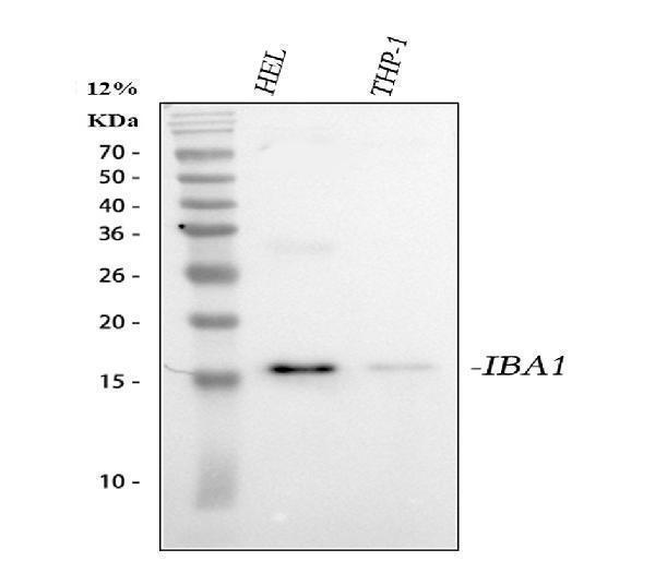
Western blot analysis of Iba1/AIF1 using anti-Iba1/AIF1 antibody (A01394).
Electrophoresis was performed on a 5-20% SDS-PAGE gel at 70V (Stacking gel) / 90V (Resolving gel) for 2-3 hours. The sample well of each lane was loaded with 30 ug of sample under reducing conditions.
Lane 1: human HEL whole cell lysates,
Lane 2: human THP-1 whole cell lysates.
After electrophoresis, proteins were transferred to a nitrocellulose membrane at 150 mA for 50-90 minutes. Blocked the membrane with 5% non-fat milk/TBS for 1.5 hour at RT. The membrane was incubated with rabbit anti-Iba1/AIF1 antigen affinity purified polyclonal antibody (Catalog # A01394) at 0.5 μg/mL overnight at 4°C, then washed with TBS-0.1%Tween 3 times with 5 minutes each and probed with a goat anti-rabbit IgG-HRP secondary antibody at a dilution of 1:5000 for 1.5 hour at RT. The signal is developed using an Enhanced Chemiluminescent detection (ECL) kit (Catalog # EK1002) with Tanon 5200 system. A specific band was detected for Iba1/AIF1 at approximately 17 kDa. The expected band size for Iba1/AIF1 is at 17 kDa.

Click image to see more details
IHC analysis of Iba1/AIF1 using anti-Iba1/AIF1 antibody (A01394).
Iba1/AIF1 was detected in a paraffin-embedded section of human spleen tissue. Heat mediated antigen retrieval was performed in EDTA buffer (pH 8.0, epitope retrieval solution). The tissue section was blocked with 10% goat serum. The tissue section was then incubated with 2 μg/ml rabbit anti-Iba1/AIF1 Antibody (A01394) overnight at 4°C. Peroxidase Conjugated Goat Anti-rabbit IgG was used as secondary antibody and incubated for 30 minutes at 37°C. The tissue section was developed using HRP Conjugated Rabbit IgG Super Vision Assay Kit (Catalog # SV0002) with DAB as the chromogen.

Click image to see more details
IHC analysis of Iba1/AIF1 using anti-Iba1/AIF1 antibody (A01394).
Iba1/AIF1 was detected in a paraffin-embedded section of mouse brain tissue. Heat mediated antigen retrieval was performed in EDTA buffer (pH 8.0, epitope retrieval solution). The tissue section was blocked with 10% goat serum. The tissue section was then incubated with 2 μg/ml rabbit anti-Iba1/AIF1 Antibody (A01394) overnight at 4°C. Peroxidase Conjugated Goat Anti-rabbit IgG was used as secondary antibody and incubated for 30 minutes at 37°C. The tissue section was developed using HRP Conjugated Rabbit IgG Super Vision Assay Kit (Catalog # SV0002) with DAB as the chromogen.

Click image to see more details
IHC analysis of Iba1/AIF1 using anti-Iba1/AIF1 antibody (A01394).
Iba1/AIF1 was detected in a paraffin-embedded section of rat brain tissue. Heat mediated antigen retrieval was performed in EDTA buffer (pH 8.0, epitope retrieval solution). The tissue section was blocked with 10% goat serum. The tissue section was then incubated with 2 μg/ml rabbit anti-Iba1/AIF1 Antibody (A01394) overnight at 4°C. Peroxidase Conjugated Goat Anti-rabbit IgG was used as secondary antibody and incubated for 30 minutes at 37°C. The tissue section was developed using HRP Conjugated Rabbit IgG Super Vision Assay Kit (Catalog # SV0002) with DAB as the chromogen.

Click image to see more details
IHC analysis of Iba1/AIF1 using anti-Iba1/AIF1 antibody (A01394).
Iba1/AIF1 was detected in a paraffin-embedded section of pig brain tissue. Heat mediated antigen retrieval was performed in EDTA buffer (pH 8.0, epitope retrieval solution). The tissue section was blocked with 10% goat serum. The tissue section was then incubated with 2 μg/ml rabbit anti-Iba1/AIF1 Antibody (A01394) overnight at 4°C. Peroxidase Conjugated Goat Anti-rabbit IgG was used as secondary antibody and incubated for 30 minutes at 37°C. The tissue section was developed using HRP Conjugated Rabbit IgG Super Vision Assay Kit (Catalog # SV0002) with DAB as the chromogen.
Specific Publications For Anti-Iba1/AIF1 Antibody Picoband® (A01394)
Loading publications
Recommended Resources
Here are featured tools and databases that you might find useful.
- Boster's Pathways Library
- Protein Databases
- Bioscience Research Protocol Resources
- Data Processing & Analysis Software
- Photo Editing Software
- Scientific Literature Resources
- Research Paper Management Tools
- Molecular Biology Software
- Primer Design Tools
- Bioinformatics Tools
- Phylogenetic Tree Analysis
Customer Reviews
Have you used Anti-Iba1/AIF1 Antibody Picoband®?
Share your experimental results or join a short interview to earn up to $1,000 in product credits or other rewards.
1 Reviews For Anti-Iba1/AIF1 Antibody Picoband®
The antibody worked well even though we used fresh frozen brain sections cut at 20 micron thickness thaw-mounted onto microscope slide that were then post-fixed with 4% paraformaldehyde. The antibody worked equally well when used on fixed brains sectioned
Excellent
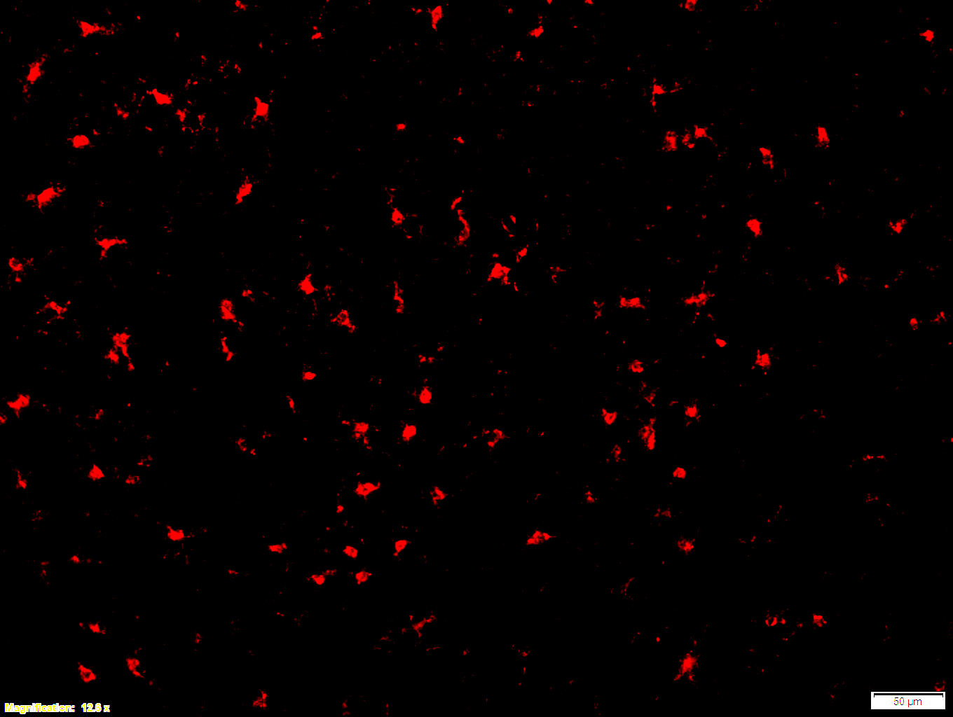
Boster bio Iba1 1:100 PFA (3-4) - cortex 12x - with scale bar
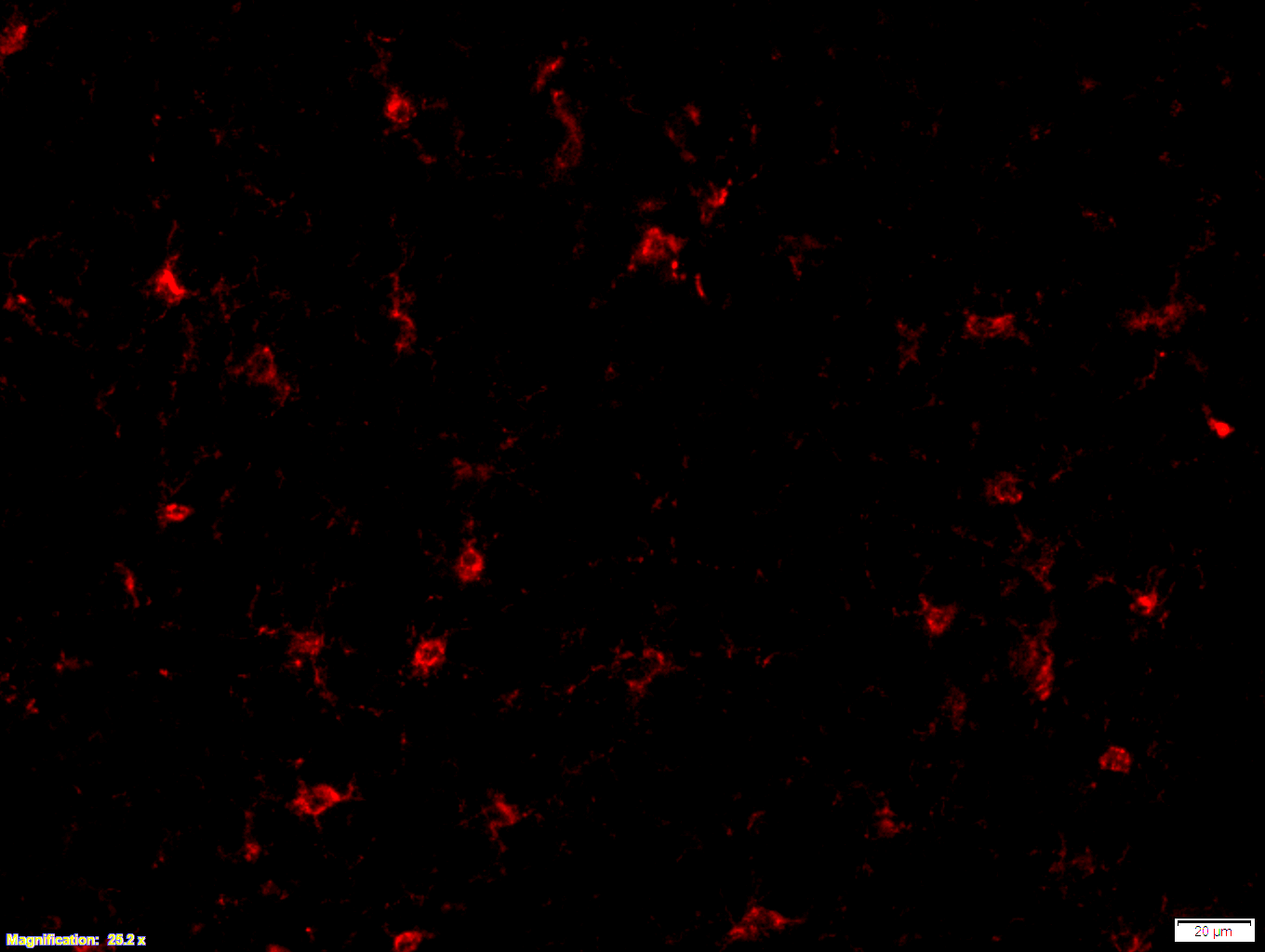
Boster bio Iba1 1:100 PFA (3-4) - cortex 25x - with scale bar
| SKU | A01394 |
|---|---|
| Application | Immunofluorescence |
| Sample | Mouse brain |
| Primary Antibody Dilution | 1:100 |
Images that were made from fresh frozen cryostat sections, were thaw-mounted onto microscope slides. Mounted sections were later post-fixed with 300 µL of 4% paraformaldehyde (PFA) in PBS at room temperature for 10 minutes. After fixation and prior to incubation with antibodies, slides were washed 3 times with 300 µL of 1X PBS with 0.01% sodium azide for 3 minutes per rinse, followed by permeabilization with 300 µL of 0.3% Triton X-100 in 1X PBS with 0.01% sodium azide for 30 minutes at room temperature, then blocking with 700 µL of 4% donkey serum diluted in 1X PBS with 0.01% sodium azide + Triton X-100 (blocking buffer) for 30 minutes at room temperature. Slides were incubated with 325 µL of Iba1 (1:100). Concentrations of 1:100 were achieved by diluting 10 µL of antibody in 1,000 µL of blocking buffer; concentrations of 1:250 were achieved by diluting 4 µL of antibody in 1,000 µL of blocking buffer. Sections were incubated overnight at 4 ˚C. On the next day, slides were washed 3 times with 300 µL of 1X PBS with 0.01% sodium azide for 3 minutes per rinse, then incubated with the secondary antibodies at room temperature, in the dark, for 1 hour. Washing step was repeated then slides were left in the dark to dry. Mounting media was added to cover slip the sections. Slides were kept in the dark at 4 ˚C prior to imaging.
Bob Speth from Nova Southeastern University
Verified customer
Submitted 2024-06-20
Customer Q&As
Have a question?
Find answers in Q&As, reviews.
Can't find your answer?
Submit your question
15 Customer Q&As for Anti-Iba1/AIF1 Antibody Picoband®
Question
Our team were satisfied with the WB result of your anti-Iba1/AIF1 antibody. However we have seen positive staining in lymphocyte cytoskeleton using this antibody. Is that expected? Could you tell me where is AIF1 supposed to be expressed?
A. Moore
Verified customer
Asked: 2020-04-16
Answer
According to literature, lymphocyte does express AIF1. Generally AIF1 expresses in cytoplasm, cytoskeleton. Regarding which tissues have AIF1 expression, here are a few articles citing expression in various tissues:
Erythroleukemia, Pubmed ID: 23186163
Glial tumor, Pubmed ID: 14702039
Liver, Pubmed ID: 24275569
Lymphocyte, Pubmed ID: 8912632
Prostate, Pubmed ID: 15489334
Boster Scientific Support
Answered: 2020-04-16
Question
I have a question about product A01394, anti-Iba1/AIF1 antibody. I was wondering if it would be possible to conjugate this antibody with biotin. I would need it to be without BSA or sodium azide. I am planning on using a buffer exchange of sodium azide with PBS only. Would there be problems for me to conjugate the antibody and store it in -20 degrees in small aliquots?
Verified Customer
Verified customer
Asked: 2020-04-09
Answer
We suggest not storing this antibody with PBS buffer only in -20 degrees. If you want to store it in -20 degrees it is best to add some cryoprotectant like glycerol. If you want carrier free A01394 anti-Iba1/AIF1 antibody, we can provide it to you in a special formula with trehalose and/or glycerol. These molecules will not interfere with conjugation chemistry and provide a good level of protection for the antibody from degradation. Please be sure to specify this in your purchase order.
Boster Scientific Support
Answered: 2020-04-09
Question
Does A01394 anti-Iba1/AIF1 antibody work on parafin embedded sections? If so, which fixation method do you recommend we use (PFA, paraformaldehyde, other)?
Verified Customer
Verified customer
Asked: 2020-03-12
Answer
It shows on the product datasheet, A01394 anti-Iba1/AIF1 antibody as been validated on WB. It is best to use PFA for fixation because it has better tissue penetration ability. PFA needs to be prepared fresh before use. Long term stored PFA turns into formalin, as the PFA molecules congregate and become formalin.
Boster Scientific Support
Answered: 2020-03-12
Question
Is this A01394 anti-Iba1/AIF1 antibody reactive to the isotypes of AIF1?
Verified Customer
Verified customer
Asked: 2020-02-26
Answer
The immunogen of A01394 anti-Iba1/AIF1 antibody is A synthetic peptide corresponding to a sequence at the C-terminus of human Iba1 (99-133aa ETFSYPDFLRMMLGKRSAILKMILMYEEKAREKEK), different from the related mouse sequence by seven amino acids, and from the related rat sequence by six amino acids. Could you tell me which isotype you are interested in so I can help see if the immunogen is part of this isotype?
Boster Scientific Support
Answered: 2020-02-26
Question
We are currently using anti-Iba1/AIF1 antibody A01394 for human tissue, and we are satisfied with the IHC results. The species of reactivity given in the datasheet says human. Is it likely that the antibody can work on zebrafish tissues as well?
Verified Customer
Verified customer
Asked: 2020-02-07
Answer
The anti-Iba1/AIF1 antibody (A01394) has not been validated for cross reactivity specifically with zebrafish tissues, though there is a good chance of cross reactivity. We have an innovator award program that if you test this antibody and show it works in zebrafish you can get your next antibody for free. Please contact me if I can help you with anything.
Boster Scientific Support
Answered: 2020-02-07
Question
Do you have a BSA free version of anti-Iba1/AIF1 antibody A01394 available?
Verified Customer
Verified customer
Asked: 2020-01-13
Answer
I appreciate your recent telephone inquiry. I can confirm that some lots of this anti-Iba1/AIF1 antibody A01394 are BSA free. For now, these lots are available and we can make a BSA free formula for you free of charge. It will take 3 extra days to prepare. If you require this antibody BSA free again in future, please do not hesitate to contact me and I will be pleased to check which lots we have in stock that are BSA free.
Boster Scientific Support
Answered: 2020-01-13
Question
Our lab used your anti-Iba1/AIF1 antibody for IHC on glial tumor a few years ago. I am using human, and We want to use the antibody for WB next. I would like examining glial tumor as well as erythroleukemia in our next experiment. Could you please give me some suggestion on which antibody would work the best for WB?
Verified Customer
Verified customer
Asked: 2019-10-07
Answer
I looked at the website and datasheets of our anti-Iba1/AIF1 antibody and I see that A01394 has been validated on human in both IHC and WB. Thus A01394 should work for your application. Our Boster satisfaction guarantee will cover this product for WB in human even if the specific tissue type has not been validated. We do have a comprehensive range of products for WB detection and you can check out our website bosterbio.com to find out more information about them.
Boster Scientific Support
Answered: 2019-10-07
Question
Does anti-Iba1/AIF1 antibody A01394 work for WB with prostate?
Verified Customer
Verified customer
Asked: 2019-09-17
Answer
According to the expression profile of prostate, AIF1 is highly expressed in prostate. So, it is likely that anti-Iba1/AIF1 antibody A01394 will work for WB with prostate.
Boster Scientific Support
Answered: 2019-09-17
Question
I have attached the WB image, lot number and protocol we used for prostate using anti-Iba1/AIF1 antibody A01394. Please let me know if you require anything else.
Verified Customer
Verified customer
Asked: 2019-08-19
Answer
Thank you very much for the data. Our lab team are working to resolve this as quickly as possible, and we appreciate your patience and understanding! You have provided everything we needed. Please let me know if there is anything you need in the meantime.
Boster Scientific Support
Answered: 2019-08-19
Question
I would like to test anti-Iba1/AIF1 antibody A01394 on human prostate for research purposes, then I may be interested in using anti-Iba1/AIF1 antibody A01394 for diagnostic purposes as well. Is the antibody suitable for diagnostic purposes?
Verified Customer
Verified customer
Asked: 2019-07-10
Answer
The products we sell, including anti-Iba1/AIF1 antibody A01394, are only intended for research use. They would not be suitable for use in diagnostic work. If you have the means to develop a product into diagnostic use, and are interested in collaborating with us and develop our product into an IVD product, please contact us for more discussions.
Boster Scientific Support
Answered: 2019-07-10
Question
We appreciate helping with my inquiry over the phone. Here are the WB image, lot number and protocol we used for prostate using anti-Iba1/AIF1 antibody A01394. Let me know if you need anything else.
Verified Customer
Verified customer
Asked: 2019-06-24
Answer
We appreciate the data. You have provided everything we needed. Our lab team are working to resolve your inquiry as quickly as possible, and we appreciate your patience and understanding! Please let me know if there is anything you need in the meantime.
Boster Scientific Support
Answered: 2019-06-24
Question
Is a blocking peptide available for product anti-Iba1/AIF1 antibody (A01394)?
Verified Customer
Verified customer
Asked: 2019-06-04
Answer
We do provide the blocking peptide for product anti-Iba1/AIF1 antibody (A01394). If you would like to place an order for it please contact support@bosterbio.com and make a special request.
Boster Scientific Support
Answered: 2019-06-04
Question
I was wanting to use your anti-Iba1/AIF1 antibody for WB for human prostate on frozen tissues, but I want to know if it has been validated for this particular application. Has this antibody been validated and is this antibody a good choice for human prostate identification?
R. Wu
Verified customer
Asked: 2017-09-20
Answer
You can see on the product datasheet, A01394 anti-Iba1/AIF1 antibody has been tested for IHC, WB on human tissues. We have an innovator award program that if you test this antibody and show it works in human prostate in IHC-frozen, you can get your next antibody for free.
Boster Scientific Support
Answered: 2017-09-20
Question
I see that the anti-Iba1/AIF1 antibody A01394 works with WB, what is the protocol used to produce the result images on the product page?
V. Krishna
Verified customer
Asked: 2017-06-14
Answer
You can find protocols for WB on the "support/technical resources" section of our navigation menu. If you have any further questions, please send an email to support@bosterbio.com
Boster Scientific Support
Answered: 2017-06-14
Question
We have seen staining in human prostate. Any tips? Is anti-Iba1/AIF1 antibody supposed to stain prostate positively?
F. Anderson
Verified customer
Asked: 2014-07-28
Answer
Based on literature prostate does express AIF1. Based on Uniprot.org, AIF1 is expressed in metanephric glomerulus, lymphocyte, glial tumor, prostate, erythroleukemia, liver, among other tissues. Regarding which tissues have AIF1 expression, here are a few articles citing expression in various tissues:
Erythroleukemia, Pubmed ID: 23186163
Glial tumor, Pubmed ID: 14702039
Liver, Pubmed ID: 24275569
Lymphocyte, Pubmed ID: 8912632
Prostate, Pubmed ID: 15489334
Boster Scientific Support
Answered: 2014-07-28
