Click image to see more details
-

-
-
-
-
+2
Product Info Summary
| SKU: | M01394-4 |
|---|---|
| Size: | 100 µl/vial |
| Reactive Species: | Human, Mouse, Rat |
| Host: | Rabbit |
| Application: | IF, IHC, ICC, WB |
Customers Who Bought This Also Bought

Product info
Product Name
Anti-Iba1 Rabbit Monoclonal Antibody
SKU/Catalog Number
M01394-4
Size
100 µl/vial
Form
Liquid
Description
Boster Bio Anti-Iba1 Rabbit Monoclonal Antibody catalog # M01394-4. Tested in WB, IHC, ICC/IF applications. This antibody reacts with Human, Mouse, Rat.
Storage & Handling
Store at -20°C for one year. For short term storage and frequent use, store at 4°C for up to one month. Avoid repeated freeze-thaw cycles.
Cite This Product
Anti-Iba1 Rabbit Monoclonal Antibody (Boster Biological Technology, Pleasanton CA, USA, Catalog # M01394-4)
Host
Rabbit
Contents
Rabbit IgG in stabilizing components, phosphate buffered saline, pH 7.4, 150mM NaCl, 0.02% sodium azide and 50% glycerol.
*This antibody is supplied in a stabilized formulation.
Compatibility with conjugation reactions depends on the chemistry of the conjugation method used.
For conjugation methods that are not compatible with the stabilizing components present in this formulation, a carrier-free antibody format is required.
Clonality
Monoclonal
Clone Number
31A74
Isotype
IgG
Immunogen
A synthesized peptide derived from human Iba1
Reactive Species
M01394-4 is reactive to AIF1 in Human, Mouse, Rat
Observed Molecular Weight
17 kDa
Antibody Validation
Boster validates all antibodies on WB, IHC, ICC, Immunofluorescence, and ELISA with known positive control and negative samples to ensure specificity and high affinity, including thorough antibody incubations.
Application & Images
Applications
M01394-4 is guaranteed for IF, IHC, ICC, WB Boster Guarantee
Recommend Dilution
WB 1:500-2000
IHC 1:50-200
ICC/IF 1:50-200
Tested application
Suggested blocking solution with 5% non-fat milk or BSA; (*)Recommended protein loading: 20-40 µg per lane
Use TE buffer pH 9.0 for antigen retrieval; (*) citrate buffer pH 6.0 is an alternative.
Validation Images & Assay Conditions
Click image to see more details
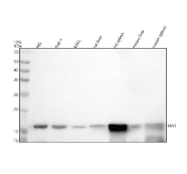
Western blot analysis of IBA1/AIF1 using anti-IBA1/AIF1 antibody (M01394-4).
Electrophoresis was performed on a 12% SDS-PAGE gel at 80V (Stacking gel) / 120V (Resolving gel) for 2 hours. The sample well of each lane was loaded with 30 ug of sample under reducing conditions.
Lane 1: human HEL whole cell lysates,
Lane 2: human THP-1 whole cell lysates,
Lane 3: human K562 whole cell lysates,
Lane 4: rat liver tissue lysates,
Lane 5: rat spleen tissue lysates,
Lane 6: mouse liver tissue lysates,
Lane 7: mouse spleen tissue lysates.
After electrophoresis, proteins were transferred to a nitrocellulose membrane at 150 mA for 50-90 minutes. Blocked the membrane with 5% non-fat milk/TBS for 1.5 hour at RT. The membrane was incubated with rabbit anti-IBA1/AIF1 antigen affinity purified monoclonal antibody (M01394-4) at 1: 500 overnight at 4°C, then washed with TBS-0.1%Tween 3 times with 5 minutes each and probed with a goat anti-rabbit IgG-HRP secondary antibody at a dilution of 1:5000 for 1.5 hour at RT. The signal is developed using an ECL Plus Western Blotting Substrate (Catalog # AR1196-200) with Tanon 5200 system. A specific band was detected for IBA1/AIF1 at approximately 17 kDa. The expected band size for IBA1/AIF1 is at 17 kDa.

Click image to see more details
IHC analysis of IBA1/AIF1 using anti-IBA1/AIF1 antibody (M01394-4).
IBA1/AIF1 was detected in a paraffin-embedded section of human spleen tissue. Heat mediated antigen retrieval was performed in EDTA buffer (pH 8.0, epitope retrieval solution). The tissue section was blocked with 10% goat serum. The tissue section was then incubated with 1:50 rabbit anti-IBA1/AIF1 Antibody (M01394-4) overnight at 4°C. Peroxidase Conjugated Goat Anti-rabbit IgG was used as secondary antibody and incubated for 30 minutes at 37°C. The tissue section was developed using HRP Conjugated Rabbit IgG Super Vision Assay Kit (Catalog # SV0002) with DAB as the chromogen.

Click image to see more details
IHC analysis of IBA1/AIF1 using anti-IBA1/AIF1 antibody (M01394-4).
IBA1/AIF1 was detected in a paraffin-embedded section of human tonsil tissue. Heat mediated antigen retrieval was performed in EDTA buffer (pH 8.0, epitope retrieval solution). The tissue section was blocked with 10% goat serum. The tissue section was then incubated with 1:50 rabbit anti-IBA1/AIF1 Antibody (M01394-4) overnight at 4°C. Peroxidase Conjugated Goat Anti-rabbit IgG was used as secondary antibody and incubated for 30 minutes at 37°C. The tissue section was developed using HRP Conjugated Rabbit IgG Super Vision Assay Kit (Catalog # SV0002) with DAB as the chromogen.

Click image to see more details
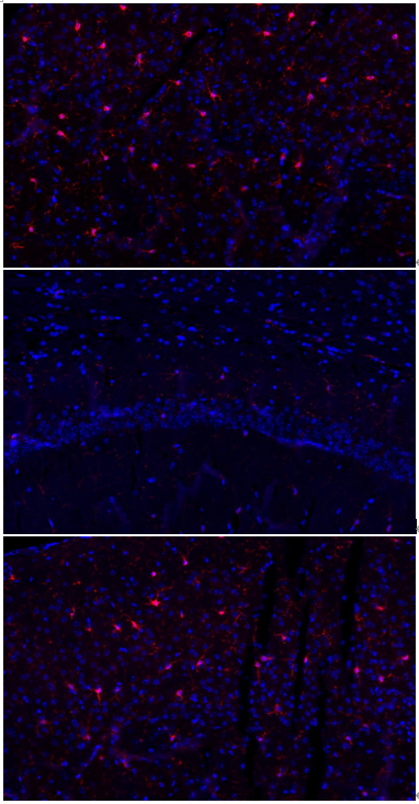
IHC analysis of IBA1/AIF1 using anti-IBA1/AIF1 antibody (M01394-4).
IBA1/AIF1 was detected in a paraffin-embedded section of mouse brain tissue. Heat mediated antigen retrieval was performed in EDTA buffer (pH 8.0, epitope retrieval solution). The tissue section was blocked with 10% goat serum. The tissue section was then incubated with 1:50 rabbit anti-IBA1/AIF1 Antibody (M01394-4) overnight at 4°C. Peroxidase Conjugated Goat Anti-rabbit IgG was used as secondary antibody and incubated for 30 minutes at 37°C. The tissue section was developed using HRP Conjugated Rabbit IgG Super Vision Assay Kit (Catalog # SV0002) with DAB as the chromogen.

Click image to see more details
IHC analysis of IBA1/AIF1 using anti-IBA1/AIF1 antibody (M01394-4).
IBA1/AIF1 was detected in a paraffin-embedded section of rat brain tissue. Heat mediated antigen retrieval was performed in EDTA buffer (pH 8.0, epitope retrieval solution). The tissue section was blocked with 10% goat serum. The tissue section was then incubated with 1:50 rabbit anti-IBA1/AIF1 Antibody (M01394-4) overnight at 4°C. Peroxidase Conjugated Goat Anti-rabbit IgG was used as secondary antibody and incubated for 30 minutes at 37°C. The tissue section was developed using HRP Conjugated Rabbit IgG Super Vision Assay Kit (Catalog # SV0002) with DAB as the chromogen.

Click image to see more details
IF analysis of Alpha-Smooth Muscle Actin using anti-Alpha-Smooth Muscle Actin antibody (MA1106).
Alpha-Smooth Muscle Actin was detected in a paraffin-embedded section of mouse cerebral infarction tissue. tissue . Heat mediated antigen retrieval was performed in EDTA buffer (pH 8.0, epitope retrieval solution). The tissue section was blocked with 10% goat serum. The tissue section was then incubated with 1:100 mouse anti-Alpha-Smooth Muscle Actin Antibody (MA1106) overnight at 4°C. Goat Anti-Rabbit IgG (H+L) Secondary Antibody, Fluoro594 Conjugated (BA1142) was used as secondary antibody incubated with 1:500 and incubated for 30 minutes at 37°C. The section was counterstained with DAPI. Visualize using a fluorescence microscope and filter sets appropriate for the label used.
Specific Publications For Anti-Iba1 Rabbit Monoclonal Antibody (M01394-4)
Loading publications
Recommended Resources
Here are featured tools and databases that you might find useful.
- Boster's Pathways Library
- Protein Databases
- Bioscience Research Protocol Resources
- Data Processing & Analysis Software
- Photo Editing Software
- Scientific Literature Resources
- Research Paper Management Tools
- Molecular Biology Software
- Primer Design Tools
- Bioinformatics Tools
- Phylogenetic Tree Analysis
Customer Reviews
Have you used Anti-Iba1 Rabbit Monoclonal Antibody?
Share your experimental results or join a short interview to earn up to $1,000 in product credits or other rewards.
1 Reviews For Anti-Iba1 Rabbit Monoclonal Antibody
In the IF experiment using Anti-IBA-1 antibody (Cat# M01394-4), the antibody clearly and specifically labeled microglial cells in mouse cerebral infarction tissue, showing distinct staining and well-defined cellular morphology.
Excellent
| SKU | M01394-4 |
|---|---|
| Application | Immunofluorescence |
| Sample | mouse brain tissue |
| Sample Processing Description | Mouse cerebral infarction model; brain tissues were collected, fixed in formaldehyde for 48 hours, and then sagittally paraffin-embedded. |
| Other Reagents | Goat serum, DAPI Staining Solution, Antifade fluorescence mounting medium. |
| Primary Antibody | Iba1 Rabbit Monoclonal Antibody |
| Primary Incubation | 1:100, overnight at 4 ℃ |
| Secondary Antibody | Goat Anti-Rabbit IgG (H+L) Secondary Antibody, Fluoro594 Conjugated (BA1142, Boster) |
| Secondary Incubation | 45 min at 37℃ |
| Detection | Imaging system:Leica DMi3000 |
| Results Summary | IBA-1 is a well-established marker of microglia in the nervous system. Microglia are the primary immune effector cells in the central nervous system and respond rapidly to CNS injury by proliferating, upregulating or re-expressing MHC antigens, migrating, and adopting a phagocyte-like morphology. In this study, IBA-1 was used to label microglia in cerebral infarction samples, showing clear staining and accurate cellular morphology. |
Yu Lei, School of Life Sciences, Wuhan University
Verified customer
Submitted 2026-02-26
Customer Q&As
Have a question?
Find answers in Q&As, reviews.
Can't find your answer?
Submit your question
