Click image to see more details
-

-
-
-
-
+2
Product Info Summary
| SKU: | M01705 |
|---|---|
| Size: | 100ug |
| Reactive Species: | Mouse |
| Host: | Mouse |
| Application: | ELISA, Flow Cytometry, IP, IF, IHC, WB |
Customers Who Bought This Also Bought

Product info
Product Name
Anti-IDO1/Ido Monoclonal Antibody
SKU/Catalog Number
M01705
Size
100ug
Form
Liquid (sterile filtered)
Description
Boster Bio Anti-IDO1/Ido Monoclonal Antibody (Catalog # M01705). Tested in ELISA, Flow Cytometry, IF, IHC, WB applications. This antibody reacts with Mouse.
Storage & Handling
Store vial at -20°C prior to opening. Aliquot contents and freeze at -20°C or below for extended storage. Avoid cycles of freezing and thawing. Centrifuge product if not completely clear after standing at room temperature. This product is stable for several weeks at 4°C as an undiluted liquid. Dilute only prior to immediate use. Expiration date is one (1) year from date of opening. (Ship on dry ice.)
Cite This Product
Anti-IDO1/Ido Monoclonal Antibody (Boster Biological Technology, Pleasanton CA, USA, Catalog # M01705)
Host
Mouse
Contents
0.02 M Potassium Phosphate, 0.15 M Sodium Chloride, pH 7.2, 0.01% (w/v) Sodium Azide
Clonality
Monoclonal
Clone Number
Clone: 2E2.6 IgG1
Isotype
IgG1
Immunogen
IDO1 antibody was produced in mouse by repeated immunizations with mouse recombinant IDO1 protein followed by hybridoma development.
Reactive Species
M01705 is reactive to IDO1 in Mouse
Calculated molecular weight
45.6 kDa
Background of IDO1
Anti-IDO-1 antibody recognizes indoleamine 2, 3-dioxygenase1 (IDO1) is a 41-42 kD intracellular enzyme that catabolizes tryptophan into kynurenine. IDO1 modulates levels of the amino acid tryptophan, which is vital for cell growth, but is also involved in the suppression of the immune response. IDO1 effects on immune suppression are due to decreased tryptophan availability and the generation of tryptophan metabolites, resulting in negative effects on T lymphocytes, including proliferation, function and survival. IDO1 may be involved in the suppression of the immune response to tumors, and blocking the IDO1 pathway may be a potential target for immuno and cancer therapy. IDO1 is expressed in a wide variety of tissues and can be upregulated by interferon gamma and other inflammatory cytokines.
Antibody Validation
Boster validates all antibodies on WB, IHC, ICC, Immunofluorescence, and ELISA with known positive control and negative samples to ensure specificity and high affinity, including thorough antibody incubations.
Application & Images
Applications
M01705 is guaranteed for ELISA, Flow Cytometry, IP, IF, IHC, WB Boster Guarantee
Recommend Dilution
| Application | Dilution | Species |
|---|---|---|
| Anti-IDO1 antibody has been tested for use in ELISA | Western Blot | IF, IHC, and Flow Cytometry. Specific conditions for reactivity should be optimized by the end user. |
Validation Images & Assay Conditions
Click image to see more details
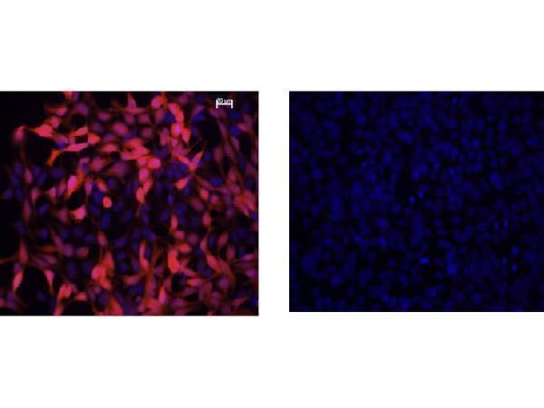
Immunofluorescence Microscopy of Mouse Anti-IDO1 Antibody. Cells: HEK293 cells. Fixation: 0.5% PFA. Expressing: mouse IDO-1 (left) and mouse IDO-2 (right). Primary antibody: IDO1 (2E2) monoclonal antibody. Antigen retrieval: not required. Secondary antibody: mouse secondary antibody at 1:10,000 for 45 min at RT. Localization: IDO-1 is located in the cytosol. Staining: IDO1 as red fluorescent signal with bis-benzimide nuclear counterstain (blue).

Click image to see more details
Immunohistochemistry of Mouse anti-IDO1 antibody. Tissue: epididymis from wild-type (left) or IDO1 null mice (right). Fixation: paraffin-embedded. Primary antibody: IDO1 (2E2) monoclonal antibody. Secondary antibody: Peroxidase mouse secondary antibody at 1:10,000 for 45 min at RT. Localization: IDO-1 is located in the cytosol. Staining: IDO 1 as precipitated brown signal.

Click image to see more details
Immunohistochemistry of Mouse Anti-IDO1 Antibody. Tissue: epididymis from wild-type (left) or IDO1 null mice (right). Fixation: frozen sections. Antigen retrieval: not required. Primary antibody: IDO1 (2E2) monoclonal antibody. Secondary antibody: Peroxidase mouse secondary antibody at 1:10,000 for 45 min at RT. Localization: IDO-1 is located in the cytosol. Staining: IDO 1 as precipitated brown signal.

Click image to see more details
Western Blot of Mouse Anti-IDO1 Antibody. Extracts from 293HEK Cells expressing: Lane 1: Control Vector. Lane 2: His-tagged mouse IDO1. Lane 3: mouse IDO1. Lane 4: His-tagged mouse IDO2. Lane 5: mouse IDO2. Lane 6: Epididymis from IDO null. Lane 7: wild type mice. Primary antibody: IDO-1(2E2) monoclonal antibody. Secondary antibody: IRDye800™ mouse secondary antibody at 1:10,000 for 45 min at RT. Block: 1xPBST overnight at 4°C. Predicted/Observed size: 41-42 kDa/41-42 kDa for IDO-1. Other band(s): none.

Click image to see more details
Flow Cytometry of Mouse Anti-IDO1 antibody. Cells: HEK293 cells. Expresing: mouse IDO-1(blue) and mouse IDO-2 (red). Primary antibody: IDO1 (2E2) monoclonal antibody. Secondary antibody: Biotin mouse secondary antibody at 1:10,000 for 45 min at RT and streptavidin PE at 1:5,000 for 30 min at RT.

Click image to see more details
Western Blot of mouse anti-IDO1 antibody. Lane 1: HEK293 control vector. Lane 2: HEK293 expressing mouse IDO1. Lane 3: HEK293 expressing mouse IDO2. Load: 35 µg per lane. Primary antibody: IDO 1 antibody at 1:400 for overnight at 4°C. Secondary antibody: IRDye800™ mouse secondary antibody at 1:10,000 for 45 min at RT. Block: 5% BLOTTO overnight at 4°C. Predicted/Observed size: 45.6 kDa, ~44 kDa for IDO1. Other band(s): non-specifics.
Specific Publications For Anti-IDO1/Ido Monoclonal Antibody (M01705)
Loading publications
Recommended Resources
Here are featured tools and databases that you might find useful.
- Boster's Pathways Library
- Protein Databases
- Bioscience Research Protocol Resources
- Data Processing & Analysis Software
- Photo Editing Software
- Scientific Literature Resources
- Research Paper Management Tools
- Molecular Biology Software
- Primer Design Tools
- Bioinformatics Tools
- Phylogenetic Tree Analysis
Customer Reviews
Have you used Anti-IDO1/Ido Monoclonal Antibody?
Share your experimental results or join a short interview to earn up to $1,000 in product credits or other rewards.
0 Reviews For Anti-IDO1/Ido Monoclonal Antibody
Customer Q&As
Have a question?
Find answers in Q&As, reviews.
Can't find your answer?
Submit your question
1 Customer Q&As for Anti-IDO1/Ido Monoclonal Antibody
Question
What is the suggested antibody dilution ratio used for IHC-P staining using M01705 and dilution used for IHC-P staining data in the datasheet as well?
Verified customer
Asked: 2019-03-04
Answer
We have the IHC-P tests done through collaboration for the Anti-IDO1/Ido Monoclonal Antibody M01705. Unfortunately, the collaborator didn’t share the exact dilution used, instead they stated "Use at an assay dependent dilution."
Boster Scientific Support
Answered: 2019-03-04
