Click image to see more details
Product Info Summary
| SKU: | A00070-3 |
|---|---|
| Size: | 100 μg/vial |
| Reactive Species: | Human |
| Host: | Rabbit |
| Application: | ELISA, WB |
Customers Who Bought This Also Bought

Product info
Product Name
Anti-IGF1R Antibody Picoband®
SKU/Catalog Number
A00070-3
Size
100 μg/vial
Form
Lyophilized
Description
Boster Bio Anti-IGF1R Antibody Picoband® catalog # A00070-3. Tested in WB, ELISA applications. This antibody reacts with Human. The brand Picoband indicates this is a premium antibody that guarantees superior quality, high affinity, and strong signals with minimal background in Western blot applications. Only our best-performing antibodies are designated as Picoband, ensuring unmatched performance.
Storage & Handling
At -20°C for one year from date of receipt. After reconstitution, at 4°C for one month. It can also be aliquotted and stored frozen at -20°C for six months. Avoid repeated freezing and thawing.
Cite This Product
Anti-IGF1R Antibody Picoband® (Boster Biological Technology, Pleasanton CA, USA, Catalog # A00070-3)
Host
Rabbit
Contents
Each vial contains 4 mg Trehalose, 0.9 mg NaCl, 0.2 mg Na2HPO4.
Clonality
Polyclonal
Immunogen
E.coli-derived human IGF1R recombinant protein (Position: Q44-R960). Human IGF1R shares 95.9% and 95.8% amino acid (aa) sequence identity with mouse and rat IGF1R, respectively.
Reactive Species
A00070-3 is reactive to IGF1R in Human
Observed Molecular Weight
110,200 kDa
Calculated molecular weight
154.8 kDa
Background of IGF1R
IGF1R(Insulin-like Growth Factor 1(IGF-1) Receptor) is a protein found on the surface of human cells. It is a transmembrane receptor that is activated by a hormone called Insulin-like growth factor 1(IGF-1) and by a related hormone called IGF-2. It belongs to the large class of tyrosine kinase receptors. The IGF1R gene is mapped on 15q26.3. IGF-1 plays an important role in growth and continues to have anabolic effects in adults - meaning that it can induce hypertrophy of skeletal muscle and other target tissues. Using a yeast 2-hybrid system, Dey et al.(1998) identified a regulatory subunit of phosphatidylinositol(PI) 3-kinase, PIK3R3, as a binding partner of IGF1R. Functional interaction between BRCA1 and SP1 in the regulation of the IGF1R gene was studied in Schneider cells, a Drosophila cell line which lacks endogenous SP1. In these cells, BRCA1 suppressed 45% of the SP1-induced trans-activation of the IGF1R promoter. Overexpression of the Grb10-binding fragment of Gigyf1 resulted in a significant increase in Igf1-stimulated Igf1r tyrosine phosphorylation. Like the insulin receptor, the IGF-1 receptor is a receptor tyrosine kinase - meaning it signals by causing the addition of a phosphate molecule on particular tyrosines. IGF-1 activates the Insulin receptor at approximately 0.1x the potency of insulin. Part of this signaling may be via IGF1R-InsulinReceptor heterodimers.
Antibody Validation
Boster validates all antibodies on WB, IHC, ICC, Immunofluorescence, and ELISA with known positive control and negative samples to ensure specificity and high affinity, including thorough antibody incubations.
Application & Images
Applications
A00070-3 is guaranteed for ELISA, WB Boster Guarantee
Assay Dilutions Recommendation
The recommendations below provide a starting point for assay optimization. The actual working concentration varies and should be decided by the user.
Western blot, 0.25-0.5 μg/ml, Human
ELISA, 0.1-0.5 μg/ml, -
Positive Control
WB: human Hela whole cell, human MCF-7 whole cell, human SH-SY5Y whole cell, human A549 whole cell
Validation Images & Assay Conditions
Click image to see more details
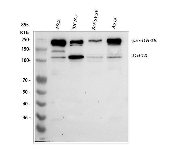
Western blot analysis of IGF1R using anti-IGF1R antibody (A00070-3).
Electrophoresis was performed on a 5-20% SDS-PAGE gel at 70V (Stacking gel) / 90V (Resolving gel) for 2-3 hours. The sample well of each lane was loaded with 30 ug of sample under reducing conditions.
Lane 1: human Hela whole cell lysates,
Lane 2: human MCF-7 whole cell lysates,
Lane 3: human SH-SY5Y whole cell lysates,
Lane 4: human A549 whole cell lysates.
After electrophoresis, proteins were transferred to a nitrocellulose membrane at 150 mA for 50-90 minutes. Blocked the membrane with 5% non-fat milk/TBS for 1.5 hour at RT. The membrane was incubated with rabbit anti-IGF1R antigen affinity purified polyclonal antibody (Catalog # A00070-3) at 0.5 μg/mL overnight at 4°C, then washed with TBS-0.1%Tween 3 times with 5 minutes each and probed with a goat anti-rabbit IgG-HRP secondary antibody at a dilution of 1:5000 for 1.5 hour at RT. The signal is developed using an Enhanced Chemiluminescent detection (ECL) kit (Catalog # EK1002) with Tanon 5200 system. A specific band was detected for IGF1R at approximately 110,200 kDa. The expected band size for IGF1R is at 155 kDa.
Specific Publications For Anti-IGF1R Antibody Picoband® (A00070-3)
Loading publications
Recommended Resources
Here are featured tools and databases that you might find useful.
- Boster's Pathways Library
- Protein Databases
- Bioscience Research Protocol Resources
- Data Processing & Analysis Software
- Photo Editing Software
- Scientific Literature Resources
- Research Paper Management Tools
- Molecular Biology Software
- Primer Design Tools
- Bioinformatics Tools
- Phylogenetic Tree Analysis
Customer Reviews
Have you used Anti-IGF1R Antibody Picoband®?
Share your experimental results or join a short interview to earn up to $1,000 in product credits or other rewards.
0 Reviews For Anti-IGF1R Antibody Picoband®
Customer Q&As
Have a question?
Find answers in Q&As, reviews.
Can't find your answer?
Submit your question
