
Click image to see more details
Product Info Summary
| SKU: | PB9034 |
|---|---|
| Size: | 100 μg/vial |
| Reactive Species: | Mouse |
| Host: | Rabbit |
| Application: | ELISA, WB |
Customers Who Bought This Also Bought

Product info
Product Name
Anti-IL6 Antibody Picoband®
SKU/Catalog Number
PB9034
PB0060 is an alternative SKU for this antibody, used in previous lots.
Size
100 μg/vial
Form
Lyophilized
Description
Boster Bio Anti-IL6 Antibody Picoband® catalog # PB9034. Tested in ELISA, WB applications. This antibody reacts with Mouse. The brand Picoband indicates this is a premium antibody that guarantees superior quality, high affinity, and strong signals with minimal background in Western blot applications. Only our best-performing antibodies are designated as Picoband, ensuring unmatched performance.
Storage & Handling
Store at -20˚C for one year from date of receipt. After reconstitution, at 4˚C for one month. It can also be aliquotted and stored frozen at -20˚C for six months. Avoid repeated freeze-thaw cycles.
Cite This Product
Anti-IL6 Antibody Picoband® (Boster Biological Technology, Pleasanton CA, USA, Catalog # PB9034)
Host
Rabbit
Contents
Each vial contains antibody formulated with stabilizing components, 0.9 mg NaCl, 0.2 mg Na2HPO4, and 0.05 mg NaN3.
*This antibody is supplied in a stabilized formulation.
Compatibility with conjugation reactions depends on the chemistry of the conjugation method used.
For conjugation methods that are not compatible with the stabilizing components present in this formulation, a carrier-free antibody format is required.
Clonality
Polyclonal
Isotype
Rabbit IgG
Immunogen
E.coli-derived mouse IL-6 recombinant protein (Position: F25-T211). Mouse IL-6 shares 40% and 85% amino acid (aa) sequences identity with human and rat IL-6, respectively.
Cross-reactivity
No cross-reactivity with other proteins
Reactive Species
PB9034 is reactive to Il6 in Mouse
Observed Molecular Weight
30 kDa
Calculated molecular weight
24.4 kDa
Background of Il6
Interleukin-6 (IL-6) is a protein that in humans is encoded by the IL6 gene. IL-6 is an interleukin that acts as both a pro-inflammatory and anti-inflammatory cytokine. It is secreted by T cells and macrophages to stimulate immune response to trauma, especially burns or other tissue damage leading to inflammation. IL-6 is one of the most important mediators of fever and of the acute phase response. IL-6 is also essential for hybridoma growth and is found in many supplemental cloning media such as briclone. Bowcock et al. (1988) assigned the IL6 gene to chromosome 7p21. By in situ hybridization and Southern blot analysis of mouse-human hybrid cell lines, Sutherland et al. (1988) mapped the IL-6 gene to chromosome 7p15.
Antibody Validation
Boster validates all antibodies on WB, IHC, ICC, Immunofluorescence, and ELISA with known positive control and negative samples to ensure specificity and high affinity, including thorough antibody incubations.
Application & Images
Applications
PB9034 is guaranteed for ELISA, WB Boster Guarantee
Recommend Dilution
| Application | Dilution | Species |
|---|---|---|
| ELISA | 0.1-0.5μg/ml | - |
| Western blot | 0.1-0.5μg/ml | Mouse |
Tested application
Suggested blocking solution with 5% non-fat milk or BSA; (*)Recommended protein loading: 20-40 µg per lane
Validation Images & Assay Conditions
Click image to see more details
Western blot analysis of IL6 using anti-IL6 antibody (PB9034).
Electrophoresis was performed on a 5-20% SDS-PAGE gel at 70V (Stacking gel) / 90V (Resolving gel) for 2-3 hours. The sample well of each lane was loaded with 30 ug of sample under reducing conditions.
Lane 1: mouse J774A.1 whole cell lysates,
Lane 2: mouse RAW264.7 whole cell lysates,
Lane 3: mouse ANA-1 whole cell lysates.
After electrophoresis, proteins were transferred to a nitrocellulose membrane at 150 mA for 50-90 minutes. Blocked the membrane with 5% non-fat milk/TBS for 1.5 hour at RT. The membrane was incubated with rabbit anti-IL6 antigen affinity purified polyclonal antibody (Catalog # PB9034) at 0.5 μg/mL overnight at 4°C, then washed with TBS-0.1%Tween 3 times with 5 minutes each and probed with a goat anti-rabbit IgG-HRP secondary antibody at a dilution of 1:5000 for 1.5 hour at RT. The signal is developed using an Enhanced Chemiluminescent detection (ECL) kit (Catalog # EK1002) with Tanon 5200 system. A specific band was detected for IL6 at approximately 30 kDa. The expected band size for IL6 is at 24 kDa.

Click image to see more details
Western blot analysis of IL6 using anti-IL6 antibody (PB9034).
Electrophoresis was performed on a 5-20% SDS-PAGE gel at 70V (Stacking gel) / 90V (Resolving gel) for 2-3 hours. The sample well of each lane was loaded with 30 ug of sample under reducing conditions.
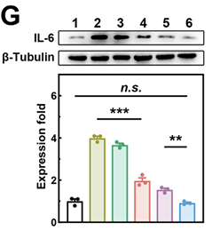
Lane 1-6: mouse PACs whole cell lysates.
After electrophoresis, proteins were transferred to a nitrocellulose membrane at 150 mA for 50-90 minutes. Blocked the membrane with 5% non-fat milk/TBS for 1.5 hour at RT. The membrane was incubated with rabbit anti-IL6 antigen affinity purified polyclonal antibody (Catalog # PB9034) at 1:1000 overnight at 4°C, then washed with TBS-0.1%Tween 3 times with 5 minutes each and probed with a goat anti-rabbit IgG-HRP secondary antibody at a dilution of 1:5000 for 1.5 hour at RT. The signal is developed using an Enhanced Chemiluminescent detection (ECL) kit (Catalog # EK1002) with Tanon 5200 system. The expected band size for IL6 is at 24 kDa.
Specific Publications For Anti-IL6 Antibody Picoband® (PB9034)
Loading publications
Recommended Resources
Here are featured tools and databases that you might find useful.
- Boster's Pathways Library
- Protein Databases
- Bioscience Research Protocol Resources
- Data Processing & Analysis Software
- Photo Editing Software
- Scientific Literature Resources
- Research Paper Management Tools
- Molecular Biology Software
- Primer Design Tools
- Bioinformatics Tools
- Phylogenetic Tree Analysis
Customer Reviews
Have you used Anti-IL6 Antibody Picoband®?
Share your experimental results or join a short interview to earn up to $1,000 in product credits or other rewards.
1 Reviews For Anti-IL6 Antibody Picoband®
Pancreatic IL-6 levels measured using the Boster Anti-IL-6 antibody (PB9034) demonstrated high specificity and sensitivity in WB analysis, providing reliable evidence for evaluating the anti-inflammatory effect of the nanodrug.
Excellent
| SKU | PB9034 |
|---|---|
| Application | Western Blot |
| Sample | mouse PACs cells |
| Sample Processing Description | After centrifugation, the supernatant was collected. A small aliquot of the protein solution was taken for BCA quantification, and the remaining protein solution was mixed with an equal volume of loading buffer, then denatured in a 100 °C water bath for 5 minutes. |
| Other Reagents | Protein-Free Rapid Blocking Buffer |
| Primary Antibody | Anti-IL6 Antibody Picoband® |
| Primary Incubation | 1:1000, overnight at 4 ℃ |
| Secondary Antibody | HRP Conjugated AffiniPure Goat Anti-rabbit IgG (H+L) |
| Secondary Incubation | 1 h in RT |
| Detection | Substrate: ECL substrate, Imaging system:ChemiDoc MP |
| Results Summary | The levels of IL-6 and IL-1β in the pancreas are key indicators for evaluating whether the nanodrug exerts anti-inflammatory effects. Initially, antibodies from two domestic and international suppliers were used for WB experiments, but their specificity was relatively poor. Subsequently, the antibody from Boster was adopted, which demonstrated strong specificity, high titer, and cost-effectiveness. |
Jiahui Yan, College of Chemistry and Chemical Engineering, Ocean University of China
Verified customer
Submitted 2026-02-27
Customer Q&As
Have a question?
Find answers in Q&As, reviews.
Can't find your answer?
Submit your question
7 Customer Q&As for Anti-IL6 Antibody Picoband®
Question
I would like to inquire about Anti-IL6 Antibody, since I will performed Western blot with rabbit muscle tissue. At what percentage the sequence identity between immunogen of these antibodies and rabbit
Verified Customer
Verified customer
Asked: 2020-04-03
Answer
The immunogen of PB9034 Anti-IL6 Antibody is full length human IL-6 that shares 40% sequence homology with rabbit IL-6.
Boster Scientific Support
Answered: 2020-04-03
Question
We are currently using anti-IL6 antibody PB9034 for mouse tissue, and we are happy with the ELISA results. The species of reactivity given in the datasheet says mouse. Is it possible that the antibody can work on monkey tissues as well?
Verified Customer
Verified customer
Asked: 2020-02-10
Answer
The anti-IL6 antibody (PB9034) has not been tested for cross reactivity specifically with monkey tissues, though there is a good chance of cross reactivity. We have an innovator award program that if you test this antibody and show it works in monkey you can get your next antibody for free. Please contact me if I can help you with anything.
Boster Scientific Support
Answered: 2020-02-10
Question
Could this PB9034 Anti-IL6 Antibody worked on formalin fixed and paraffin embedded tissue?
Verified Customer
Verified customer
Asked: 2020-01-13
Answer
The PB9034 Anti-IL6 Antibody has been tested successfully on fixed paraffin sections.
Boster Scientific Support
Answered: 2020-01-13
Question
We were content with the WB result of your anti-IL6 antibody. However we have seen positive staining in left coronary artery secreted. using this antibody. Is that expected? Could you tell me where is IL6 supposed to be expressed?
Verified Customer
Verified customer
Asked: 2019-12-31
Answer
Based on literature, left coronary artery does express IL6. Generally IL6 expresses in secreted. Regarding which tissues have IL6 expression, here are a few articles citing expression in various tissues:
Fibroblast, Pubmed ID: 3758081
Lung, Pubmed ID: 15489334
Boster Scientific Support
Answered: 2019-12-31
Question
We have observed staining in mouse fibroblast. Do you have any suggestions? Is anti-IL6 antibody supposed to stain fibroblast positively?
Verified Customer
Verified customer
Asked: 2019-11-12
Answer
From what I have seen in literature fibroblast does express IL6. From what I have seen in Uniprot.org, IL6 is expressed in left coronary artery, fibroblast, lung, among other tissues. Regarding which tissues have IL6 expression, here are a few articles citing expression in various tissues:
Fibroblast, Pubmed ID: 3758081
Lung, Pubmed ID: 15489334
Boster Scientific Support
Answered: 2019-11-12
Question
I am interested in using your anti-IL6 antibody for negative regulation of interleukin-1-mediated signaling pathway studies. Has this antibody been tested with western blotting on nih whole cell lysate? We would like to see some validation images before ordering.
N. Patel
Verified customer
Asked: 2018-04-10
Answer
We appreciate your inquiry. This PB9034 anti-IL6 antibody is tested on hepa whole cell lysate, nih whole cell lysate. It is guaranteed to work for ELISA, WB in mouse. Our Boster guarantee will cover your intended experiment even if the sample type has not been be directly tested.
Boster Scientific Support
Answered: 2018-04-10
Question
We ordered your anti-IL6 antibody for ELISA on lung a few years ago. I am using mouse, and We intend to use the antibody for WB next. We want examining lung as well as fibroblast in our next experiment. Could you please give me some suggestion on which antibody would work the best for WB?
M. Carter
Verified customer
Asked: 2014-10-31
Answer
I looked at the website and datasheets of our anti-IL6 antibody and it seems that PB9034 has been tested on mouse in both ELISA and WB. Thus PB9034 should work for your application. Our Boster satisfaction guarantee will cover this product for WB in mouse even if the specific tissue type has not been validated. We do have a comprehensive range of products for WB detection and you can check out our website bosterbio.com to find out more information about them.
Boster Scientific Support
Answered: 2014-10-31
