Click image to see more details
Product Info Summary
| SKU: | A02062-1 |
|---|---|
| Size: | 100 μg/vial |
| Reactive Species: | Human, Mouse, Rat |
| Host: | Rabbit |
| Application: | ELISA, WB |
Customers Who Bought This Also Bought

Product info
Product Name
Anti-IL17F Antibody Picoband®
SKU/Catalog Number
A02062-1
Size
100 μg/vial
Form
Lyophilized
Description
Boster Bio Anti-IL17F Antibody Picoband® catalog # A02062-1. Tested in ELISA, WB applications. This antibody reacts with Human, Mouse, Rat. The brand Picoband indicates this is a premium antibody that guarantees superior quality, high affinity, and strong signals with minimal background in Western blot applications. Only our best-performing antibodies are designated as Picoband, ensuring unmatched performance.
Storage & Handling
Store at -20˚C for one year from date of receipt. After reconstitution, at 4˚C for one month. It can also be aliquotted and stored frozen at -20˚C for six months. Avoid repeated freeze-thaw cycles.
Cite This Product
Anti-IL17F Antibody Picoband® (Boster Biological Technology, Pleasanton CA, USA, Catalog # A02062-1)
Host
Rabbit
Contents
Each vial contains antibody formulated with stabilizing components, 0.9 mg NaCl, 0.2 mg Na2HPO4, and 0.05 mg NaN3.
*This antibody is supplied in a stabilized formulation.
Compatibility with conjugation reactions depends on the chemistry of the conjugation method used.
For conjugation methods that are not compatible with the stabilizing components present in this formulation, a carrier-free antibody format is required.
Clonality
Polyclonal
Isotype
Rabbit IgG
Immunogen
E. coli-derived human IL17F recombinant protein (Position: R31-Q163).
Cross-reactivity
No cross-reactivity with other proteins.
Reactive Species
A02062-1 is reactive to IL17F in Human, Mouse, Rat
Observed Molecular Weight
27-30kDa
Calculated molecular weight
18.0 kDa
Background of IL17F
Interleukin 17F, also called IL17F is involved in the regulation of normal versus aberrant T-cell responses. This gene is mapped to 6p12.2. The protein encoded by this gene is a cytokine that shares sequence similarity with IL17. This cytokine is expressed by activated T cells, and has been shown to stimulate the production of several other cytokines, including IL6, IL8, and CSF2/GM_CSF. This cytokine is also found to inhibit the angiogenesis of endothelial cells and induce endothelial cells to produce IL2, TGFB1/TGFB, and monocyte chemoattractant protein-1. It is suggested that targeting IL17 and IL17F or antagonizing IL17R might mitigate neutrophil-mediated inflammation in CF.
Antibody Validation
Boster validates all antibodies on WB, IHC, ICC, Immunofluorescence, and ELISA with known positive control and negative samples to ensure specificity and high affinity, including thorough antibody incubations.
Application & Images
Applications
A02062-1 is guaranteed for ELISA, WB Boster Guarantee
Recommend Dilution
| Application | Dilution | Species |
|---|---|---|
| Western blot | 0.1-0.5μg/ml | Human, Mouse, Rat |
| ELISA | 0.1-0.5μg/ml | - |
Tested application
Suggested blocking solution with 5% non-fat milk or BSA; (*)Recommended protein loading: 20-40 µg per lane
Validation Images & Assay Conditions
Click image to see more details
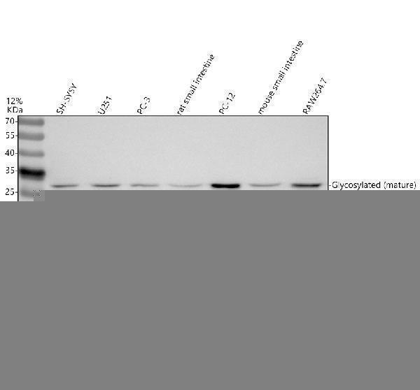
Western blot analysis of IL17F using anti-IL17F antibody (A02062-1).
Electrophoresis was performed on a 12% SDS-PAGE gel at 80V (Stacking gel) / 120V (Resolving gel) for 2 hours. The sample well of each lane was loaded with 30 ug of sample under reducing conditions.
Lane 1: human SH-SY5Y whole cell lysates,
Lane 2: human U251 whole cell lysates,
Lane 3: human PC-3 whole cell lysates,
Lane 4: rat small intestine lysates,
Lane 5: rat PC-12 whole cell lysates,
Lane 6: mouse small intestine lysates,
Lane 7: mouse RAW264.7 whole cell lysates.
After electrophoresis, proteins were transferred to a nitrocellulose membrane at 150 mA for 50-90 minutes. Blocked the membrane with 5% non-fat milk/TBS for 1.5 hour at RT. The membrane was incubated with rabbit anti-IL17F antigen affinity purified polyclonal antibody (A02062-1) at 0.5 μg/mL overnight at 4°C, then washed with TBS-0.1%Tween 3 times with 5 minutes each and probed with a goat anti-rabbit IgG-HRP secondary antibody (Catalog # BA1054) at a dilution of 1:5000 for 1.5 hour at RT. The signal is developed using an ECL Plus Western Blotting Substrate (Catalog # AR1196-200) with Tanon 5200 system. A specific band was detected for IL17F at approximately 27-30 kDa. The expected band size for IL17F is at 18 kDa.
Specific Publications For Anti-IL17F Antibody Picoband® (A02062-1)
Loading publications
Recommended Resources
Here are featured tools and databases that you might find useful.
- Boster's Pathways Library
- Protein Databases
- Bioscience Research Protocol Resources
- Data Processing & Analysis Software
- Photo Editing Software
- Scientific Literature Resources
- Research Paper Management Tools
- Molecular Biology Software
- Primer Design Tools
- Bioinformatics Tools
- Phylogenetic Tree Analysis
Customer Reviews
Have you used Anti-IL17F Antibody Picoband®?
Share your experimental results or join a short interview to earn up to $1,000 in product credits or other rewards.
0 Reviews For Anti-IL17F Antibody Picoband®
Customer Q&As
Have a question?
Find answers in Q&As, reviews.
Can't find your answer?
Submit your question
