Click image to see more details
Product Info Summary
| SKU: | M00165-3 |
|---|---|
| Size: | 100 μg/vial |
| Reactive Species: | Mouse, Rat |
| Host: | Mouse |
| Application: | Flow Cytometry, IF, ICC, WB |
Customers Who Bought This Also Bought

Product info
Product Name
Anti-IRF3 Antibody Picoband® (monoclonal, 3B4)
SKU/Catalog Number
M00165-3
Size
100 μg/vial
Form
Lyophilized
Description
Boster Bio Anti-IRF3 Antibody Picoband® (monoclonal, 3B4) catalog # M00165-3. Tested in Flow Cytometry, IF, ICC, WB applications. This antibody reacts with Mouse, Rat. The brand Picoband indicates this is a premium antibody that guarantees superior quality, high affinity, and strong signals with minimal background in Western blot applications. Only our best-performing antibodies are designated as Picoband, ensuring unmatched performance.
Storage & Handling
Store at -20˚C for one year from date of receipt. After reconstitution, at 4˚C for one month. It can also be aliquotted and stored frozen at -20˚C for six months. Avoid repeated freeze-thaw cycles.
Cite This Product
Anti-IRF3 Antibody Picoband® (monoclonal, 3B4) (Boster Biological Technology, Pleasanton CA, USA, Catalog # M00165-3)
Host
Mouse
Contents
Each vial contains 4mg Trehalose, 0.9mg NaCl and 0.2mg Na2HPO4.
Clonality
Monoclonal
Clone Number
3B4
Isotype
Mouse IgG2a
Immunogen
E.coli-derived mouse IRF3 recombinant protein (Position: M1-I419).
Cross-reactivity
No cross-reactivity with other proteins.
Reactive Species
M00165-3 is reactive to Irf3 in Mouse, Rat
Observed Molecular Weight
50-55 kDa
Calculated molecular weight
46.9 kDa
Background of Irf3
IRF3 (interferon regulatory factor 3) is a member of the interferon regulatory transcription factor (IRF) family. The IRF3 gene is mapped on 19q13.33. IRF3 is found in an inactive cytoplasmic form that upon serine/threonine phosphorylation forms a complex with CREBBP. IRF3 plays an important role in the innate immune system's response to viral infection. Aggregated MAVS have been found to activate IRF3 dimerization. Although IRF3 increased transcriptional activity from an ISRE-containing promoter, expression of IRF3 as a Gal4 fusion protein did not activate expression of a chloramphenicol acetyltransferase (CAT) reporter gene containing repeats of the Gal4-binding sites. Translocation of IRF3 was accompanied by an increase in serine and threonine phosphorylation. The transcriptional activators CREBBP and EP300 coimmunoprecipitated with IRF3 only subsequent to viral infection, and the authors stated that these are also subunits of DRAF1.
Antibody Validation
Boster validates all antibodies on WB, IHC, ICC, Immunofluorescence, and ELISA with known positive control and negative samples to ensure specificity and high affinity, including thorough antibody incubations.
Application & Images
Applications
M00165-3 is guaranteed for Flow Cytometry, IF, ICC, WB Boster Guarantee
Assay Dilutions Recommendation
The recommendations below provide a starting point for assay optimization. The actual working concentration varies and should be decided by the user.
Western blot, 0.25-0.5μg/ml, Mouse, Rat
Immunocytochemistry/Immunofluorescence, 5μg/ml, Mouse
Flow Cytometry (Fixed), 1-3μg/1x106 cells, Mouse
Positive Control
WB: rat brain tissue, rat C6 whole cell, mouse lung tissue, mouse brain tissue, mouse NIH/3T3 whole cell
ICC/IF: RM-1 cell
FCM: HEPA1-6 cell
Validation Images & Assay Conditions
Click image to see more details
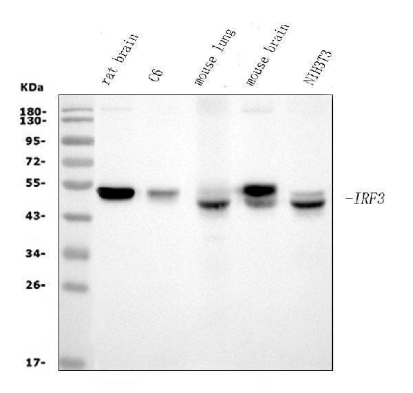
Western blot analysis of IRF3 using anti-IRF3 antibody (M00165-3).
Electrophoresis was performed on a 5-20% SDS-PAGE gel at 70V (Stacking gel) / 90V (Resolving gel) for 2-3 hours. The sample well of each lane was loaded with 30ug of sample under reducing conditions.
Lane 1: rat brain tissue lysates,
Lane 2: rat C6 whole cell lysates,
Lane 3: mouse lung tissue lysates,
Lane 4: mouse brain tissue lysates,
Lane 5: mouse NIH/3T3 whole cell lysates.
After Electrophoresis, proteins were transferred to a Nitrocellulose membrane at 150mA for 50-90 minutes. Blocked the membrane with 5% Non-fat Milk/ TBS for 1.5 hour at RT. The membrane was incubated with mouse anti-IRF3 antigen affinity purified monoclonal antibody (Catalog # M00165-3) at 0.5 μg/mL overnight at 4°C, then washed with TBS-0.1%Tween 3 times with 5 minutes each and probed with a goat anti-mouse IgG-HRP secondary antibody at a dilution of 1:10000 for 1.5 hour at RT. The signal is developed using an Enhanced Chemiluminescent detection (ECL) kit (Catalog # EK1001) with Tanon 5200 system. A specific band was detected for IRF3 at approximately 50-55KD. The expected band size for IRF3 is at 50-55KD.

Click image to see more details
IF analysis of IRF3 using anti-IRF3 antibody (M00165-3) .
IRF3 was detected in an immunocytochemical section of mouse NIH/3T3 cells. The cells were fixed with 4% paraformaldehyde for 10 minutes and then treated with a membrane permeabilization agent (AR0205) for 5 minutes.The cells were blocked with 10% goat serum. And then incubated with mouse anti-IRF3 Antibody (M00165-3) at a dilution of 10 ug/mL overnight at 4°C. DyLight®488 Conjugated Goat Anti-mouse IgG (BA1126) was used as secondary antibody at 1:500 dilution and incubated for 30 minutes at 37°C. The section was counterstained with DAPI. Visualize using a fluorescence microscope and filter sets appropriate for the label used.

Click image to see more details
IF analysis of IRF3 using anti-IRF3 antibody (M00165-3).
IRF3 was detected in immunocytochemical section of RM-1 cells. Enzyme antigen retrieval was performed using IHC enzyme antigen retrieval reagent (AR0022) for 15 mins. The cells were blocked with 10% goat serum. And then incubated with 5μg/mL mouse anti-IRF3 Antibody (M00165-3) overnight at 4°C. DyLight®488 Conjugated Goat Anti-Mouse IgG (BA1126) was used as secondary antibody at 1:100 dilution and incubated for 30 minutes at 37°C. The section was counterstained with DAPI. Visualize using a fluorescence microscope and filter sets appropriate for the label used.

Click image to see more details
Flow Cytometry analysis of HEPA1-6 cells using anti-IRF3 antibody (M00165-3).
Overlay histogram showing HEPA1-6 cells stained with M00165-3 (Blue line). To facilitate intracellular staining, cells were fixed with 4% paraformaldehyde and permeabilized with permeabilization buffer. The cells were blocked with 10% normal goat serum. And then incubated with mouse anti- IRF3 Antibody (M00165-3, 1μg/1x106 cells) for 30 min at 20°C. DyLight®488 conjugated goat anti-mouse IgG (BA1126, 5-10μg/1x106 cells) was used as secondary antibody for 30 minutes at 20°C. Isotype control antibody (Green line) was mouse IgG (1μg/1x106) used under the same conditions. Unlabelled sample without incubation with primary antibody and secondary antibody (Red line) was used as a blank control.
Specific Publications For Anti-IRF3 Antibody Picoband® (monoclonal, 3B4) (M00165-3)
Loading publications
Recommended Resources
Here are featured tools and databases that you might find useful.
- Boster's Pathways Library
- Protein Databases
- Bioscience Research Protocol Resources
- Data Processing & Analysis Software
- Photo Editing Software
- Scientific Literature Resources
- Research Paper Management Tools
- Molecular Biology Software
- Primer Design Tools
- Bioinformatics Tools
- Phylogenetic Tree Analysis
Customer Reviews
Have you used Anti-IRF3 Antibody Picoband® (monoclonal, 3B4)?
Share your experimental results or join a short interview to earn up to $1,000 in product credits or other rewards.
0 Reviews For Anti-IRF3 Antibody Picoband® (monoclonal, 3B4)
Customer Q&As
Have a question?
Find answers in Q&As, reviews.
Can't find your answer?
Submit your question
