Click image to see more details
Product Info Summary
| SKU: | RP1056 |
|---|---|
| Size: | 100 μg/vial |
| Reactive Species: | Human |
| Host: | Rabbit |
| Application: | Flow Cytometry, IF, ICC, WB |
Customers Who Bought This Also Bought

Product info
Product Name
Anti-KAT13A/SRC1/NCOA1 Antibody Picoband®
SKU/Catalog Number
RP1056
PB0305 is an alternative SKU for this antibody, used in previous lots.
Size
100 μg/vial
Form
Lyophilized
Description
Boster Bio Anti-KAT13A/SRC1/NCOA1 Antibody catalog # RP1056. Tested in Flow Cytometry, IF, ICC, WB applications. This antibody reacts with Human. The brand Picoband indicates this is a premium antibody that guarantees superior quality, high affinity, and strong signals with minimal background in Western blot applications. Only our best-performing antibodies are designated as Picoband, ensuring unmatched performance.
Storage & Handling
Store at -20˚C for one year from date of receipt. After reconstitution, at 4˚C for one month. It can also be aliquotted and stored frozen at -20˚C for six months. Avoid repeated freeze-thaw cycles.
Cite This Product
Anti-KAT13A/SRC1/NCOA1 Antibody Picoband® (Boster Biological Technology, Pleasanton CA, USA, Catalog # RP1056)
Host
Rabbit
Contents
Each vial contains antibody formulated with stabilizing components, 0.9 mg NaCl, 0.2 mg Na2HPO4, and 0.05 mg NaN3.
*This antibody is supplied in a stabilized formulation.
Compatibility with conjugation reactions depends on the chemistry of the conjugation method used.
For conjugation methods that are not compatible with the stabilizing components present in this formulation, a carrier-free antibody format is required.
Clonality
Polyclonal
Isotype
Rabbit IgG
Immunogen
E.coli-derived human KAT13A recombinant protein (Position: H614-Q826). Human KAT13A shares 92% amino acid (aa) sequence identity with mouse KAT13A.
Cross-reactivity
No cross-reactivity with other proteins
Reactive Species
RP1056 is reactive to NCOA1 in Human
Observed Molecular Weight
157 kDa
Calculated molecular weight
156.8 kDa
Background of NCOA1
The nuclear receptor coactivator 1 (NCOA1), also known as SRC1, is a transcriptional coregulatory protein that contains several nuclear receptor interacting domains and an intrinsic histone acetyltransferase activity. NCOA1 is recruited to DNA promotion sites by ligand-activated nuclear receptors. NCOA1, in turn, acylates histones, which makes downsteam DNA more accessible to transcription. Hence, NCOA1 assists nuclear receptors in the upregulation of DNA expression. It has been found that NCOA1 can enhance the transcriptional activity of ligand-bound PGR but does not alter the basal activity of the target promoter. It also enhances estrogen receptor, glucocorticoid receptor, thyroid hormone receptor, and retinoid X receptor transcriptional activities through their cognate DNA response elements in the presence of hormone. What’s more, SRC1 may play a role as a bridging molecule between nuclear hormone receptors and general transcription factors.
Antibody Validation
Boster validates all antibodies on WB, IHC, ICC, Immunofluorescence, and ELISA with known positive control and negative samples to ensure specificity and high affinity, including thorough antibody incubations.
Application & Images
Applications
RP1056 is guaranteed for Flow Cytometry, IF, ICC, WB Boster Guarantee
Recommend Dilution
| Application | Dilution | Species |
|---|---|---|
| Western blot | 0.1-0.5μg/ml | Human |
| Immunocytochemistry/Immunofluorescence | 2μg/ml | Human |
| Flow Cytometry (Fixed) | 1-3μg/1x106 cells | Human |
Tested application
Suggested blocking solution with 5% non-fat milk or BSA; (*)Recommended protein loading: 20-40 µg per lane
Validation Images & Assay Conditions
Click image to see more details
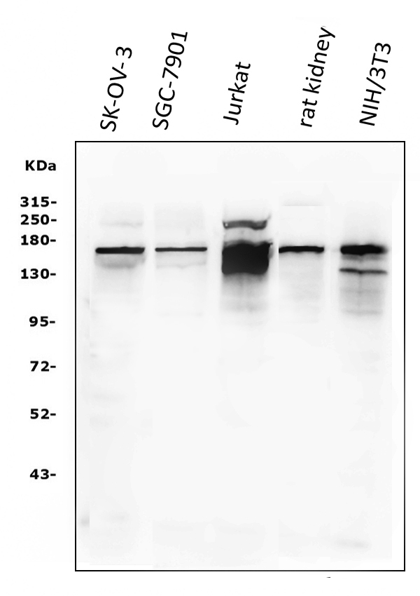
Western blot analysis of KAT13A/SRC1 using anti-KAT13A/SRC1 antibody (RP1056).
Electrophoresis was performed on a 5-20% SDS-PAGE gel at 70V (Stacking gel) / 90V (Resolving gel) for 2-3 hours. The sample well of each lane was loaded with 50ug of sample under reducing conditions.
Lane 1: SK-OV-3 whole cell lysates,
Lane 2: SGC-7901 whole cell lysates,
Lane 3: Jurkat whole cell lysates,
Lane 4: rat kidney tissue lysates,
Lane 5: NIH/3T3 whole cell lysates.
After Electrophoresis, proteins were transferred to a Nitrocellulose membrane at 150mA for 50-90 minutes. Blocked the membrane with 5% Non-fat Milk/ TBS for 1.5 hour at RT. The membrane was incubated with rabbit anti-KAT13A/SRC1 antigen affinity purified polyclonal antibody (Catalog # RP1056) at 0.5 μg/mL overnight at 4°C, then washed with TBS-0.1%Tween 3 times with 5 minutes each and probed with a goat anti-rabbit IgG-HRP secondary antibody at a dilution of 1:5000 for 1.5 hour at RT. The signal is developed using an Enhanced Chemiluminescent detection (ECL) kit (Catalog # EK1002) with Tanon 5200 system. A specific band was detected for KAT13A/SRC1 at approximately 157KD. The expected band size for KAT13A/SRC1 is at 157KD.

Click image to see more details
IF analysis of KAT13A/SRC1 using anti-KAT13A/SRC1 antibody (RP1056).
KAT13A/SRC1 was detected in immunocytochemical section of U20S cells. Enzyme antigen retrieval was performed using IHC enzyme antigen retrieval reagent (AR0022) for 15 mins. The cells were blocked with 10% goat serum. And then incubated with 2μg/mL rabbit anti-KAT13A/SRC1 Antibody (RP1056) overnight at 4°C. DyLight®488 Conjugated Goat Anti-Rabbit IgG (BA1127) was used as secondary antibody at 1:100 dilution and incubated for 30 minutes at 37°C. The section was counterstained with DAPI. Visualize using a fluorescence microscope and filter sets appropriate for the label used.

Click image to see more details
Flow Cytometry analysis of SiHa cells using anti-KAT13A/SRC1 antibody (RP1056).
Overlay histogram showing SiHa cells stained with RP1056 (Blue line). To facilitate intracellular staining, cells were fixed with 4% paraformaldehyde and permeabilized with permeabilization buffer. The cells were blocked with 10% normal goat serum. And then incubated with rabbit anti-KAT13A/SRC1 Antibody (RP1056,1μg/1x106 cells) for 30 min at 20°C. DyLight®488 conjugated goat anti-rabbit IgG (BA1127, 5-10μg/1x106 cells) was used as secondary antibody for 30 minutes at 20°C. Isotype control antibody (Green line) was rabbit IgG (1μg/1x106) used under the same conditions. Unlabelled sample without incubation with primary antibody and secondary antibody (Red line) was used as a blank control.
Specific Publications For Anti-KAT13A/SRC1/NCOA1 Antibody Picoband® (RP1056)
Loading publications
Recommended Resources
Here are featured tools and databases that you might find useful.
- Boster's Pathways Library
- Protein Databases
- Bioscience Research Protocol Resources
- Data Processing & Analysis Software
- Photo Editing Software
- Scientific Literature Resources
- Research Paper Management Tools
- Molecular Biology Software
- Primer Design Tools
- Bioinformatics Tools
- Phylogenetic Tree Analysis
Customer Reviews
Have you used Anti-KAT13A/SRC1/NCOA1 Antibody Picoband®?
Share your experimental results or join a short interview to earn up to $1,000 in product credits or other rewards.
0 Reviews For Anti-KAT13A/SRC1/NCOA1 Antibody Picoband®
Customer Q&As
Have a question?
Find answers in Q&As, reviews.
Can't find your answer?
Submit your question
1 Customer Q&As for Anti-KAT13A/SRC1/NCOA1 Antibody Picoband®
Question
We are currently using anti-KAT13A/SRC1/NCOA1 antibody RP1056 for human tissue, and we are satisfied with the WB results. The species of reactivity given in the datasheet says human. Is it likely that the antibody can work on primate tissues as well?
K. Parker
Verified customer
Asked: 2016-03-30
Answer
The anti-KAT13A/SRC1/NCOA1 antibody (RP1056) has not been validated for cross reactivity specifically with primate tissues, but there is a good chance of cross reactivity. We have an innovator award program that if you test this antibody and show it works in primate you can get your next antibody for free. Please contact me if I can help you with anything.
Boster Scientific Support
Answered: 2016-03-30
