Click image to see more details
-

-
-
-
-
+2
Product Info Summary
| SKU: | RP1082 |
|---|---|
| Size: | 100μg/vial |
| Reactive Species: | Human, Monkey, Mouse, Rat |
| Host: | Rabbit |
| Application: | Flow Cytometry, IF, IHC, ICC, WB |
Customers Who Bought This Also Bought

Product info
Product Name
Anti-KAT13D/CLOCK Antibody Picoband®
SKU/Catalog Number
RP1082
Size
100μg/vial
Form
Lyophilized
Description
Boster Bio Anti-KAT13D/CLOCK Picoband® Antibody catalog # RP1082. Tested in Flow Cytometry, IF, IHC, ICC, WB applications. This antibody reacts with Human, Monkey, Mouse, Rat. The brand Picoband indicates this is a premium antibody that guarantees superior quality, high affinity, and strong signals with minimal background in Western blot applications. Only our best-performing antibodies are designated as Picoband, ensuring unmatched performance.
Storage & Handling
Store at -20˚C for one year from date of receipt. After reconstitution, at 4˚C for one month. It can also be aliquotted and stored frozen at -20˚C for six months. Avoid repeated freeze-thaw cycles.
Cite This Product
Anti-KAT13D/CLOCK Antibody Picoband® (Boster Biological Technology, Pleasanton CA, USA, Catalog # RP1082)
Host
Rabbit
Contents
Each vial contains 4 mg Trehalose, 0.9 mg NaCl and 0.2 mg Na2HPO4.
Clonality
Polyclonal
Immunogen
A synthetic peptide corresponding to a sequence at the N-terminus of human KAT13D/CLOCK , different from the related mouse sequence by one amino acid, and identical to the related rat sequence.
Cross-reactivity
No cross reactivity with other proteins
Reactive Species
RP1082 is reactive to CLOCK in Human, Monkey, Mouse, Rat
Observed Molecular Weight
110 kDa
Calculated molecular weight
95.3 kDa
Background of CLOCK
Clock (Circadian Locomotor Output Cycles Kaput) is also known as KAT13D. The protein encoded by this gene plays a central role in the regulation of circadian rhythms. This protein encodes a transcription factor of the basic helix-loop-helix (bHLH) family and contains DNA binding histone acetyltransferase activity. And the encoded protein forms a heterodimer with ARNTL (BMAL1) that binds E-box enhancer elements upstream of Period (PER1, PER2, PER3) and Cryptochrome (CRY1, CRY2) genes and activates transcription of these genes. PER and CRY proteins heterodimerize and repress their own transcription by interacting in a feedback loop with CLOCK/ARNTL complexes. Polymorphisms in this gene may be associated with behavioral changes in certain populations and with obesity and metabolic syndrome. Alternative splicing results in multiple transcript variants.
Antibody Validation
Boster validates all antibodies on WB, IHC, ICC, Immunofluorescence, and ELISA with known positive control and negative samples to ensure specificity and high affinity, including thorough antibody incubations.
Application & Images
Applications
RP1082 is guaranteed for Flow Cytometry, IF, IHC, ICC, WB Boster Guarantee
Assay Dilutions Recommendation
The recommendations below provide a starting point for assay optimization. The actual working concentration varies and should be decided by the user.
Western blot, 0.1-0.5μg/ml, Human, Monkey, Mouse, Rat
Immunohistochemistry (Paraffin-embedded Section), 2-5μg/ml, Human, Mouse, Rat
Immunocytochemistry/Immunofluorescence, 5 μg/ml, Human
Flow Cytometry(Fixed), 1-3 μg/1x106 cells, Human
Positive Control
WB: human Hela whole cell, human 293T whole cell, human PC-3 whole cell, monkey COS-7 whole cellrat L6 whole cell, mouse NIH/3T3 whole cell
IHC: human pancrese cancer tissue, mouse brain tissue, rat brain tissue
ICC/IF: U2OS cell
FCM: Hela cell
Validation Images & Assay Conditions
Click image to see more details
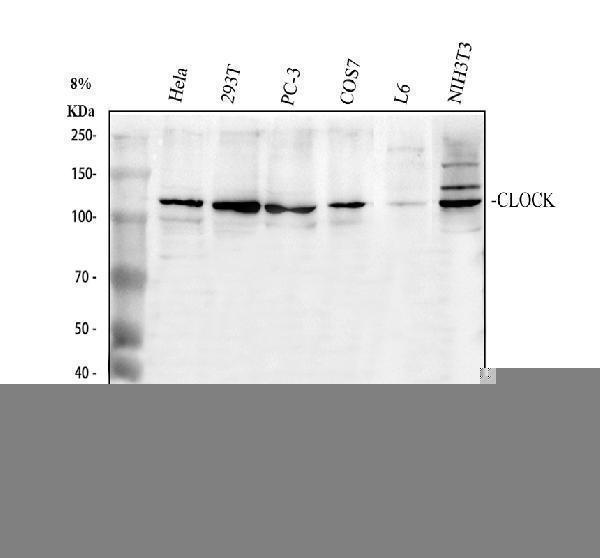
Western blot analysis of KAT13D/CLOCK using anti-KAT13D/CLOCK antibody (RP1082).
Electrophoresis was performed on a 8% SDS-PAGE gel at 80V (Stacking gel) / 120V (Resolving gel) for 2 hours. The sample well of each lane was loaded with 30 ug of sample under reducing conditions.
Lane 1: human Hela whole cell lysates,
Lane 2: human 293T whole cell lysates,
Lane 3: human PC-3 whole cell lysates,
Lane 4: monkey COS-7 whole cell lysates,
Lane 5: rat L6 whole cell lysates,
Lane 6: mouse NIH/3T3 whole cell lysates.
After electrophoresis, proteins were transferred to a nitrocellulose membrane at 150 mA for 50-90 minutes. Blocked the membrane with 5% non-fat milk/TBS for 1.5 hour at RT. The membrane was incubated with rabbit anti-KAT13D/CLOCK antigen affinity purified polyclonal antibody (RP1082) at 0.5 μg/mL overnight at 4°C, then washed with TBS-0.1%Tween 3 times with 5 minutes each and probed with a goat anti-rabbit IgG-HRP secondary antibody (Catalog # BA1054) at a dilution of 1:5000 for 1.5 hour at RT. The signal is developed using an ECL Plus Western Blotting Substrate (Catalog # AR1196-200) with Tanon 5200 system. A specific band was detected for KAT13D/CLOCK at approximately 110 kDa. The expected band size for KAT13D/CLOCK is at 95 kDa.

Click image to see more details
IHC analysis of KAT13D/CLOCK using anti-KAT13D/CLOCK antibody (RP1082).
KAT13D/CLOCK was detected in a paraffin-embedded section of human pancrese cancer tissue. Heat mediated antigen retrieval was performed in EDTA buffer (pH 8.0, epitope retrieval solution). The tissue section was blocked with 10% goat serum. The tissue section was then incubated with 2 μg/ml rabbit anti-KAT13D/CLOCK Antibody (RP1082) overnight at 4°C. Peroxidase Conjugated Goat Anti-rabbit IgG was used as secondary antibody and incubated for 30 minutes at 37°C. The tissue section was developed using HRP Conjugated Rabbit IgG Super Vision Assay Kit (Catalog # SV0002) with DAB as the chromogen.

Click image to see more details
IHC analysis of KAT13D/CLOCK using anti-KAT13D/CLOCK antibody (RP1082).
KAT13D/CLOCK was detected in a paraffin-embedded section of mouse brain tissue. Heat mediated antigen retrieval was performed in EDTA buffer (pH 8.0, epitope retrieval solution). The tissue section was blocked with 10% goat serum. The tissue section was then incubated with 2 μg/ml rabbit anti-KAT13D/CLOCK Antibody (RP1082) overnight at 4°C. Peroxidase Conjugated Goat Anti-rabbit IgG was used as secondary antibody and incubated for 30 minutes at 37°C. The tissue section was developed using HRP Conjugated Rabbit IgG Super Vision Assay Kit (Catalog # SV0002) with DAB as the chromogen.

Click image to see more details
IHC analysis of KAT13D/CLOCK using anti-KAT13D/CLOCK antibody (RP1082).
KAT13D/CLOCK was detected in a paraffin-embedded section of rat brain tissue. Heat mediated antigen retrieval was performed in EDTA buffer (pH 8.0, epitope retrieval solution). The tissue section was blocked with 10% goat serum. The tissue section was then incubated with 2 μg/ml rabbit anti-KAT13D/CLOCK Antibody (RP1082) overnight at 4°C. Peroxidase Conjugated Goat Anti-rabbit IgG was used as secondary antibody and incubated for 30 minutes at 37°C. The tissue section was developed using HRP Conjugated Rabbit IgG Super Vision Assay Kit (Catalog # SV0002) with DAB as the chromogen.

Click image to see more details
IF analysis of KAT13D/CLOCK using anti-KAT13D/CLOCK antibody (RP1082) and anti-Tubulin Alpha antibody (M03989-3).
KAT13D/CLOCK was detected in immunocytochemical section of U2OS cell. Enzyme antigen retrieval was performed using IHC enzyme antigen retrieval reagent (AR0022) for 15 mins. The cells were blocked with 10% goat serum. And then incubated with 5 μg/mL rabbit anti-KAT13D/CLOCK Antibody (RP1082) and mouse anti-Tubulin Alpha antibody (M03989-3) overnight at 4°C. DyLight®488 Conjugated Goat Anti-Rabbit IgG (BA1127) and Cy3 Conjugated Goat Anti-Mouse IgG (BA1031) were used as secondary antibody at 1:500 dilution and incubated for 30 minutes at 37°C. The section was counterstained with DAPI. Visualize using a fluorescence microscope and filter sets appropriate for the label used.

Click image to see more details
Flow Cytometry analysis of Hela cells using anti-KAT13D/CLOCK antibody (RP1082).
Overlay histogram showing Hela cells stained with RP1082 (Blue line). To facilitate intracellular staining, cells were fixed with 4% paraformaldehyde and permeabilized with permeabilization buffer. The cells were blocked with 10% normal goat serum. And then incubated with rabbit anti-KAT13D/CLOCK Antibody (RP1082, 1 μg/1x106 cells) for 30 min at 20°C. DyLight®488 conjugated goat anti-rabbit IgG (BA1127, 5-10 μg/1x106 cells) was used as secondary antibody for 30 minutes at 20°C. Isotype control antibody (Green line) was rabbit IgG (1 μg/1x106) used under the same conditions. Unlabelled sample without incubation with primary antibody and secondary antibody (Red line) was used as a blank control.
Specific Publications For Anti-KAT13D/CLOCK Antibody Picoband® (RP1082)
Loading publications
Recommended Resources
Here are featured tools and databases that you might find useful.
- Boster's Pathways Library
- Protein Databases
- Bioscience Research Protocol Resources
- Data Processing & Analysis Software
- Photo Editing Software
- Scientific Literature Resources
- Research Paper Management Tools
- Molecular Biology Software
- Primer Design Tools
- Bioinformatics Tools
- Phylogenetic Tree Analysis
Customer Reviews
Have you used Anti-KAT13D/CLOCK Antibody Picoband®?
Share your experimental results or join a short interview to earn up to $1,000 in product credits or other rewards.
0 Reviews For Anti-KAT13D/CLOCK Antibody Picoband®
Customer Q&As
Have a question?
Find answers in Q&As, reviews.
Can't find your answer?
Submit your question
5 Customer Q&As for Anti-KAT13D/CLOCK Antibody Picoband®
Question
Please see the WB image, lot number and protocol we used for brain using anti-KAT13D/CLOCK antibody RP1082. Please let me know if you require anything else.
Verified Customer
Verified customer
Asked: 2019-12-18
Answer
Thank you very much for the data. Our lab team are working to resolve this as quickly as possible, and we appreciate your patience and understanding! You have provided everything we needed. Please let me know if there is anything you need in the meantime.
Boster Scientific Support
Answered: 2019-12-18
Question
Would anti-KAT13D/CLOCK antibody RP1082 work for ICC with brain?
Verified Customer
Verified customer
Asked: 2019-10-30
Answer
According to the expression profile of brain, CLOCK is highly expressed in brain. So, it is likely that anti-KAT13D/CLOCK antibody RP1082 will work for ICC with brain.
Boster Scientific Support
Answered: 2019-10-30
Question
We are currently using anti-KAT13D/CLOCK antibody RP1082 for rat tissue, and we are happy with the ICC results. The species of reactivity given in the datasheet says human, mouse, rat. Is it likely that the antibody can work on horse tissues as well?
M. Banerjee
Verified customer
Asked: 2019-08-12
Answer
The anti-KAT13D/CLOCK antibody (RP1082) has not been validated for cross reactivity specifically with horse tissues, but there is a good chance of cross reactivity. We have an innovator award program that if you test this antibody and show it works in horse you can get your next antibody for free. Please contact me if I can help you with anything.
Boster Scientific Support
Answered: 2019-08-12
Question
I see that the anti-KAT13D/CLOCK antibody RP1082 works with ICC, what is the protocol used to produce the result images on the product page?
Verified Customer
Verified customer
Asked: 2019-07-17
Answer
You can find protocols for ICC on the "support/technical resources" section of our navigation menu. If you have any further questions, please send an email to support@bosterbio.com
Boster Scientific Support
Answered: 2019-07-17
Question
Would RP1082 anti-KAT13D/CLOCK antibody work on parafin embedded sections? If so, which fixation method do you recommend we use (PFA, paraformaldehyde, other)?
Verified Customer
Verified customer
Asked: 2018-01-04
Answer
As indicated on the product datasheet, RP1082 anti-KAT13D/CLOCK antibody as been tested on ICC. It is best to use PFA for fixation because it has better tissue penetration ability. PFA needs to be prepared fresh before use. Long term stored PFA turns into formalin, as the PFA molecules congregate and become formalin.
Boster Scientific Support
Answered: 2018-01-04
