Click image to see more details
Product Info Summary
| SKU: | A03994-2 |
|---|---|
| Size: | 100 μl/vial |
| Reactive Species: | Human, Mouse |
| Host: | Rabbit |
| Application: | ELISA, IP, IF, IHC, ICC, WB |
Customers Who Bought This Also Bought

Product info
Product Name
Anti-JMJD1A/KDM3A Antibody
SKU/Catalog Number
A03994-2
Size
100 μl/vial
Form
Liquid
Description
Boster Bio Anti-JMJD1A/KDM3A Antibody catalog # A03994-2. Tested in WB, IHC, ICC, IF, IP, ELISA applications. This antibody reacts with Human, Mouse.
Storage & Handling
12 months from date of receipt,-20℃ as supplied. 6 months 2 to 8℃ after reconstitution. Avoid repeated freezing and thawing.
Cite This Product
Anti-JMJD1A/KDM3A Antibody (Boster Biological Technology, Pleasanton CA, USA, Catalog # A03994-2)
Host
Rabbit
Contents
500 μg/ml antibody with PBS, 0.02% NaN3, 1 mg stabilizing protein and 50% glycerol
*This antibody is supplied in a stabilized formulation.
Compatibility with conjugation reactions depends on the chemistry of the conjugation method used.
For conjugation methods that are not compatible with the stabilizing components present in this formulation, a carrier-free antibody format is required.
Clonality
Polyclonal
Immunogen
E.coli-derived human KDM3A recombinant protein (Position: T4-T359).
Reactive Species
A03994-2 is reactive to KDM3A in Human, Mouse
Calculated molecular weight
147.3 kDa
Background of KDM3A
The jumonji domain containing 1A protein (JMJD1A) was originally discovered as a testes specific gene, but has been found to be expressed in numerous tissues. JMJD1A is a histone demethylase and specifically demethylates mono- and dimethyl-H3K9. It has also been found to be a novel interaction partner with ER71, a transcription factor expressed in the testes of adult mice and during embryogenesis. JMJD1A is also a downstream gene of STAT3, a protein that has been found to be important in the maintenance of mouse embryonic stem (mES) cells, and decreased JMJD1A expression correlated with the differentiation of cultured mES cells following the removal of the cytokine LIF. These findings suggest that JMJD1A might be a critical signaling molecule underlying the maintenance of pluripotency in embryonic stem cells. At least two isoforms of JMJD1A are known to exist.
Antibody Validation
Boster validates all antibodies on WB, IHC, ICC, Immunofluorescence, and ELISA with known positive control and negative samples to ensure specificity and high affinity, including thorough antibody incubations.
Application & Images
Applications
A03994-2 is guaranteed for ELISA, IP, IF, IHC, ICC, WB Boster Guarantee
Recommend Dilution
| Application | Dilution | Species |
|---|---|---|
| Western blot | 1:500-2000 | |
| Immunohistochemistry | 1:50-400 | |
| Immunocytochemistry/Immunofluorescence | 1:50-400 | |
| Immunoprecipitation | 1:50 | |
| ELISA | 1:100-1000 |
Validation Images & Assay Conditions
Click image to see more details
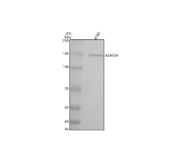
Western blot analysis of JMJD1A/KDM3A using anti-JMJD1A/KDM3A antibody (A03994-2).
Electrophoresis was performed on a 10% SDS-PAGE gel at 80V (Stacking gel) / 120V (Resolving gel) for 2 hours. The sample well of each lane was loaded with 30 ug of sample under reducing conditions.
Lane 1: human K562 whole cell lysates.
After electrophoresis, proteins were transferred to a nitrocellulose membrane at 150 mA for 50-90 minutes. Blocked the membrane with 5% non-fat milk/TBS for 1.5 hour at RT. The membrane was incubated with rabbit anti-JMJD1A/KDM3A antigen affinity purified polyclonal antibody (A03994-2) at 1:1000 overnight at 4°C, then washed with TBS-0.1%Tween 3 times with 5 minutes each and probed with a goat anti-rabbit IgG-HRP secondary antibody at a dilution of 1:5000 for 1.5 hour at RT. The signal is developed using an ECL Plus Western Blotting Substrate (Catalog # AR1196-200) with Tanon 5200 system. A specific band was detected for JMJD1A/KDM3A at approximately 150 kDa. The expected band size for JMJD1A/KDM3A is at 147 kDa.

Click image to see more details
IHC analysis of JMJD1A/KDM3A using anti-JMJD1A/KDM3A antibody (A03994-2).
JMJD1A/KDM3A was detected in a paraffin-embedded section of human stomach cancer tissue. Heat mediated antigen retrieval was performed in EDTA buffer (pH 8.0, epitope retrieval solution). The tissue section was blocked with 10% goat serum. The tissue section was then incubated with 2 μg/ml rabbit anti-JMJD1A/KDM3A Antibody (A03994-2) overnight at 4°C. Peroxidase Conjugated Goat Anti-rabbit IgG was used as secondary antibody and incubated for 30 minutes at 37°C. The tissue section was developed using HRP Conjugated Rabbit IgG Super Vision Assay Kit (Catalog # SV0002) with DAB as the chromogen.
Specific Publications For Anti-JMJD1A/KDM3A Antibody (A03994-2)
Loading publications
Recommended Resources
Here are featured tools and databases that you might find useful.
- Boster's Pathways Library
- Protein Databases
- Bioscience Research Protocol Resources
- Data Processing & Analysis Software
- Photo Editing Software
- Scientific Literature Resources
- Research Paper Management Tools
- Molecular Biology Software
- Primer Design Tools
- Bioinformatics Tools
- Phylogenetic Tree Analysis
Customer Reviews
Have you used Anti-JMJD1A/KDM3A Antibody?
Share your experimental results or join a short interview to earn up to $1,000 in product credits or other rewards.
0 Reviews For Anti-JMJD1A/KDM3A Antibody
Customer Q&As
Have a question?
Find answers in Q&As, reviews.
Can't find your answer?
Submit your question
