Click image to see more details
Product Info Summary
| SKU: | M03214 |
|---|---|
| Size: | 100 μl/vial |
| Reactive Species: | Human, Mouse, Rat |
| Host: | Rabbit |
| Application: | Flow Cytometry, IP, IF, IHC, ICC, WB |
Customers Who Bought This Also Bought

Product info
Product Name
Anti-Laminin gamma 2/LAMC2 Antibody (Monoclonal, 33L12)
SKU/Catalog Number
M03214
Size
100 μl/vial
Form
Liquid
Description
Boster Bio Anti-Laminin gamma 2/LAMC2 Antibody (Monoclonal, 33L12) catalog # M03214. Tested in WB, IHC, IF, ICC/IF, IP, Flow Cytometry applications. This antibody reacts with Human, Mouse, Rat.
Storage & Handling
Store at -20°C for one year. For short term storage and frequent use, store at 4°C for up to one month. Avoid repeated freeze-thaw cycles.
Cite This Product
Anti-Laminin gamma 2/LAMC2 Antibody (Monoclonal, 33L12) (Boster Biological Technology, Pleasanton CA, USA, Catalog # M03214)
Host
Rabbit
Contents
Rabbit IgG in stabilizing components, phosphate buffered saline, pH 7.4, 150mM NaCl, 0.02% sodium azide and 50% glycerol.
This antibody is supplied in a stabilized formulation.
Compatibility with conjugation reactions depends on the chemistry of the conjugation method used.
For conjugation methods that are not compatible with the stabilizing components present in this formulation, a carrier-free antibody format is required.
Clonality
Monoclonal
Clone Number
33L12
Immunogen
Recombinant protein within human LAMC2 aa 552-1191.
Reactive Species
M03214 is reactive to LAMC2 in Human, Mouse, Rat
Observed Molecular Weight
140 kDa
Calculated molecular weight
131.0 kDa
Background of LAMC2
Laminin gamma2, Laminin subunit gamma-2, is a protein that in humans is encoded by the LAMC2 gene. Laminins, a family of extracellular matrix glycoproteins, are the major noncollagenous constituent of basement membranes. They have been implicated in a wide variety of biological processes including cell adhesion, differentiation, migration, signaling, neurite outgrowth and metastasis. Laminins are composed of 3 non identical chains: laminin alpha, beta and gamma(formerly A, B1, and B2, respectively) and they form a cruciform structure consisting of 3 short arms, each formed by a different chain, and a long arm composed of all 3 chains. Each laminin chain is a multidomain protein encoded by a distinct gene. Several isoforms of each chain have been described. Different alpha, beta and gamma chain isomers combine to give rise to different heterotrimeric laminin isoforms which are designated by Arabic numerals in the order of their discovery, i.e. alpha1beta1gamma1 heterotrimer is laminin 1. The biological functions of the different chains and trimer molecules are largely unknown, but some of the chains have been shown to differ with respect to their tissue distribution, presumably reflecting diverse functions in vivo. This gene encodes the gamma chain isoform laminin, gamma 2. The gamma 2 chain, formerly though to be a truncated version of beta chain(B2t), is highly homologous to the gamma 1 chain; however, it lacks domain VI, and domains V, IV and III are shorter. It is expressed in several fetal tissues but differently from gamma 1, and is specifically localized to epithelial cells in skin, lung and kidney. The gamma 2 chain together with alpha 3 and beta 3 chains constitute laminin 5(earlier known as kalinin), which is an integral part of the anchoring filaments that connect epithelial cells to the underlying basement membrane. The epithelium-specific expression of the gamma 2 chain implied its role as an epithelium attachment molecule, and mutations in this gene have been associated with junctional epidermolysis bullosa, a skin disease characterized.
Antibody Validation
Boster validates all antibodies on WB, IHC, ICC, Immunofluorescence, and ELISA with known positive control and negative samples to ensure specificity and high affinity, including thorough antibody incubations.
Application & Images
Applications
M03214 is guaranteed for Flow Cytometry, IP, IF, IHC, ICC, WB Boster Guarantee
Recommend Dilution
| Application | Dilution | Species |
|---|---|---|
| Western blot | 1:500-2000 | |
| Immunohistochemistry | 1:50-200 | |
| Immunofluorescence | 1:50-200 | |
| Immunocytochemistry/Immunofluorescence | 1:50-200 | |
| ImmunoPrecipitation | 1:50 | |
| Flow Cytometry (Fixed) | 1:50-200 |
Tested application
Suggested blocking solution with 5% non-fat milk or BSA; (*)Recommended protein loading: 20-40 µg per lane
Use TE buffer pH 9.0 for antigen retrieval; (*) citrate buffer pH 6.0 is an alternative.
Validation Images & Assay Conditions
Click image to see more details
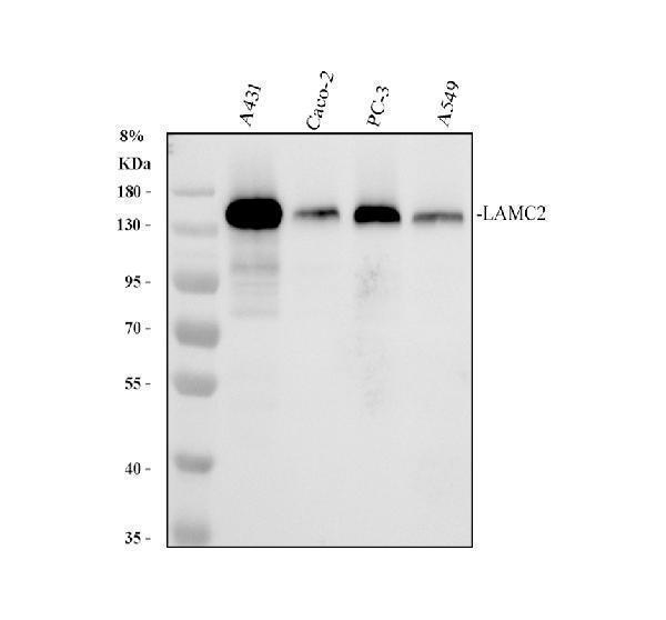
Western blot analysis of LAMC2/MRC1 using anti-LAMC2/MRC1 antibody (M03214).
Electrophoresis was performed on a 8% SDS-PAGE gel at 80V (Stacking gel) / 120V (Resolving gel) for 2 hours. The sample well of each lane was loaded with 30 ug of sample under reducing conditions.
Lane 1: human A431 whole cell lysates,
Lane 2: human CACO-2 whole cell lysates,
Lane 3: human PC-3 whole cell lysates,
Lane 4: human A549 whole cell lysates.
After electrophoresis, proteins were transferred to a nitrocellulose membrane at 150 mA for 50-90 minutes. Blocked the membrane with 5% non-fat milk/TBS for 1.5 hour at RT. The membrane was incubated with rabbit anti-LAMC2/MRC1 antigen affinity purified monoclonal antibody (M03214) at 1:1000 overnight at 4°C, then washed with TBS-0.1%Tween 3 times with 5 minutes each and probed with a goat anti-rabbit IgG-HRP secondary antibody at a dilution of 1:5000 for 1.5 hour at RT. The signal is developed using an ECL Plus Western Blotting Substrate (Catalog # AR1196-200) with Tanon 5200 system. A specific band was detected for LAMC2/MRC1 at approximately 140 kDa. The expected band size for LAMC2/MRC1 is at 131 kDa.

Click image to see more details
IHC analysis of LAMC2/MRC1 using anti-LAMC2/MRC1 antibody (M03214).
LAMC2/MRC1 was detected in a paraffin-embedded section of human lung cancer tissue. Heat mediated antigen retrieval was performed in EDTA buffer (pH 8.0, epitope retrieval solution). The tissue section was blocked with 10% goat serum. The tissue section was then incubated with 1:200 rabbit anti-LAMC2/MRC1 Antibody (M03214) overnight at 4°C. Peroxidase Conjugated Goat Anti-rabbit IgG was used as secondary antibody and incubated for 30 minutes at 37°C. The tissue section was developed using HRP Conjugated Rabbit IgG Super Vision Assay Kit (Catalog # SV0002) with DAB as the chromogen.

Click image to see more details
IHC analysis of Laminin gamma 2/LAMC2 using anti-Laminin gamma 2/LAMC2 antibody (M03214).
Laminin gamma 2/LAMC2 was detected in a paraffin-embedded section of human pancreas cancer tissue. Heat mediated antigen retrieval was performed in EDTA buffer (pH 8.0, epitope retrieval solution). The tissue section was blocked with 10% goat serum. The tissue section was then incubated with 1:50 rabbit anti-Laminin gamma 2/LAMC2 Antibody (M03214) overnight at 4°C. Peroxidase Conjugated Goat Anti-rabbit IgG was used as secondary antibody and incubated for 30 minutes at 37°C. The tissue section was developed using HRP Conjugated Rabbit IgG Super Vision Assay Kit (Catalog # SV0002) with DAB as the chromogen.

Click image to see more details
IHC analysis of Laminin gamma 2/LAMC2 using anti-Laminin gamma 2/LAMC2 antibody (M03214).
Laminin gamma 2/LAMC2 was detected in a paraffin-embedded section of human colon cancer tissue. Heat mediated antigen retrieval was performed in EDTA buffer (pH 8.0, epitope retrieval solution). The tissue section was blocked with 10% goat serum. The tissue section was then incubated with 1:50 rabbit anti-Laminin gamma 2/LAMC2 Antibody (M03214) overnight at 4°C. Peroxidase Conjugated Goat Anti-rabbit IgG was used as secondary antibody and incubated for 30 minutes at 37°C. The tissue section was developed using HRP Conjugated Rabbit IgG Super Vision Assay Kit (Catalog # SV0002) with DAB as the chromogen.
Specific Publications For Anti-Laminin gamma 2/LAMC2 Antibody (Monoclonal, 33L12) (M03214)
Loading publications
Recommended Resources
Here are featured tools and databases that you might find useful.
- Boster's Pathways Library
- Protein Databases
- Bioscience Research Protocol Resources
- Data Processing & Analysis Software
- Photo Editing Software
- Scientific Literature Resources
- Research Paper Management Tools
- Molecular Biology Software
- Primer Design Tools
- Bioinformatics Tools
- Phylogenetic Tree Analysis
Customer Reviews
Have you used Anti-Laminin gamma 2/LAMC2 Antibody (Monoclonal, 33L12)?
Share your experimental results or join a short interview to earn up to $1,000 in product credits or other rewards.
0 Reviews For Anti-Laminin gamma 2/LAMC2 Antibody (Monoclonal, 33L12)
Customer Q&As
Have a question?
Find answers in Q&As, reviews.
Can't find your answer?
Submit your question
