Click image to see more details
-

-
-
-
-
+22
Product Info Summary
| SKU: | PB10075 |
|---|---|
| Size: | 100 μg/vial |
| Reactive Species: | Human, Mouse, Rat |
| Host: | Rabbit |
| Application: | Flow Cytometry, IF, IHC, ICC, WB |
Customers Who Bought This Also Bought

Product info
Product Name
Anti-LDHA Antibody Picoband®
SKU/Catalog Number
PB10075
Size
100 μg/vial
Form
Lyophilized
Description
Boster Bio Anti-LDHA Antibody Picoband® catalog # PB10075. Tested in Flow Cytometry, IF, IHC, ICC, WB applications. This antibody reacts with Human, Mouse, Rat. The brand Picoband indicates this is a premium antibody that guarantees superior quality, high affinity, and strong signals with minimal background in Western blot applications. Only our best-performing antibodies are designated as Picoband, ensuring unmatched performance.
Storage & Handling
Store at -20˚C for one year from date of receipt. After reconstitution, at 4˚C for one month. It can also be aliquotted and stored frozen at -20˚C for six months. Avoid repeated freeze-thaw cycles.
Cite This Product
Anti-LDHA Antibody Picoband® (Boster Biological Technology, Pleasanton CA, USA, Catalog # PB10075)
Host
Rabbit
Contents
Each vial contains antibody formulated with stabilizing components, 0.9 mg NaCl, 0.2 mg Na2HPO4, and 0.05 mg NaN3.
*This antibody is supplied in a stabilized formulation.
Compatibility with conjugation reactions depends on the chemistry of the conjugation method used.
For conjugation methods that are not compatible with the stabilizing components present in this formulation, a carrier-free antibody format is required.
Clonality
Polyclonal
Isotype
Rabbit IgG
Immunogen
E. coli-derived human LDHA recombinant protein (Position: A2-R106). Human LDHA shares 94.3% amino acid (aa) sequence identity with both mouse and rat LDHA.
Cross-reactivity
No cross-reactivity with other proteins
Reactive Species
PB10075 is reactive to LDHA in Human, Mouse, Rat
Observed Molecular Weight
37 kDa
Calculated molecular weight
36.7 kDa
Background of LDHA
Lactate dehydrogenase A, also known as LDHA, is an enzyme which in humans is encoded by the LDHA gene. The protein encoded by this gene catalyzes the conversion of L-lactate and NAD to pyruvate and NADH in the final step of anaerobic glycolysis. The protein is found predominantly in muscle tissue and belongs to the lactate dehydrogenase family. Mutations in this gene have been linked to exertional myoglobinuria. Multiple transcript variants encoding different isoforms have been found for this gene. The human genome contains several non-transcribed pseudogenes of this gene.
Antibody Validation
Boster validates all antibodies on WB, IHC, ICC, Immunofluorescence, and ELISA with known positive control and negative samples to ensure specificity and high affinity, including thorough antibody incubations.
Application & Images
Applications
PB10075 is guaranteed for Flow Cytometry, IF, IHC, ICC, WB Boster Guarantee
Recommend Dilution
| Application | Dilution | Species |
|---|---|---|
| Western blot | 0.1-0.5μg/ml | Human, Mouse, Rat |
| Immunohistochemistry (Paraffin-embedded Section) | 0.5-1μg/ml | Human, Mouse, Rat |
| Immunocytochemistry/Immunofluorescence | 2μg/ml | Human |
| Flow Cytometry (Fixed) | 1-3μg/1x106 cells | Human |
Tested application
Suggested blocking solution with 5% non-fat milk or BSA; (*)Recommended protein loading: 20-40 µg per lane
Use TE buffer pH 9.0 for antigen retrieval; (*) citrate buffer pH 6.0 is an alternative.
Validation Images & Assay Conditions
Click image to see more details
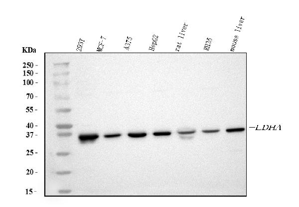
Western blot analysis of LDHA using anti-LDHA antibody (PB10075).
Electrophoresis was performed on a 5-20% SDS-PAGE gel at 70V (Stacking gel) / 90V (Resolving gel) for 2-3 hours. The sample well of each lane was loaded with 30 ug of sample under reducing conditions.
Lane 1: human 293T whole cell lysates,
Lane 2: human MCF-7 whole cell lysates,
Lane 3: human A375 whole cell lysates,
Lane 4: human HepG2 whole cell lysates,
Lane 5: rat liver tissue lysates,
Lane 6: rat RH35 whole cell lysates,
Lane 7: mouse liver tissue lysates.
After electrophoresis, proteins were transferred to a nitrocellulose membrane at 150 mA for 50-90 minutes. Blocked the membrane with 5% non-fat milk/TBS for 1.5 hour at RT. The membrane was incubated with rabbit anti-LDHA antigen affinity purified polyclonal antibody (Catalog # PB10075) at 0.5 μg/mL overnight at 4°C, then washed with TBS-0.1%Tween 3 times with 5 minutes each and probed with a goat anti-rabbit IgG-HRP secondary antibody at a dilution of 1:5000 for 1.5 hour at RT. The signal is developed using an Enhanced Chemiluminescent detection (ECL) kit (Catalog # EK1002) with Tanon 5200 system. A specific band was detected for LDHA at approximately 37 kDa. The expected band size for LDHA is at 37 kDa.

Click image to see more details
Western blot analysis of LDHA using anti-LDHA antibody (PB10075).
Electrophoresis was performed on a 10% SDS-PAGE gel at 80V (Stacking gel) / 120V (Resolving gel) for 2 hours. The sample well of each lane was loaded with 30 ug of sample under reducing conditions.
Lane 1: human MSTO-211H- WT whole cell lysates,
Lane 2: human MSTO-211H-LDHA KO whole cell lysates.
After electrophoresis, proteins were transferred to a nitrocellulose membrane at 150 mA for 50-90 minutes. Blocked the membrane with 5% non-fat milk/TBS for 1.5 hour at RT. Then the membrane was incubated with rabbit anti-LDHA antigen affinity purified polyclonal antibody (PB10075) at 0.5 μg/mL overnight at 4°C, then washed with TBS-0.1%Tween 3 times with 5 minutes each and probed with a goat anti-rabbit IgG-HRP secondary antibody (Catalog # BA1054) at a dilution of 1:5000 for 1.5 hour at RT. The signal is developed using an ECL Plus Western Blotting Substrate (Catalog # AR1196-200) with Tanon 5200 system. A specific band was detected for LDHA at approximately 37 kDa. The expected band size for LDHA is at 37 kDa.

Click image to see more details
IHC analysis of LDHA using anti-LDHA antibody (PB10075).
LDHA was detected in a paraffin-embedded section of mouse intestine tissue. Heat mediated antigen retrieval was performed in EDTA buffer (pH 8.0, epitope retrieval solution). The tissue section was blocked with 10% goat serum. The tissue section was then incubated with 1 μg/ml rabbit anti-LDHA Antibody (PB10075) overnight at 4°C. Biotinylated goat anti-rabbit IgG was used as secondary antibody and incubated for 30 minutes at 37°C. The tissue section was developed using Strepavidin-Biotin-Complex (SABC) (Catalog # SA1022) with DAB as the chromogen.

Click image to see more details
IHC analysis of LDHA using anti-LDHA antibody (PB10075).
LDHA was detected in a paraffin-embedded section of rat intestine tissue. Heat mediated antigen retrieval was performed in EDTA buffer (pH 8.0, epitope retrieval solution). The tissue section was blocked with 10% goat serum. The tissue section was then incubated with 1 μg/ml rabbit anti-LDHA Antibody (PB10075) overnight at 4°C. Biotinylated goat anti-rabbit IgG was used as secondary antibody and incubated for 30 minutes at 37°C. The tissue section was developed using Strepavidin-Biotin-Complex (SABC) (Catalog # SA1022) with DAB as the chromogen.

Click image to see more details
IHC analysis of LDHA using anti-LDHA antibody (PB10075).
LDHA was detected in a paraffin-embedded section of human mammary cancer tissue. Heat mediated antigen retrieval was performed in EDTA buffer (pH 8.0, epitope retrieval solution). The tissue section was blocked with 10% goat serum. The tissue section was then incubated with 1 μg/ml rabbit anti-LDHA Antibody (PB10075) overnight at 4°C. Biotinylated goat anti-rabbit IgG was used as secondary antibody and incubated for 30 minutes at 37°C. The tissue section was developed using Strepavidin-Biotin-Complex (SABC) (Catalog # SA1022) with DAB as the chromogen.

Click image to see more details
IHC analysis of LDHAusing anti-LDHA antibody (PB10075).
LDHA was detected in a paraffin-embedded section of mouse muscle skeletal tissue. Heat mediated antigen retrieval was performed in EDTA buffer (pH 8.0, epitope retrieval solution). The tissue section was blocked with 10% goat serum. The tissue section was then incubated with 1 μg/ml rabbit anti-LDHA Antibody (PB10075) overnight at 4°C. HRP-AffiniPure Goat Anti-Rabbit IgG was used as secondary antibody and incubated for 30 minutes at 37°C. The tissue section was developed using HRP Conjugated Rabbit IgG Super Vision Assay Kit (Catalog # SV0002) with DAB as the chromogen.

Click image to see more details
IHC analysis of LDHA using anti-LDHA antibody (PB10075).
LDHA was detected in a paraffin-embedded section of rat muscle skeletal tissue. Heat mediated antigen retrieval was performed in EDTA buffer (pH 8.0, epitope retrieval solution). The tissue section was blocked with 10% goat serum. The tissue section was then incubated with 1 μg/ml rabbit anti-LDHA Antibody (PB10075) overnight at 4°C. HRP-AffiniPure Goat Anti-Rabbit IgG was used as secondary antibody and incubated for 30 minutes at 37°C. The tissue section was developed using HRP Conjugated Rabbit IgG Super Vision Assay Kit (Catalog # SV0002) with DAB as the chromogen.

Click image to see more details
IHC analysis of LDHA using anti-LDHA antibody (PB10075).
LDHA was detected in a paraffin-embedded section of human stomach cancer tissue. Heat mediated antigen retrieval was performed in EDTA buffer (pH 8.0, epitope retrieval solution). The tissue section was blocked with 10% goat serum. The tissue section was then incubated with 1 μg/ml rabbit anti-LDHA Antibody (PB10075) overnight at 4°C. HRP-AffiniPure Goat Anti-Rabbit IgG was used as secondary antibody and incubated for 30 minutes at 37°C. The tissue section was developed using HRP Conjugated Rabbit IgG Super Vision Assay Kit (Catalog # SV0002) with DAB as the chromogen.

Click image to see more details
IHC analysis of LDHA using anti-LDHA antibody (PB10075).
LDHA was detected in a paraffin-embedded section of human thyroid cancer tissue. Heat mediated antigen retrieval was performed in EDTA buffer (pH 8.0, epitope retrieval solution). The tissue section was blocked with 10% goat serum. The tissue section was then incubated with 1 μg/ml rabbit anti-LDHA Antibody (PB10075) overnight at 4°C. HRP-AffiniPure Goat Anti-Rabbit IgG was used as secondary antibody and incubated for 30 minutes at 37°C. The tissue section was developed using HRP Conjugated Rabbit IgG Super Vision Assay Kit (Catalog # SV0002) with DAB as the chromogen.

Click image to see more details
IHC analysis of LDHA using anti-LDHA antibody (PB10075).
LDHA was detected in a paraffin-embedded section of human pancreas cancer tissue. Heat mediated antigen retrieval was performed in EDTA buffer (pH 8.0, epitope retrieval solution). The tissue section was blocked with 10% goat serum. The tissue section was then incubated with 1 μg/ml rabbit anti-LDHA Antibody (PB10075) overnight at 4°C. HRP-AffiniPure Goat Anti-Rabbit IgG was used as secondary antibody and incubated for 30 minutes at 37°C. The tissue section was developed using HRP Conjugated Rabbit IgG Super Vision Assay Kit (Catalog # SV0002) with DAB as the chromogen.

Click image to see more details
IF analysis of LDHA using anti-LDHA antibody (PB10075).
LDHA was detected in immunocytochemical section of U20S cells. Enzyme antigen retrieval was performed using IHC enzyme antigen retrieval reagent (AR0022) for 15 mins. The cells were blocked with 10% goat serum. And then incubated with 2μg/mL rabbit anti-LDHA Antibody (PB10075) overnight at 4°C. DyLight®594 Conjugated Goat Anti-Rabbit IgG (BA1142) was used as secondary antibody at 1:100 dilution and incubated for 30 minutes at 37°C. The section was counterstained with DAPI. Visualize using a fluorescence microscope and filter sets appropriate for the label used.

Click image to see more details
IF analysis of LDHA using anti-LDHA antibody (PB10075).
LDHA was detected in a paraffin-embedded section of mouse muscle skeletal tissue. Heat mediated antigen retrieval was performed in EDTA buffer (pH 8.0, epitope retrieval solution). The tissue section was blocked with 10% goat serum. The tissue section was then incubated with 25 μg/mL rabbit anti-LDHA Antibody (PB10075) overnight at 4°C. DyLight®594 Conjugated Goat Anti-Rabbit IgG (BA1142) was used as secondary antibody at 1:100 dilution and incubated for 30 minutes at 37°C. The section was counterstained with DAPI. Visualize using a fluorescence microscope and filter sets appropriate for the label used.

Click image to see more details
IF analysis of LDHA using anti-LDHA antibody (PB10075).
LDHA was detected in a paraffin-embedded section of rat muscle skeletal tissue. Heat mediated antigen retrieval was performed in EDTA buffer (pH 8.0, epitope retrieval solution). The tissue section was blocked with 10% goat serum. The tissue section was then incubated with 25 μg/mL rabbit anti-LDHA Antibody (PB10075) overnight at 4°C. DyLight®594 Conjugated Goat Anti-Rabbit IgG (BA1142) was used as secondary antibody at 1:100 dilution and incubated for 30 minutes at 37°C. The section was counterstained with DAPI. Visualize using a fluorescence microscope and filter sets appropriate for the label used.

Click image to see more details
IF analysis of LDHA using anti-LDHA antibody (PB10075).
LDHA was detected in a paraffin-embedded section of human liver cancer tissue. Heat mediated antigen retrieval was performed in EDTA buffer (pH 8.0, epitope retrieval solution). The tissue section was blocked with 10% goat serum. The tissue section was then incubated with 25 μg/mL rabbit anti-LDHA Antibody (PB10075) overnight at 4°C. DyLight®594 Conjugated Goat Anti-Rabbit IgG (BA1142) was used as secondary antibody at 1:100 dilution and incubated for 30 minutes at 37°C. The section was counterstained with DAPI. Visualize using a fluorescence microscope and filter sets appropriate for the label used.

Click image to see more details
IF analysis of LDHA using anti-LDHA antibody (PB10075).
LDHA was detected in a paraffin-embedded section of human stomach cancer tissue. Heat mediated antigen retrieval was performed in EDTA buffer (pH 8.0, epitope retrieval solution). The tissue section was blocked with 10% goat serum. The tissue section was then incubated with 25 μg/mL rabbit anti-LDHA Antibody (PB10075) overnight at 4°C. DyLight®594 Conjugated Goat Anti-Rabbit IgG (BA1142) was used as secondary antibody at 1:100 dilution and incubated for 30 minutes at 37°C. The section was counterstained with DAPI. Visualize using a fluorescence microscope and filter sets appropriate for the label used.

Click image to see more details
IF analysis of LDHA using anti-LDHA antibody (PB10075).
LDHA was detected in a paraffin-embedded section of human pancreas cancer tissue. Heat mediated antigen retrieval was performed in EDTA buffer (pH 8.0, epitope retrieval solution). The tissue section was blocked with 10% goat serum. The tissue section was then incubated with 25 μg/mL rabbit anti-LDHA Antibody (PB10075) overnight at 4°C. DyLight®594 Conjugated Goat Anti-Rabbit IgG (BA1142) was used as secondary antibody at 1:100 dilution and incubated for 30 minutes at 37°C. The section was counterstained with DAPI. Visualize using a fluorescence microscope and filter sets appropriate for the label used.

Click image to see more details
Flow Cytometry analysis of A549 cells using anti-LDHA antibody (PB10075).
Overlay histogram showing A549 cells stained with PB10075 (Blue line). To facilitate intracellular staining, cells were fixed with 4% paraformaldehyde and permeabilized with permeabilization buffer. The cells were blocked with 10% normal goat serum. And then incubated with rabbit anti-LDHA Antibody (PB10075,1μg/1x106 cells) for 30 min at 20°C. DyLight®488 conjugated goat anti-rabbit IgG (BA1127, 5-10μg/1x106 cells) was used as secondary antibody for 30 minutes at 20°C. Isotype control antibody (Green line) was rabbit IgG (1μg/1x106) used under the same conditions. Unlabelled sample without incubation with primary antibody and secondary antibody (Red line) was used as a blank control.

Click image to see more details
TPX2 regulated proliferation, apoptosis, and aerobic glycolysis in glioma cells. a – l LN229 and U251 cells were introduced with si-NC or si-TPX2. a The transfection efficiency of si-TPX2 was checked with RT-qPCR assay in LN229 and U251 cells. b , c The cell viability of LN229 and U251 cells was determined with MTT assay. d The apoptosis rate of transfected LN229 and U251 cells was represented by flow cytometry assay. e The western blot assay was used to assay the expression levels of Bcl-2 and Bax in LN229 and U251 cells. f The activity of caspase-3 was detected with a caspase-3 assay kit. g – i The glucose, lactate, and ATP production levels were shown. j The protein expression levels of HK2 and LDHA were estimated by western blot assay in LN229 and U251 cells. k , l LDHA enzyme activity and ROS content were evaluated in LN229 and U251 cells post-transfection. * P < 0.05
Index in PubMed under a CC BY license. PMID: 32774168

Click image to see more details
The influences of circPOSTN silencing on proliferation, apoptosis and aerobic glycolysis of glioma cells. a – l LN229 and U251 cells were transfected with si-circPOSTN or si-NC. a The interference efficiency of si-circPOSTN was analyzed with RT-qPCR assay in LN229 and U251 cells. b , c Effect of circPOSTN silencing on the cell viability of LN229 and U251 cells was assessed with MTT assay. d The apoptosis rate was computed with flow cytometry assay in transfected LN229 and U251 cells. e The western blot assay showed the expression levels of Bcl-2 and Bax in LN229 and U251 cells. f The caspase-3 activity was measured with a caspase-3 assay kit. g – i The concentration of glucose and lactate in the culture medium, as well as ATP production level were measured with a series of kits, respectively. j The protein expression levels of HK2 and LDHA were determined with western blot assay in transfected LN229 and U251 cells. k – l LDHA enzyme activity and ROS accumulation were evaluated in LN229 and U251 cells post-transfection with lactate dehydrogenase activity detection kit and reactive oxygen species assay kit, respectively. * P < 0.05
Index in PubMed under a CC BY license. PMID: 32774168

Click image to see more details
CircPOSTN silencing inhibited aerobic glycolysis of glioma cells via regulating miR-361-5p. a – l LN229 and U251 cells were transfected with si-NC, si-circPOSTN, si-circPOSTN + anti-miR-NC, or si-circPOSTN + anti-miR-361-5p. a – f The concentration of glucose and lactate, as well as cellular ATP level were detected with different kits. g , h The protein expression levels of HK2 and LDHA in LN229 and U251 cells were measured with western blot assay. i – l The enzyme activity of LDHA and ROS level were measured in transfected LN229 and U251 cells. * P < 0.05
Index in PubMed under a CC BY license. PMID: 32774168

Click image to see more details
Associations between LDHA expression and ferroptosis-related genes in EC. (A) Connection of LDHA to ferroptosis-related genes in and TCGA-UCEC cohort. (B) Connection of LDHA to FANCD2 and TFRC in TCGA-UCEC cohort. (C) Connection of LDHA to FANCD2 and TFRC in . (D) The differential expression of ferroptosis-related genes between high and low LDHA groups in the TCGA-UCEC cohort. (E) Hub genes of expression association and differential expression. (F, G) The Kaplan–Meier curve of hub genes. (H, I) The changes of ferroptosis-related genes after LDHA knockdown in EC cells. * P <0.05, ** P <0.01, *** P <0.001.
Index in PubMed under a CC BY license. PMID: 39582531

Click image to see more details
Associations between LDHA expression and m6A-related genes in EC. (A) Connection of LDHA to m6A-related genes in and TCGA-UCEC cohort. (B) Connection of LDHA to ALKBH5 and HNRNPC in TCGA-UCEC cohort. (C) Connection of LDHA to ALKBH5 and HNRNPC in . (D) The differential expression of m6A-related genes between high and low LDHA groups in the TCGA-UCEC cohort. (E) Hub genes of expression association and differential expression. (F) The Kaplan–Meier curve of hub genes. * P <0.05, *** P <0.001.
Index in PubMed under a CC BY license. PMID: 39582531

Click image to see more details
Influence of LDHA knockdown on EC functions. (A) LDHA protein level after LDHA knockdown detected by Western Blot. (B, C) Cell proliferation after LDHA knockdown detected by CCK-8 assay. (D, E) Cell proliferation after LDHA knockdown detected by cell clone formation assay. (F, G) . Cell apoptosis after LDHA knockdown detected by flow cytometry. (H, I) Cell migration after LDHA knockdown was detected by the Transwell assay. (J, K) Cell invasion after LDHA knockdown was detected by the Transwell assay. * P <0.05.
Index in PubMed under a CC BY license. PMID: 39582531

Click image to see more details
Associations between LDHA and tumor immune infiltrating cells. (A) Correlation of LDHA to stromal cells and immune cells calculated by the ESTIMATE method. (B) Relationship between LDHA expression and infiltration levels of immune cells. (C) Enrichment scores of immune cells in the high LDHA group and low LDHA group. (D) Infiltration levels of immune cells in WT LDHA group and mutated LDHA group. (E) The survival curves of patients with different combinations of LDHA and immune cells. WT, wild type. * P <0.05, ** P <0.01, *** P <0.001.
Index in PubMed under a CC BY license. PMID: 39582531

Click image to see more details
Enrichment analysis of LDHA-related genes in EC. (A) LDHA-related genes in TCGA-UCEC cohort detected by the LinkedOmics database. The top 50 co-expression genes positively (B) and negatively (C) associated with LDHA in the TCGA-UCEC cohort. (D–F) Enrichment analysis of (GO) terms for LDHA-related genes. (G) Enrichment analysis of KEGG terms for LDHA-related genes.
Index in PubMed under a CC BY license. PMID: 39582531

Click image to see more details
The LDHA expression in endometrial cancer. (A) The LDHA expression summarized in the TCGA-UCEC cohort. (B) LDHA expression in paired tumor/normal EC tissues based on TCGA-UCEC cohort. (C) ROC curve analysis of LDHA. (D) The LDHA mRNA in endometrial cells detected by qRT-PCR. (E) LDHA protein stained in normal endometrial tissues by the HPA database. (F) LDHA protein stained in EC tissues by the HPA database. (G) The relationship between LDHA and clinicopathologic features. * P <0.05, ** P <0.01, *** P <0.001.
Index in PubMed under a CC BY license. PMID: 39582531
Specific Publications For Anti-LDHA Antibody Picoband® (PB10075)
Loading publications
Recommended Resources
Here are featured tools and databases that you might find useful.
- Boster's Pathways Library
- Protein Databases
- Bioscience Research Protocol Resources
- Data Processing & Analysis Software
- Photo Editing Software
- Scientific Literature Resources
- Research Paper Management Tools
- Molecular Biology Software
- Primer Design Tools
- Bioinformatics Tools
- Phylogenetic Tree Analysis
Customer Reviews
Have you used Anti-LDHA Antibody Picoband®?
Share your experimental results or join a short interview to earn up to $1,000 in product credits or other rewards.
0 Reviews For Anti-LDHA Antibody Picoband®
Customer Q&As
Have a question?
Find answers in Q&As, reviews.
Can't find your answer?
Submit your question
16 Customer Q&As for Anti-LDHA Antibody Picoband®
Question
We need using your anti-LDHA antibody for pyruvate metabolism studies. Has this antibody been tested with western blotting on jurkat whole cell lysates? We would like to see some validation images before ordering.
Verified Customer
Verified customer
Asked: 2020-04-21
Answer
We appreciate your inquiry. This PB10075 anti-LDHA antibody is validated on jurkat whole cell lysates, a549 cells. It is guaranteed to work for Flow Cytometry, IHC-P, IHC-F, ICC, WB in human, mouse, rat. Our Boster guarantee will cover your intended experiment even if the sample type has not been be directly tested.
Boster Scientific Support
Answered: 2020-04-21
Question
Is a blocking peptide available for product anti-LDHA antibody (PB10075)?
Verified Customer
Verified customer
Asked: 2019-11-25
Answer
We do provide the blocking peptide for product anti-LDHA antibody (PB10075). If you would like to place an order for it please contact support@bosterbio.com and make a special request.
Boster Scientific Support
Answered: 2019-11-25
Question
I have a question about product PB10075, anti-LDHA antibody. I was wondering if it would be possible to conjugate this antibody with biotin. I would need it to be without BSA or sodium azide. I am planning on using a buffer exchange of sodium azide with PBS only. Would there be problems for me to conjugate the antibody and store it in -20 degrees in small aliquots?
Verified Customer
Verified customer
Asked: 2019-10-01
Answer
It is not recommended storing this antibody with PBS buffer only in -20 degrees. If you want to store it in -20 degrees it is best to add some cryoprotectant like glycerol. If you want carrier free PB10075 anti-LDHA antibody, we can provide it to you in a special formula with trehalose and/or glycerol. These molecules will not interfere with conjugation chemistry and provide a good level of protection for the antibody from degradation. Please be sure to specify this in your purchase order.
Boster Scientific Support
Answered: 2019-10-01
Question
Does PB10075 anti-LDHA antibody work on parafin embedded sections? If so, which fixation method do you recommend we use (PFA, paraformaldehyde, other)?
Verified Customer
Verified customer
Asked: 2019-07-25
Answer
As indicated on the product datasheet, PB10075 anti-LDHA antibody as been tested on ICC. It is best to use PFA for fixation because it has better tissue penetration ability. PFA needs to be prepared fresh before use. Long term stored PFA turns into formalin, as the PFA molecules congregate and become formalin.
Boster Scientific Support
Answered: 2019-07-25
Question
We have tried in the past anti-LDHA antibody for IHC-P on liver last year. I am using human, and We intend to use the antibody for IHC-F next. I would like examining liver as well as left coronary artery in our next experiment. Do you have any suggestion on which antibody would work the best for IHC-F?
Verified Customer
Verified customer
Asked: 2019-07-24
Answer
I looked at the website and datasheets of our anti-LDHA antibody and it seems that PB10075 has been tested on human in both IHC-P and IHC-F. Thus PB10075 should work for your application. Our Boster satisfaction guarantee will cover this product for IHC-F in human even if the specific tissue type has not been validated. We do have a comprehensive range of products for IHC-F detection and you can check out our website bosterbio.com to find out more information about them.
Boster Scientific Support
Answered: 2019-07-24
Question
We are interested in to test anti-LDHA antibody PB10075 on mouse gastric carcinoma for research purposes, then I may be interested in using anti-LDHA antibody PB10075 for diagnostic purposes as well. Is the antibody suitable for diagnostic purposes?
Verified Customer
Verified customer
Asked: 2019-07-18
Answer
The products we sell, including anti-LDHA antibody PB10075, are only intended for research use. They would not be suitable for use in diagnostic work. If you have the means to develop a product into diagnostic use, and are interested in collaborating with us and develop our product into an IVD product, please contact us for more discussions.
Boster Scientific Support
Answered: 2019-07-18
Question
Our team were content with the WB result of your anti-LDHA antibody. However we have been able to see positive staining in liver cytoplasm. using this antibody. Is that expected? Could you tell me where is LDHA supposed to be expressed?
Verified Customer
Verified customer
Asked: 2019-06-19
Answer
Based on literature, liver does express LDHA. Generally LDHA expresses in cytoplasm. Regarding which tissues have LDHA expression, here are a few articles citing expression in various tissues:
Cervix carcinoma, and Erythroleukemia, Pubmed ID: 23186163
Leukemic T-cell, Pubmed ID: 19690332
Liver, Pubmed ID: 24275569
Lymphoblast, Pubmed ID: 14654843
Umbilical cord, Pubmed ID: 14702039
Boster Scientific Support
Answered: 2019-06-19
Question
I have attached the WB image, lot number and protocol we used for gastric carcinoma using anti-LDHA antibody PB10075. Please let me know if you require anything else.
H. Li
Verified customer
Asked: 2018-09-11
Answer
Thank you very much for the data. Our lab team are working to resolve this as quickly as possible, and we appreciate your patience and understanding! You have provided everything we needed. Please let me know if there is anything you need in the meantime.
Boster Scientific Support
Answered: 2018-09-11
Question
Does anti-LDHA antibody PB10075 work for ICC with gastric carcinoma?
Verified Customer
Verified customer
Asked: 2018-08-02
Answer
According to the expression profile of gastric carcinoma, LDHA is highly expressed in gastric carcinoma. So, it is likely that anti-LDHA antibody PB10075 will work for ICC with gastric carcinoma.
Boster Scientific Support
Answered: 2018-08-02
Question
Thank you for helping with my inquiry over the phone. Here are the WB image, lot number and protocol we used for gastric carcinoma using anti-LDHA antibody PB10075. Let me know if you need anything else.
K. Yang
Verified customer
Asked: 2017-08-17
Answer
We appreciate the data. You have provided everything we needed. Our lab team are working to resolve your inquiry as quickly as possible, and we appreciate your patience and understanding! Please let me know if there is anything you need in the meantime.
Boster Scientific Support
Answered: 2017-08-17
Question
Is this PB10075 anti-LDHA antibody reactive to the isotypes of LDHA?
L. Anderson
Verified customer
Asked: 2017-01-18
Answer
The immunogen of PB10075 anti-LDHA antibody is E. coli-derived human LDHA recombinant protein (Position: A2-R106). Human LDHA shares 94.3% amino acid (aa) sequence identity with both mouse and rat LDHA. Could you tell me which isotype you are interested in so I can help see if the immunogen is part of this isotype?
Boster Scientific Support
Answered: 2017-01-18
Question
I see that the anti-LDHA antibody PB10075 works with ICC, what is the protocol used to produce the result images on the product page?
W. Johnson
Verified customer
Asked: 2017-01-03
Answer
You can find protocols for ICC on the "support/technical resources" section of our navigation menu. If you have any further questions, please send an email to support@bosterbio.com
Boster Scientific Support
Answered: 2017-01-03
Question
We have seen staining in rat gastric carcinoma. Any tips? Is anti-LDHA antibody supposed to stain gastric carcinoma positively?
N. Thomas
Verified customer
Asked: 2016-06-16
Answer
From literature gastric carcinoma does express LDHA. From Uniprot.org, LDHA is expressed in left coronary artery, umbilical cord, gastric carcinoma, bone marrow, b-cell lymphoma, renal cell carcinoma, lymphoblast, leukemic t-cell, cervix carcinoma erythroleukemia, liver, among other tissues. Regarding which tissues have LDHA expression, here are a few articles citing expression in various tissues:
Cervix carcinoma, and Erythroleukemia, Pubmed ID: 23186163
Leukemic T-cell, Pubmed ID: 19690332
Liver, Pubmed ID: 24275569
Lymphoblast, Pubmed ID: 14654843
Umbilical cord, Pubmed ID: 14702039
Boster Scientific Support
Answered: 2016-06-16
Question
I was wanting to use your anti-LDHA antibody for ICC for mouse gastric carcinoma on frozen tissues, but I want to know if it has been validated for this particular application. Has this antibody been validated and is this antibody a good choice for mouse gastric carcinoma identification?
E. Johnson
Verified customer
Asked: 2016-04-04
Answer
As indicated on the product datasheet, PB10075 anti-LDHA antibody has been validated for Flow Cytometry, IHC-P, IHC-F, ICC, WB on human, mouse, rat tissues. We have an innovator award program that if you test this antibody and show it works in mouse gastric carcinoma in IHC-frozen, you can get your next antibody for free.
Boster Scientific Support
Answered: 2016-04-04
Question
Do you have a BSA free version of anti-LDHA antibody PB10075 available?
L. Kulkarni
Verified customer
Asked: 2015-04-09
Answer
Thanks for your recent telephone inquiry. I can confirm that some lots of this anti-LDHA antibody PB10075 are BSA free. For now, these lots are available and we can make a BSA free formula for you free of charge. It will take 3 extra days to prepare. If you require this antibody BSA free again in future, please do not hesitate to contact me and I will be pleased to check which lots we have in stock that are BSA free.
Boster Scientific Support
Answered: 2015-04-09
Question
We are currently using anti-LDHA antibody PB10075 for rat tissue, and we are content with the IHC-F results. The species of reactivity given in the datasheet says human, mouse, rat. Is it true that the antibody can work on goat tissues as well?
R. Zhao
Verified customer
Asked: 2014-07-18
Answer
The anti-LDHA antibody (PB10075) has not been validated for cross reactivity specifically with goat tissues, but there is a good chance of cross reactivity. We have an innovator award program that if you test this antibody and show it works in goat you can get your next antibody for free. Please contact me if I can help you with anything.
Boster Scientific Support
Answered: 2014-07-18
