Click image to see more details
Product Info Summary
| SKU: | RP1075 |
|---|---|
| Size: | 100 μg/vial |
| Reactive Species: | Human, Mouse, Rat |
| Host: | Rabbit |
| Application: | WB |
Customers Who Bought This Also Bought

Product info
Product Name
Anti-liver Arginase/ARG1 Antibody Picoband®
SKU/Catalog Number
RP1075
PB0525 is an alternative SKU for this antibody, used in previous lots.
Size
100 μg/vial
Form
Lyophilized
Description
Boster Bio Anti-liver Arginase/ARG1 Antibody catalog # RP1075. Tested in WB applications. This antibody reacts with Human, Mouse, Rat. The brand Picoband indicates this is a premium antibody that guarantees superior quality, high affinity, and strong signals with minimal background in Western blot applications. Only our best-performing antibodies are designated as Picoband, ensuring unmatched performance.
Storage & Handling
Store at -20˚C for one year from date of receipt. After reconstitution, at 4˚C for one month. It can also be aliquotted and stored frozen at -20˚C for six months. Avoid repeated freeze-thaw cycles.
Cite This Product
Anti-liver Arginase/ARG1 Antibody Picoband® (Boster Biological Technology, Pleasanton CA, USA, Catalog # RP1075)
Host
Rabbit
Contents
Each vial contains antibody formulated with stabilizing components, 0.9mg NaCl, 0.2mg Na2HPO4, 0.01mg NaN3.
*This antibody is supplied in a stabilized formulation.
Compatibility with conjugation reactions depends on the chemistry of the conjugation method used.
For conjugation methods that are not compatible with the stabilizing components present in this formulation, a carrier-free antibody format is required.
Clonality
Polyclonal
Isotype
Rabbit IgG
Immunogen
A synthetic peptide corresponding to a sequence at the N-terminus of human liver Arginase, different from the related mouse sequence by four amino acids, and from the related rat sequence by five amino acids.
Cross-reactivity
No cross-reactivity with other proteins
Reactive Species
RP1075 is reactive to ARG1 in Human, Mouse, Rat
Observed Molecular Weight
38 kDa
Calculated molecular weight
34.7 kDa
Background of ARG1
ARG1 (arginase, live) is a cytosolic enzyme and expressed predominantly in the liver as a component of the urea cycle. The isoform encoded by ARG1, referred to as the liver, or A-I, isoform, contributes 98% of the arginase activity in liver but is also present in red cells. Using a rat liver ARG1 cDNA clone to probe a human liver cDNA library, Haraguchi et al. (1987) isolated and characterized a cDNA corresponding to the ARG1 gene. The ARG1 gene is mapped on 6q23.2 and the arginase gene contains 8 exons. By immunologic studies, 90% of the arginase in red blood cell and liver was precipitated by the antibody, whereas only 50% of the arginase in kidney, brain, and the gastrointestinal tract reacted with it. Inherited deficiency of this enzyme results in argininemia, an autosomal recessive disorder characterized by hyperammonemia. Two transcript variants encoding different isoforms have been found for this gene.
Antibody Validation
Boster validates all antibodies on WB, IHC, ICC, Immunofluorescence, and ELISA with known positive control and negative samples to ensure specificity and high affinity, including thorough antibody incubations.
Application & Images
Applications
RP1075 is guaranteed for WB Boster Guarantee
Recommend Dilution
| Application | Dilution | Species |
|---|---|---|
| Western blot | 0.1-0.5μg/ml | Human, Mouse, Rat |
Tested application
Suggested blocking solution with 5% non-fat milk or BSA; (*)Recommended protein loading: 20-40 µg per lane
Validation Images & Assay Conditions
Click image to see more details
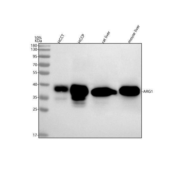
Western blot analysis of Arginase-1/ARG1 using anti-Arginase-1/ARG1 antibody (RP1075).
Electrophoresis was performed on a 5-20% SDS-PAGE gel at 70V (Stacking gel) / 90V (Resolving gel) for 2-3 hours. The sample well of each lane was loaded with 30 ug of sample under reducing conditions.
Lane 1: human hepatocellular carcinoma tumor tissue (HCCT) lysates,
Lane 2: human hepatocellular carcinoma paracancerous tissue (HCCP) lysates,
Lane 3: rat liver tissue lysates,
Lane 4: mouse liver tissue lysates.
After electrophoresis, proteins were transferred to a nitrocellulose membrane at 150 mA for 50-90 minutes. Blocked the membrane with 5% non-fat milk/TBS for 1.5 hour at RT. The membrane was incubated with rabbit anti-Arginase-1/ARG1 antigen affinity purified polyclonal antibody (RP1075) at 0.5 μg/mL overnight at 4°C, then washed with TBS-0.1%Tween 3 times with 5 minutes each and probed with a goat anti-rabbit IgG-HRP secondary antibody at a dilution of 1:5000 for 1.5 hour at RT. The signal is developed using an Enhanced Chemiluminescent detection (ECL) kit (Catalog # EK1002) with Tanon 5200 system. A specific band was detected for Arginase-1/ARG1 at approximately 38 kDa. The expected band size for Arginase-1/ARG1 is at 35 kDa.
Specific Publications For Anti-liver Arginase/ARG1 Antibody Picoband® (RP1075)
Loading publications
Recommended Resources
Here are featured tools and databases that you might find useful.
- Boster's Pathways Library
- Protein Databases
- Bioscience Research Protocol Resources
- Data Processing & Analysis Software
- Photo Editing Software
- Scientific Literature Resources
- Research Paper Management Tools
- Molecular Biology Software
- Primer Design Tools
- Bioinformatics Tools
- Phylogenetic Tree Analysis
Customer Reviews
Have you used Anti-liver Arginase/ARG1 Antibody Picoband®?
Share your experimental results or join a short interview to earn up to $1,000 in product credits or other rewards.
0 Reviews For Anti-liver Arginase/ARG1 Antibody Picoband®
Customer Q&As
Have a question?
Find answers in Q&As, reviews.
Can't find your answer?
Submit your question
4 Customer Q&As for Anti-liver Arginase/ARG1 Antibody Picoband®
Question
We were satisfied with the WB result of your anti-liver Arginase/ARG1 antibody. However we have observed positive staining in liver cytoplasm. using this antibody. Is that expected? Could you tell me where is ARG1 supposed to be expressed?
Verified Customer
Verified customer
Asked: 2020-03-18
Answer
According to literature, liver does express ARG1. Generally ARG1 expresses in cytoplasm. Regarding which tissues have ARG1 expression, here are a few articles citing expression in various tissues:
Blood, Pubmed ID: 3174433
Erythroblast, Pubmed ID: 14574404
Liver, Pubmed ID: 2241902, 3540966, 24275569
Liver, and Skeletal muscle, Pubmed ID: 15489334
Boster Scientific Support
Answered: 2020-03-18
Question
We have seen staining in human liver skeletal muscle. Any tips? Is anti-liver Arginase/ARG1 antibody supposed to stain liver skeletal muscle positively?
Verified Customer
Verified customer
Asked: 2019-10-29
Answer
From literature liver skeletal muscle does express ARG1. From Uniprot.org, ARG1 is expressed in liver, blood, erythroblast, liver skeletal muscle, among other tissues. Regarding which tissues have ARG1 expression, here are a few articles citing expression in various tissues:
Blood, Pubmed ID: 3174433
Erythroblast, Pubmed ID: 14574404
Liver, Pubmed ID: 2241902, 3540966, 24275569
Liver, and Skeletal muscle, Pubmed ID: 15489334
Boster Scientific Support
Answered: 2019-10-29
Question
We are currently using anti-liver Arginase/ARG1 antibody RP1075 for rat tissue, and we are well pleased with the WB results. The species of reactivity given in the datasheet says human, rat. Is it true that the antibody can work on monkey tissues as well?
Verified Customer
Verified customer
Asked: 2018-02-15
Answer
The anti-liver Arginase/ARG1 antibody (RP1075) has not been validated for cross reactivity specifically with monkey tissues, though there is a good chance of cross reactivity. We have an innovator award program that if you test this antibody and show it works in monkey you can get your next antibody for free. Please contact me if I can help you with anything.
Boster Scientific Support
Answered: 2018-02-15
Question
We are interested in using your anti-liver Arginase/ARG1 antibody for maternal process involved in female pregnancy studies. Has this antibody been tested with western blotting on tissue lysate? We would like to see some validation images before ordering.
J. Dhar
Verified customer
Asked: 2015-12-28
Answer
I appreciate your inquiry. This RP1075 anti-liver Arginase/ARG1 antibody is validated on liver tissue, rat liver tissue, tissue lysate. It is guaranteed to work for WB in human, rat. Our Boster guarantee will cover your intended experiment even if the sample type has not been be directly tested.
Boster Scientific Support
Answered: 2015-12-28
