This website uses cookies to ensure you get the best experience on our website.
- Table of Contents
3 Citations 16 Q&As
1 Citations 15 Q&As
1 Citations 5 Q&As
3 Citations 5 Q&As
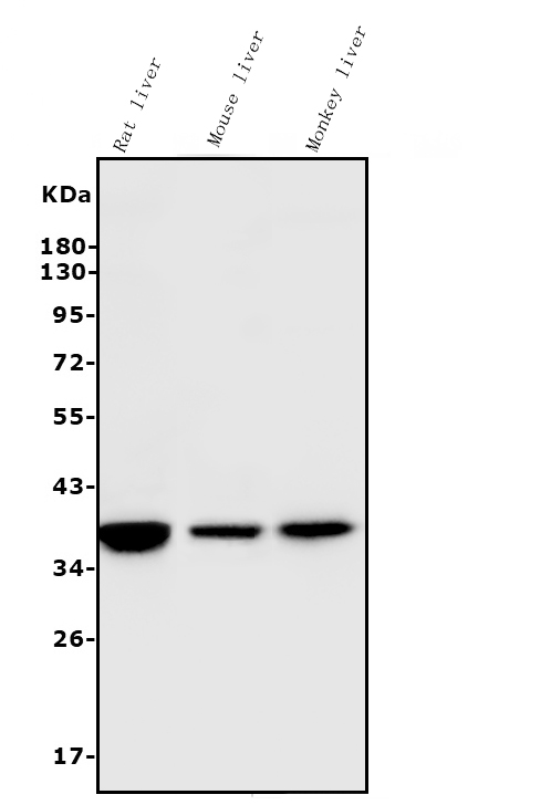
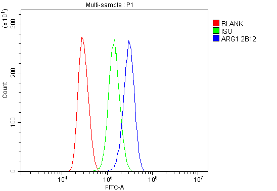
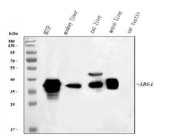
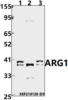
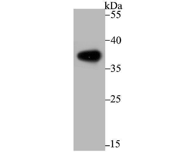
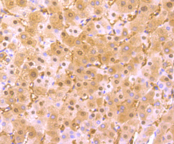
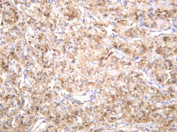
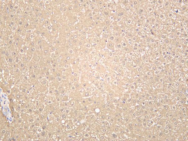
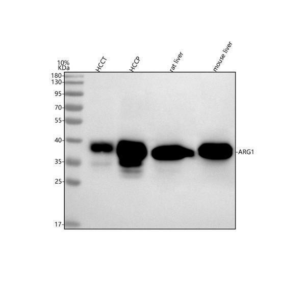
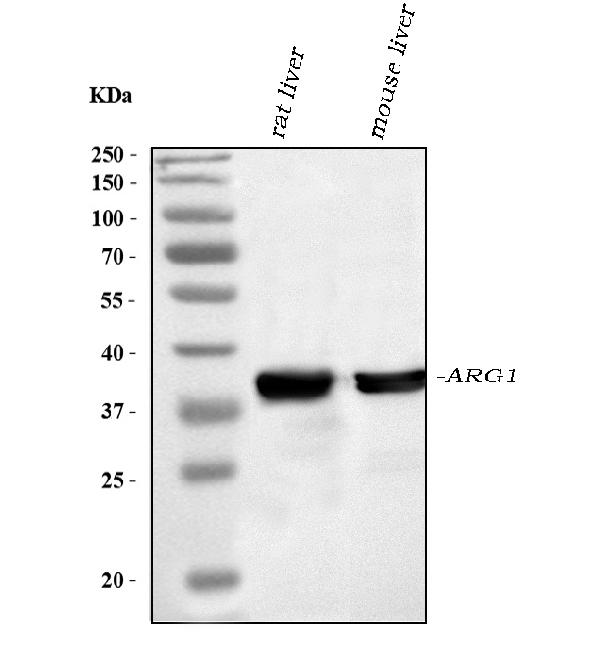
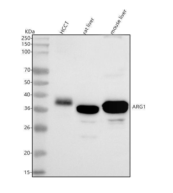
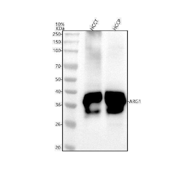
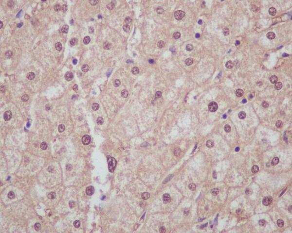
Facts about Arginase-1.
.
Human | |
---|---|
Gene Name: | ARG1 |
Uniprot: | P05089 |
Entrez: | 383 |
Belongs to: |
---|
arginase family |
AI; ARG1; Arginase 1; arginase, liver; Arginase-1; EC 3.5.3.1; Liver Arginase; Liver-type arginase; PGIF; Type I Arginase
Mass (kDA):
34.735 kDA
Human | |
---|---|
Location: | 6q23.2 |
Sequence: | 6; NC_000006.12 (131573226..131584329) |
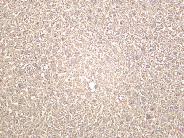
Within the immune system initially reported to be selectively expressed in granulocytes (polymorphonuclear leukocytes [PMNs]) (PubMed:15546957). Also detected in macrophages mycobacterial granulomas (PubMed:23749634). Expressed in group2 innate lymphoid cells (ILC2s) during lung disease (PubMed:27043409).
Cytoplasm. Cytoplasmic granule. Localized in azurophil granules of neutrophils (PubMed:15546957).
Boster Bio is an excellent antibody manufacturer. Boster Bio was founded in 1993. They are well-known for their signature antibodies and ELISA product lines. They've recently expanded to PCR-related molecular Biology products. They offer various services to scientists, including free technical assistance and 24 hour customer support. Read on to learn more about Boster Bio and its products.
Several markers differentiate apoptotic Arg1+ mammaphages (Arg1 macrophages) from Arg1 ones. Sessile Arg1B cells were uniquely defined by expression of Sgk1, Pmepa1, and Ms4a7, markers consistent with their M2 function and tissue homeostasis. Arg1B cells had higher levels of Cd72, Cxcl16 and tetraspannins Cd63, Cd81. These findings are consistent with previous literature reports linking peripheral TAM to M2-like cells.
Several markers were associated with Arg1 expression, including IL12/arginase ratio, and IL4/LPS+IFN-g/Lps+IFN-g ratio. Interestingly, IL12/arginase ratio was associated with Arg1+ macrophages, while IL4/LPS+IFN-g-producing cells were not. Many of these markers could also be expressed by other immune cells.
Several markers were used to differentiate Arg1+ macrophages and Arg1 microphages by analyzing their morphology, motility, and morphology. Arg1+TAM revealed a dichotomy within cellular structure: cells located outside of the tumor margin have a rounded morphology whereas cells located within the core have a stellate or elongated morphology. Interestingly, Arg1+ had a median circularity index (0.71) that was lower than the peripheral TAM (0.81).
Researchers were able distinguish between Arg1+ Arg1 microphages using these markers. These markers were found in adenoviruses. They also appeared on the amyloid precursor and central nervous system. IL-4 also increased phagosomes' ability to degrade Alzheimer amyloid fibrils. This improves cognitive deficits in Alzheimer patients.
These researchers identified a number of markers that differentiate Arg1+ macrophages from apoptotic microglia. The difference in polarity between the two types is important for disease models. Several markers were present on the surface of phagocytes. After spinal cord injury, Arg1+ and M1 cells were detected in brains. The M2 cells were unable to repair damage in aging mice.
Our study has further supported the role played by chemokine receptacles in regulation of immune responses and innate immunity. These results confirm the role of chemokine regulators in activation CD8 T cells. This mechanism is not yet fully understood but could be important in preventing spread of infectious disease. These findings are consistent with previous studies.
Lung infection causes a rise in chemokine gene expression. samples. This increases the retention and trafficking in leukocytes. Ccl1 or Xcl12 can be used as chemokine antagonists to stimulate the growth memory T cells. These findings are important because they play a role in the immune system's response to bacterial infection. However, they are just one mechanism that could be involved in innate immune response.
The findings show that over-expression of chemokine genes in boster bio are associated with a variety of diseases. These include many types of bacterial infections, such as tuberculosis. Thus, multiple targeted cytokines may help in differentiation of tuberculosis. The research is also applicable in other areas, which is a bonus. There are still many challenges in tuberculosis diagnosis. There are many improvements in this field. The latest results are compelling.
In addition to identifying genes that are associated with cancer, chemokine receptors are also involved. These chemokines are known to activate the immune system and promote tumor growth. They also facilitate angiogenesis. Normal brain tissue, as well as tumor tissue, contains chemokine receptors. GBM showed an overexpression of CXCL11, PF4V1, and CXCL8. These three genes are known to be important in cell communication, proliferation, and cellular component movement.
Boster Bio's gene infographics provide basic information on each gene. The gene infographics are available for both human and mouse chemokine gene types. You can also access gene infographics by typing the gene name in to the search bar. Researchers will find the gene informationgraphics to be a valuable source of information. Boster Bio is also available for information on specific genes.
Boster Biologicals developed the ARG1 mAb to detect biomarkers in developmental biology, cancer, inflammation, and immunelogy. Its sensitivity of up to picogram levels makes it an ideal choice for a wide range of applications. The company also offers antibodies, immunological reagents, and other products through its website, www.tebubio.com.
In a recent study, the ARG1 gene was expressed in GM-CSF-differentiated human Mphs and stimulated with LPS and IFNg for 2 days. Flow Cytometry was used to measure surface expression of Mph markers. Cells were mock-activated, while activated cells were stained with IL-10 and IL-4. Cells mock-activated with monoclonal antibodies against isotype controls were also stained.
The Arginase-1 monoclonal anti-mouse antibody recognizes Arginase1 in normal lysed human whole blood cells and human neutrophils. The antibody can be used with the eBioscience(tm), Foxp3/Transcription factor Staining Buffer set. The monoclonal antibody works with multiple chemokine assays, allowing researchers to compare gene expression within an immune system population.
The ARG1 genes is an enzyme that converts L.arginine (ornithine) into urea. It produces polyamines which are vital for cell growth and removes toxins that are produced during protein decay. It also deprives the NO synase enzyme its substrate, which reduces nitric dioxide production. The ARG1 gene is also recognized by the ARG1 monoclonal antibody, A1exF5.
Imaging Arg1eYFP mouse is a useful method to visualize mutations in the arginase protein gene. The ARG1 gene is found in the breast and spleen of humans. Arg1-eYFP mice show increased expression in these tissues and may provide a new therapeutic target for cancer. The gene is found in three major types of breast cancer.
Using a tissue microarray, images of Arg1+CD68-expressing myeloid cells have been generated. The ARG1+CD68-positive cells were identified in breast, lung, and melanoma tissue by immunofluorescence staining. The cells were scored based on the cancer stage and total CD68+ cells. The mouse tumors from mice were used to analyze the effects of ARG1 and breast cancer progression.
GM/CSF/LA induce Arg1 transcription in BMMs. PyMT-BO1 tumor cells expressing GM-CSF and LA induce Arg1 mRNA in BMMs. FACS analysis of ARG1+ cells obtained from YARG animals that were treated using CSF2-KO (or recombinant GM–CSF) was used to analyze them. Western blotting confirmed Arg1 expression in BMMs treated using GM–CSF–KO or PyMT–BO1 cancer cells.
High concentrations of GM-CSF and LA failed to induce ARG1 expression in myeloid cells. These findings suggest that ARG1 synthesis in TIMs may be dependent on local levels of these secreted substances and not directly linked to cell-cell interactions. These interactions are thought to be crucial for sculpting immune-suppressive TMEs. The next step is to identify the underlying molecular mechanisms.
PMID: 3540966 by Haraguchi Y., et al. Molecular cloning and nucleotide sequence of cDNA for human liver arginase.
PMID: 3174433 by Takiguchi M., et al. Human liver-type arginase gene: structure of the gene and analysis of the promoter region.
*More publications can be found for each product on its corresponding product page