Click image to see more details
-

-
-
-
-
+5
Product Info Summary
| SKU: | A03264 |
|---|---|
| Size: | 100 μg/vial |
| Reactive Species: | Human, Monkey, Mouse, Rat |
| Host: | Rabbit |
| Application: | ELISA, Flow Cytometry, IF, IHC, ICC, WB |
Customers Who Bought This Also Bought

Product info
Product Name
Anti-LRPPRC/GP130 Antibody Picoband®
SKU/Catalog Number
A03264
Size
100 μg/vial
Form
Lyophilized
Description
Boster Bio Anti-LRPPRC/GP130 Antibody Picoband® catalog # A03264. Tested in ELISA, Flow Cytometry, IF, IHC, ICC, WB applications. This antibody reacts with Human, Monkey, Mouse, Rat. The brand Picoband indicates this is a premium antibody that guarantees superior quality, high affinity, and strong signals with minimal background in Western blot applications. Only our best-performing antibodies are designated as Picoband, ensuring unmatched performance.
Storage & Handling
Store at -20˚C for one year from date of receipt. After reconstitution, at 4˚C for one month. It can also be aliquotted and stored frozen at -20˚C for six months. Avoid repeated freeze-thaw cycles.
Cite This Product
Anti-LRPPRC/GP130 Antibody Picoband® (Boster Biological Technology, Pleasanton CA, USA, Catalog # A03264)
Host
Rabbit
Contents
Each vial contains 4mg Trehalose, 0.9mg NaCl, 0.2mg Na2HPO4, 0.05mg NaN3.
Clonality
Polyclonal
Isotype
Rabbit IgG
Immunogen
E.coli-derived human LRPPRC/GP130 recombinant protein (Position: A1281-S1394).
Cross-reactivity
No cross-reactivity with other proteins.
Reactive Species
A03264 is reactive to LRPPRC in Human, Monkey, Mouse, Rat
Observed Molecular Weight
158 kDa
Calculated molecular weight
157.9 kDa
Background of LRPPRC
Leucine-rich PPR motif-containing protein, mitochondrial is a protein that in humans is encoded by the LRPPRC gene. It is mapped to 2p21. This gene encodes a leucine-rich protein that has multiple pentatricopeptide repeats (PPR). The precise role of this protein is unknown but studies suggest it may play a role in cytoskeletal organization, vesicular transport, or in transcriptional regulation of both nuclear and mitochondrial genes. The protein localizes primarily to mitochondria and is predicted to have an N-terminal mitochondrial targeting sequence. Mutations in this gene are associated with the French-Canadian type of Leigh syndrome.
Antibody Validation
Boster validates all antibodies on WB, IHC, ICC, Immunofluorescence, and ELISA with known positive control and negative samples to ensure specificity and high affinity, including thorough antibody incubations.
Application & Images
Applications
A03264 is guaranteed for ELISA, Flow Cytometry, IF, IHC, ICC, WB Boster Guarantee
Recommend Dilution
| Application | Dilution | Species |
|---|---|---|
| Western blot | 0.1-0.25μg/ml | Human, Mouse, Rat, Monkey |
| Immunohistochemistry (Paraffin-embedded Section) | 0.5-1μg/ml | Human, Mouse, Rat |
| Immunocytochemistry/Immunofluorescence | 2μg/ml | Human |
| Flow Cytometry (Fixed) | 1-3μg/1x106 cells | Human, Rat |
| ELISA | 0.1-0.5μg/ml | - |
Tested application
Suggested blocking solution with 5% non-fat milk or BSA; (*)Recommended protein loading: 20-40 µg per lane
Use TE buffer pH 9.0 for antigen retrieval; (*) citrate buffer pH 6.0 is an alternative.
Validation Images & Assay Conditions
Click image to see more details
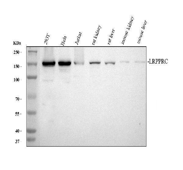
Western blot analysis of LRPPRC using anti-LRPPRC antibody (A03264).
Electrophoresis was performed on a 8% SDS-PAGE gel at 80V (Stacking gel) / 120V (Resolving gel) for 2 hours. The sample well of each lane was loaded with 30 ug of sample under reducing conditions.
Lane 1: human 293T whole cell lysates,
Lane 2: human Hela whole cell lysates,
Lane 3: human Jurkat whole cell lysates,
Lane 4: rat kidney tissue lysates,
Lane 5: rat liver tissue lysates,
Lane 6: mouse kidney tissue lysates,
Lane 7: mouse liver tissue lysates.
After electrophoresis, proteins were transferred to a nitrocellulose membrane at 150 mA for 50-90 minutes. Blocked the membrane with 5% non-fat milk/TBS for 1.5 hour at RT. The membrane was incubated with rabbit anti-LRPPRC antigen affinity purified polyclonal antibody (A03264) at 0.5 μg/mL overnight at 4°C, then washed with TBS-0.1%Tween 3 times with 5 minutes each and probed with a goat anti-rabbit IgG-HRP secondary antibody (Catalog # BA1054) at a dilution of 1:5000 for 1.5 hour at RT. The signal is developed using an ECL Plus Western Blotting Substrate (Catalog # AR1196-200) with Tanon 5200 system. A specific band was detected for LRPPRC at approximately 158 kDa. The expected band size for LRPPRC is at 158 kDa.

Click image to see more details
LRPPRC expression is associated with the prognosis of patients with UCEC. (A) Kaplan-Meier survival estimates for patients with high and low LRPPRC expression. (B) Proportions of different grades and stages in high and low LRPPRC groups. (C) Representative IHC staining of LRPPRC in paracancerous and tumor UCEC samples. (D) Univariate and multivariate Cox analyses evaluating the independent prognostic value of LRPPRC in UCEC patients.
Index in PubMed under a CC BY license. PMID: 39445012

Click image to see more details
Knockdown of LRPPRC restrained the proliferation and invasion abilities of UCEC. (A) GO analysis displaying the normalized enrichment scores (NES) of adaptive immune response and immune cell-related pathways in UCEC patients with high LRPPRC expression. Negative NES indicates a negative correlation between the pathway and LRPPRC expression. (B) GSEA analysis showing enrichment of HALLMARK signaling pathways or processes in UCEC patients with high LRPPRC expression. (C) Western blot validation of LRPPRC knockdown in HEC-1A cells. (D) Proliferation of LRPPRC knockdown versus control cells. (E–G) Clone formation, migration, and invasion abilities of indicated UCEC cells.
Index in PubMed under a CC BY license. PMID: 39445012

Click image to see more details
IHC analysis of LRPPRC using anti-LRPPRC antibody (A03264).
LRPPRC was detected in paraffin-embedded section of human rectal cancer tissue. Heat mediated antigen retrieval was performed in EDTA buffer (pH8.0, epitope retrieval solution). The tissue section was blocked with 10% goat serum. The tissue section was then incubated with 1μg/ml rabbit anti-LRPPRC Antibody (A03264) overnight at 4°C. Biotinylated goat anti-rabbit IgG was used as secondary antibody and incubated for 30 minutes at 37°C. The tissue section was developed using Strepavidin-Biotin-Complex (SABC) (Catalog # SA1022) with DAB as the chromogen.

Click image to see more details
IHC analysis of LRPPRC using anti-LRPPRC antibody (A03264).
LRPPRC was detected in paraffin-embedded section of mouse intestine tissue. Heat mediated antigen retrieval was performed in EDTA buffer (pH8.0, epitope retrieval solution). The tissue section was blocked with 10% goat serum. The tissue section was then incubated with 1μg/ml rabbit anti-LRPPRC Antibody (A03264) overnight at 4°C. Biotinylated goat anti-rabbit IgG was used as secondary antibody and incubated for 30 minutes at 37°C. The tissue section was developed using Strepavidin-Biotin-Complex (SABC) (Catalog # SA1022) with DAB as the chromogen.

Click image to see more details
IHC analysis of LRPPRC using anti-LRPPRC antibody (A03264).
LRPPRC was detected in paraffin-embedded section of rat intestine tissue. Heat mediated antigen retrieval was performed in EDTA buffer (pH8.0, epitope retrieval solution). The tissue section was blocked with 10% goat serum. The tissue section was then incubated with 1μg/ml rabbit anti-LRPPRC Antibody (A03264) overnight at 4°C. Biotinylated goat anti-rabbit IgG was used as secondary antibody and incubated for 30 minutes at 37°C. The tissue section was developed using Strepavidin-Biotin-Complex (SABC) (Catalog # SA1022) with DAB as the chromogen.

Click image to see more details
IF analysis of LRPPRC using anti-LRPPRC antibody (A03264).
LRPPRC was detected in immunocytochemical section of Hela cells. Enzyme antigen retrieval was performed using IHC enzyme antigen retrieval reagent (AR0022) for 15 mins. The cells were blocked with 10% goat serum. And then incubated with 2μg/mL rabbit anti-LRPPRC Antibody (A03264) overnight at 4°C. DyLight®488 Conjugated Goat Anti-Rabbit IgG (BA1127) was used as secondary antibody at 1:100 dilution and incubated for 30 minutes at 37°C. The section was counterstained with DAPI. Visualize using a fluorescence microscope and filter sets appropriate for the label used.

Click image to see more details
Flow Cytometry analysis of A431 cells using anti-LRPPRC antibody (A03264).
Overlay histogram showing A431 cells stained with A03264 (Blue line). To facilitate intracellular staining, cells were fixed with 4% paraformaldehyde and permeabilized with permeabilization buffer. The cells were blocked with 10% normal goat serum. And then incubated with rabbit anti-LRPPRC Antibody (A03264, 1μg/1x106 cells) for 30 min at 20°C. DyLight®488 conjugated goat anti-rabbit IgG (BA1127, 5-10μg/1x106 cells) was used as secondary antibody for 30 minutes at 20°C. Isotype control antibody (Green line) was rabbit IgG (1μg/1x106) used under the same conditions. Unlabelled sample without incubation with primary antibody and secondary antibody (Red line) was used as a blank control.

Click image to see more details
Flow Cytometry analysis of C6 cells using anti-LRPPRC antibody (A03264).
Overlay histogram showing C6 cells stained with A03264 (Blue line). To facilitate intracellular staining, cells were fixed with 4% paraformaldehyde and permeabilized with permeabilization buffer. The cells were blocked with 10% normal goat serum. And then incubated with rabbit anti-LRPPRC Antibody (A03264, 1μg/1x106 cells) for 30 min at 20°C. DyLight®488 conjugated goat anti-rabbit IgG (BA1127, 5-10μg/1x106 cells) was used as secondary antibody for 30 minutes at 20°C. Isotype control antibody (Green line) was rabbit IgG (1μg/1x106) used under the same conditions. Unlabelled sample without incubation with primary antibody and secondary antibody (Red line) was used as a blank control.
Specific Publications For Anti-LRPPRC/GP130 Antibody Picoband® (A03264)
Loading publications
Recommended Resources
Here are featured tools and databases that you might find useful.
- Boster's Pathways Library
- Protein Databases
- Bioscience Research Protocol Resources
- Data Processing & Analysis Software
- Photo Editing Software
- Scientific Literature Resources
- Research Paper Management Tools
- Molecular Biology Software
- Primer Design Tools
- Bioinformatics Tools
- Phylogenetic Tree Analysis
Customer Reviews
Have you used Anti-LRPPRC/GP130 Antibody Picoband®?
Share your experimental results or join a short interview to earn up to $1,000 in product credits or other rewards.
0 Reviews For Anti-LRPPRC/GP130 Antibody Picoband®
Customer Q&As
Have a question?
Find answers in Q&As, reviews.
Can't find your answer?
Submit your question
