Click image to see more details
-

-
-
-
-
+2
Product Info Summary
| SKU: | A01034-2 |
|---|---|
| Size: | 100 µg/vial |
| Reactive Species: | Human, Mouse, Rat |
| Host: | Rabbit |
| Application: | ELISA, Flow Cytometry, IHC, WB |
Customers Who Bought This Also Bought

Product info
Product Name
Anti-Lumican/LUM Antibody Picoband®
SKU/Catalog Number
A01034-2
Size
100 µg/vial
Form
Lyophilized
Description
Boster Bio Anti-Lumican/LUM Antibody Picoband® catalog # A01034-2. Tested in ELISA, IHC, WB, Flow Cytometry applications. This antibody reacts with Human, Mouse, Rat. The brand Picoband indicates this is a premium antibody that guarantees superior quality, high affinity, and strong signals with minimal background in Western blot applications. Only our best-performing antibodies are designated as Picoband, ensuring unmatched performance.
Storage & Handling
At -20°C for one year from date of receipt. After reconstitution, at 4°C for one month. It can also be aliquotted and stored frozen at -20°C for six months. Avoid repeated freezing and thawing.
Cite This Product
Anti-Lumican/LUM Antibody Picoband® (Boster Biological Technology, Pleasanton CA, USA, Catalog # A01034-2)
Host
Rabbit
Contents
Each vial contains 4 mg Trehalose, 0.9 mg NaCl, 0.2 mg Na2HPO4.
Clonality
Polyclonal
Isotype
IgG
Immunogen
E.coli-derivedhumanLumican/LUMrecombinantprotein(Position:Q19-N338).HumanLUMshares88.4%and86.9%aminoacid(aa)sequenceidentitywithmouseandratLUM,respectively.
Cross-reactivity
No cross reactivity with other proteins.
Reactive Species
A01034-2 is reactive to LUM in Human, Mouse, Rat
Observed Molecular Weight
70 kDa
Calculated molecular weight
38.4 kDa
Background of LUM
Lumican, also known as LUM, is an extracellular matrix protein that, in humans, is encoded by the LUM gene on chromosome 12. This gene encodes a member of the small leucine-rich proteoglycan (SLRP) family that includes decorin, biglycan, fibromodulin, keratocan, epiphycan, and osteoglycin. In these bifunctional molecules, the protein moiety binds collagen fibrils and the highly charged hydrophilic glycosaminoglycans regulate interfibrillar spacings. Lumican is the major keratan sulfate proteoglycan of the cornea but is also distributed in interstitial collagenous matrices throughout the body. Lumican may regulate collagen fibril organization and circumferential growth, corneal transparency, and epithelial cell migration and tissue repair.
Antibody Validation
Boster validates all antibodies on WB, IHC, ICC, Immunofluorescence, and ELISA with known positive control and negative samples to ensure specificity and high affinity, including thorough antibody incubations.
Application & Images
Applications
A01034-2 is guaranteed for ELISA, Flow Cytometry, IHC, WB Boster Guarantee
Assay Dilutions Recommendation
The recommendations below provide a starting point for assay optimization. The actual working concentration varies and should be decided by the user.
Western blot, 0.25-0.5 μg/ml, Human, Mouse, Rat
Immunohistochemistry(Paraffin-embedded Section), 2-5 μg/ml, Human, Mouse
Flow Cytometry (Fixed), 1-3 μg/1x106 cells, Human
ELISA, 0.1-0.5 μg/ml, -
Positive Control
WB: human Jurkat whole cell, human placenta tissue, human Caco-2 whole cell, rat liver tissue, mouse liver tissue, mouse NIH/3T3 whole cell
IHC: human esophageal squamous carcinoma tissue, human lung cancer tissue, human thyroid papillary carcinoma tissue, mouse kidney tissue
FCM: SH-SY5Y cell
Validation Images & Assay Conditions
Click image to see more details
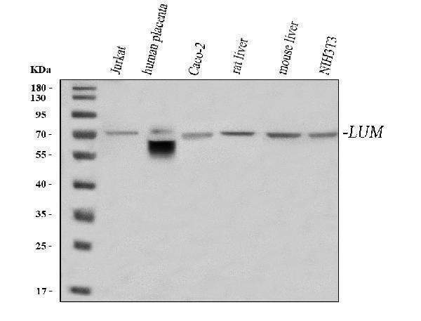
Western blot analysis of Lumican/LUM using anti-Lumican/LUM antibody (A01034-2).
Electrophoresis was performed on a 5-20% SDS-PAGE gel at 70V (Stacking gel) / 90V (Resolving gel) for 2-3 hours. The sample well of each lane was loaded with 30 ug of sample under reducing conditions.
Lane 1: human Jurkat whole cell lysates,
Lane 2: human placenta tissue lysates,
Lane 3: human Caco-2 whole cell lysates,
Lane 4: rat liver tissue lysates,
Lane 5: mouse liver tissue lysates,
Lane 6: mouse NIH/3T3 whole cell lysates.
After electrophoresis, proteins were transferred to a nitrocellulose membrane at 150 mA for 50-90 minutes. Blocked the membrane with 5% non-fat milk/TBS for 1.5 hour at RT. The membrane was incubated with rabbit anti-Lumican/LUM antigen affinity purified polyclonal antibody (Catalog # A01034-2) at 0.5 μg/mL overnight at 4°C, then washed with TBS-0.1%Tween 3 times with 5 minutes each and probed with a goat anti-rabbit IgG-HRP secondary antibody at a dilution of 1:5000 for 1.5 hour at RT. The signal is developed using an Enhanced Chemiluminescent detection (ECL) kit (Catalog # EK1002) with Tanon 5200 system. A specific band was detected for Lumican/LUM at approximately 70 kDa. The expected band size for Lumican/LUM is at 38 kDa.

Click image to see more details
IHC analysis of Lumican/LUM using anti-Lumican/LUM antibody (A01034-2).
Lumican/LUM was detected in a paraffin-embedded section of human esophageal squamous carcinoma tissue. Heat mediated antigen retrieval was performed in EDTA buffer (pH 8.0, epitope retrieval solution). The tissue section was blocked with 10% goat serum. The tissue section was then incubated with 2 μg/ml rabbit anti-Lumican/LUM Antibody (A01034-2) overnight at 4°C. Peroxidase Conjugated Goat Anti-rabbit IgG was used as secondary antibody and incubated for 30 minutes at 37°C. The tissue section was developed using HRP Conjugated Rabbit IgG Super Vision Assay Kit (Catalog # SV0002) with DAB as the chromogen.

Click image to see more details
IHC analysis of Lumican/LUM using anti-Lumican/LUM antibody (A01034-2).
Lumican/LUM was detected in a paraffin-embedded section of human lung cancer tissue. Heat mediated antigen retrieval was performed in EDTA buffer (pH 8.0, epitope retrieval solution). The tissue section was blocked with 10% goat serum. The tissue section was then incubated with 2 μg/ml rabbit anti-Lumican/LUM Antibody (A01034-2) overnight at 4°C. Peroxidase Conjugated Goat Anti-rabbit IgG was used as secondary antibody and incubated for 30 minutes at 37°C. The tissue section was developed using HRP Conjugated Rabbit IgG Super Vision Assay Kit (Catalog # SV0002) with DAB as the chromogen.

Click image to see more details
IHC analysis of Lumican/LUM using anti-Lumican/LUM antibody (A01034-2).
Lumican/LUM was detected in a paraffin-embedded section of human thyroid papillary carcinoma tissue. Heat mediated antigen retrieval was performed in EDTA buffer (pH 8.0, epitope retrieval solution). The tissue section was blocked with 10% goat serum. The tissue section was then incubated with 2 μg/ml rabbit anti-Lumican/LUM Antibody (A01034-2) overnight at 4°C. Peroxidase Conjugated Goat Anti-rabbit IgG was used as secondary antibody and incubated for 30 minutes at 37°C. The tissue section was developed using HRP Conjugated Rabbit IgG Super Vision Assay Kit (Catalog # SV0002) with DAB as the chromogen.

Click image to see more details
IHC analysis of Lumican/LUM using anti-Lumican/LUM antibody (A01034-2).
Lumican/LUM was detected in a paraffin-embedded section of mouse kidney tissue. Heat mediated antigen retrieval was performed in EDTA buffer (pH 8.0, epitope retrieval solution). The tissue section was blocked with 10% goat serum. The tissue section was then incubated with 2 μg/ml rabbit anti-Lumican/LUM Antibody (A01034-2) overnight at 4°C. Peroxidase Conjugated Goat Anti-rabbit IgG was used as secondary antibody and incubated for 30 minutes at 37°C. The tissue section was developed using HRP Conjugated Rabbit IgG Super Vision Assay Kit (Catalog # SV0002) with DAB as the chromogen.

Click image to see more details
Flow Cytometry analysis of SH-SY5Y cells using anti-Lumican/LUM antibody (A01034-2).
Overlay histogram showing SH-SY5Y cells stained with A01034-2 (Blue line). The cells were fixed with 4% paraformaldehyde and blocked with 10% normal goat serum. And then incubated with rabbit anti-Lumican/LUM Antibody (A01034-2, 1 μg/1x106 cells) for 30 min at 20°C. DyLight®488 conjugated goat anti-rabbit IgG (BA1127, 5-10 μg/1x106 cells) was used as secondary antibody for 30 minutes at 20°C. Isotype control antibody (Green line) was rabbit IgG (1 μg/1x106) used under the same conditions. Unlabelled sample (Red line) was also used as a control.
Specific Publications For Anti-Lumican/LUM Antibody Picoband® (A01034-2)
Loading publications
Recommended Resources
Here are featured tools and databases that you might find useful.
- Boster's Pathways Library
- Protein Databases
- Bioscience Research Protocol Resources
- Data Processing & Analysis Software
- Photo Editing Software
- Scientific Literature Resources
- Research Paper Management Tools
- Molecular Biology Software
- Primer Design Tools
- Bioinformatics Tools
- Phylogenetic Tree Analysis
Customer Reviews
Have you used Anti-Lumican/LUM Antibody Picoband®?
Share your experimental results or join a short interview to earn up to $1,000 in product credits or other rewards.
0 Reviews For Anti-Lumican/LUM Antibody Picoband®
Customer Q&As
Have a question?
Find answers in Q&As, reviews.
Can't find your answer?
Submit your question
