Click image to see more details
-

-
-
-
-
+2
Product Info Summary
| SKU: | A04262-2 |
|---|---|
| Size: | 100 µg/vial |
| Reactive Species: | Human, Mouse, Rat |
| Host: | Rabbit |
| Application: | ELISA, Flow Cytometry, WB |
Customers Who Bought This Also Bought

Product info
Product Name
Anti-MDH1 Antibody Picoband®
SKU/Catalog Number
A04262-2
Size
100 µg/vial
Form
Lyophilized
Description
Boster Bio Anti-MDH1 Antibody Picoband® catalog # A04262-2. Tested in ELISA, Flow Cytometry, WB applications. This antibody reacts with Human, Mouse, Rat. The brand Picoband indicates this is a premium antibody that guarantees superior quality, high affinity, and strong signals with minimal background in Western blot applications. Only our best-performing antibodies are designated as Picoband, ensuring unmatched performance.
Storage & Handling
At -20°C for one year from date of receipt. After reconstitution, at 4°C for one month. It can also be aliquotted and stored frozen at -20°C for six months. Avoid repeated freezing and thawing.
Cite This Product
Anti-MDH1 Antibody Picoband® (Boster Biological Technology, Pleasanton CA, USA, Catalog # A04262-2)
Host
Rabbit
Contents
Each vial contains 4 mg Trehalose, 0.9 mg NaCl, 0.2 mg Na2HPO4.
Clonality
Polyclonal
Isotype
Rabbit IgG
Immunogen
E.coli-derived human MDH1 recombinant protein (Position: E56-A334).
Cross-reactivity
No cross-reactivity with other proteins.
Reactive Species
A04262-2 is reactive to MDH1 in Human, Mouse, Rat
Observed Molecular Weight
36 kDa
Calculated molecular weight
36.4 kDa
Background of MDH1
Malate dehydrogenase, cytoplasmic also known as malate dehydrogenase 1 is an enzyme that in humans is encoded by the MDH1 gene. This gene encodes an enzyme that catalyzes the NAD/NADH-dependent, reversible oxidation of malate to oxaloacetate in many metabolic pathways, including the citric acid cycle. Two main isozymes are known to exist in eukaryotic cells: one is found in the mitochondrial matrix and the other in the cytoplasm. This gene encodes the cytosolic isozyme, which plays a key role in the malate-aspartate shuttle that allows malate to pass through the mitochondrial membrane to be transformed into oxaloacetate for further cellular processes. Alternatively spliced transcript variants have been found for this gene. A recent study showed that a C-terminally extended isoform is produced by use of an alternative in-frame translation termination codon via a stop codon readthrough mechanism, and that this isoform is localized in the peroxisomes. Pseudogenes have been identified on chromosomes X and 6.
Antibody Validation
Boster validates all antibodies on WB, IHC, ICC, Immunofluorescence, and ELISA with known positive control and negative samples to ensure specificity and high affinity, including thorough antibody incubations.
Application & Images
Applications
A04262-2 is guaranteed for ELISA, Flow Cytometry, WB Boster Guarantee
Recommend Dilution
| Application | Dilution | Species |
|---|---|---|
| Western blot | 0.1-0.25 μg/ml | Human, Mouse, Rat |
| Flow Cytometry (Fixed) | 1-3 μg/1x106 cells | Human |
| ELISA | 0.1-0.5 μg/ml | - |
Tested application
Suggested blocking solution with 5% non-fat milk or BSA; (*)Recommended protein loading: 20-40 µg per lane
Validation Images & Assay Conditions
Click image to see more details
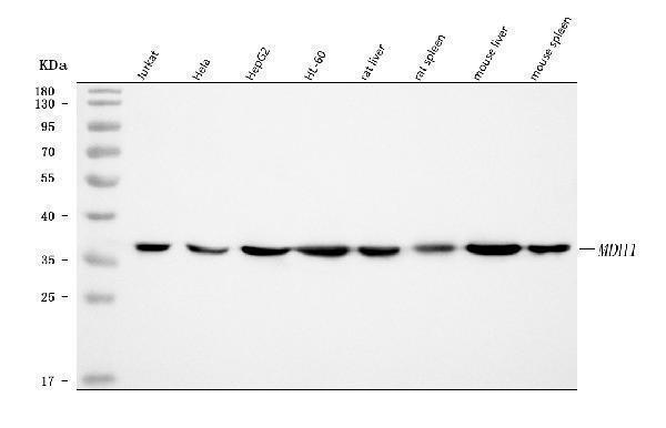
Western blot analysis of MDH1 using anti-MDH1 antibody (A04262-2).
Electrophoresis was performed on a 5-20% SDS-PAGE gel at 70V (Stacking gel) / 90V (Resolving gel) for 2-3 hours. The sample well of each lane was loaded with 30 ug of sample under reducing conditions.
Lane 1: human Jurkat whole cell lysates,
Lane 2: human Hela whole cell lysates,
Lane 3: human HepG2 whole cell lysates,
Lane 4: human HL-60 whole cell lysates,
Lane 5: rat liver tissue lysates,
Lane 6: rat spleen tissue lysates,
Lane 7: mouse liver tissue lysates,
Lane 8: mouse spleen tissue lysates.
After electrophoresis, proteins were transferred to a nitrocellulose membrane at 150 mA for 50-90 minutes. Blocked the membrane with 5% non-fat milk/TBS for 1.5 hour at RT. The membrane was incubated with rabbit anti-MDH1 antigen affinity purified polyclonal antibody (Catalog # A04262-2) at 0.25 μg/mL overnight at 4°C, then washed with TBS-0.1%Tween 3 times with 5 minutes each and probed with a goat anti-rabbit IgG-HRP secondary antibody at a dilution of 1:5000 for 1.5 hour at RT. The signal is developed using an Enhanced Chemiluminescent detection (ECL) kit (Catalog # EK1002) with Tanon 5200 system. A specific band was detected for MDH1 at approximately 36 kDa. The expected band size for MDH1 is at 59 kDa.

Click image to see more details
Correlation analysis between pyroptosis-AD hub genes and immune cell infiltration. (A) Heatmap showed the correlation and p-values of 22 immune infiltrating cells and pyroptosis-related genes. The red indicated a positive correlation, whereas the blue represented a negative correlation, and p- values were shown as * p < 0.05, ** p < 0.01, *** p < 0.001. (B) Correlation analysis between MDH1 and infiltrating immune cells. (C–H) Correlation scatter plots between the expression of MDH1 and immune cells presented significance. (I) Correlation analysis between FOXP3 and infiltrating immune cells. (J–O) Correlation scatter plots between the expression of MDH1 and immune cells presented significance.
Index in PubMed under a CC BY license. PMID: 40438507

Click image to see more details
Validation of the pyroptosis-AD hub genes at the level of RNA and protein in AD mice. (A) A hierarchical clustering heatmap based on the normalized expression of the five pyroptosis-AD genes in the combined dataset. (B) A clustering heatmap was constructed based on the normalized expression of the five pyroptosis-AD genes in the 6- and 12-month-old APP/PS1 and control mice. The 6- and 12-month-old APP/PS1 or WT mice were abbreviated as A6 and A12 or W6 and W12, respectively. (C–G) qPCR validation of mRNA expression of the pyroptosis-AD hub genes (Chmp2a, Egfr, Pkn2, Hsp90b1, and Mdh1, respectively) between the 12 months APP/PS1 and wild-type (WT) mice. Data are mean ± SEM ( n = 6 for WT, and n = 5 for APP/PS1 mice group, * p < 0.05, ** p < 0.01, unpaired two-tailed t -test). (H–K) The cell lysates from the hippocampus of APP/PS1 and WT mice were prepared and blotted with anti-Egfr, Pkn2, Hsp90b1, and Mdh1, respectively (up). The relative protein expressions of Egfr, Pkn2, Hsp90b1, and Mdh1 were calculated using Gapdh as an internal reference (below). Data are mean ± SEM ( n = 6 per group, * p < 0.05, ** p < 0.01, unpaired two-tailed t -test).
Index in PubMed under a CC BY license. PMID: 40438507

Click image to see more details
Screening and validation of candidate PRGs for the diagnosis of AD. (A) The ROC curve shows the diagnostic performance of the five feature genes in the combined dataset (training set). (B,C) ROC curves showing the diagnostic performance in the hippocampus of datasets GSE5281 (B) and GSE48350 (C) . (D–G) ROC curves show the diagnostic performance in the validation sets (entorhinal cortex of GSE5281, the hippocampus, or the temporal cortex data of GSE36980). (H,I) Differential expression of PKN2 and MDH1 in the GSE36980 (* p < 0.05, ** p < 0.01, unpaired two-tailed t -test).
Index in PubMed under a CC BY license. PMID: 40438507

Click image to see more details
Construction of lncRNA regulatory network of the pyroptosis-AD hub genes. (A) PCA of lncRNAs expression profiles of the APP/PS1 and WT mice at the age of 3, 6, and 12 months. (B) Visualization of the clustered volcano diagram for the DElncRs from six different comparisons, including APP/PS1 mice vs. WT mice at the age of 3, 6, and 12 months and comparison of APP/PS1 mice between different ages. (C) A hierarchical clustering heatmap based on the normalized expression in all samples of DElncRs. The 3-, 6-, and 12-month-old APP/PS1 or WT mice were abbreviated as A3, A6, and A12 or W3, W6, and W12, respectively. (D) The clustered heatmap was produced based on the membership scores of the six clusters obtained by time series analysis. All the DElncRs and five pyroptosis-AD hub genes were clustered into six groups. (E) Line charts showed the relative expression trend in each cluster. The five pyroptosis-AD hub genes were divided into cluster 2 (Champ2 and Mdh1), cluster 3 (Pkn2), and cluster (Egfr and Hsp90b1). The horizontal axis represents a total of nine samples in the age 3-, 6-, and 12-month groups in turn. (F) The heatmaps of correlation analysis of the five pyroptosis-AD hub genes and DElncRs. (G) Regulatory networks constructed by the five pyroptosis-AD hub genes and their top10 (show all if the numbers of lncRNA less than 10) correlated lncRNAs (the ID of lncRNAs could be queried in the NONCODE, NCBI, or Ensemble databases).
Index in PubMed under a CC BY license. PMID: 40438507

Click image to see more details
Flow Cytometry analysis of HL-60 cells using anti-MDH1 antibody (A04262-2).
Overlay histogram showing HL-60 cells stained with A04262-2 (Blue line). To facilitate intracellular staining, cells were fixed with 4% paraformaldehyde and permeabilized with permeabilization buffer. The cells were blocked with 10% normal goat serum. And then incubated with rabbit anti-MDH1 Antibody (A04262-2, 1 μg/1x106 cells) for 30 min at 20°C. DyLight®488 conjugated goat anti-rabbit IgG (BA1127, 5-10 μg/1x106 cells) was used as secondary antibody for 30 minutes at 20°C. Isotype control antibody (Green line) was rabbit IgG (1 μg/1x106) used under the same conditions. Unlabelled sample without incubation with primary antibody and secondary antibody (Red line) was used as a blank control.
Specific Publications For Anti-MDH1 Antibody Picoband® (A04262-2)
Loading publications
Recommended Resources
Here are featured tools and databases that you might find useful.
- Boster's Pathways Library
- Protein Databases
- Bioscience Research Protocol Resources
- Data Processing & Analysis Software
- Photo Editing Software
- Scientific Literature Resources
- Research Paper Management Tools
- Molecular Biology Software
- Primer Design Tools
- Bioinformatics Tools
- Phylogenetic Tree Analysis
Customer Reviews
Have you used Anti-MDH1 Antibody Picoband®?
Share your experimental results or join a short interview to earn up to $1,000 in product credits or other rewards.
0 Reviews For Anti-MDH1 Antibody Picoband®
Customer Q&As
Have a question?
Find answers in Q&As, reviews.
Can't find your answer?
Submit your question
