Click image to see more details
-

-
-
-
-
+10
Product Info Summary
| SKU: | M00775 |
|---|---|
| Size: | 100 μl |
| Reactive Species: | Human, Mouse, Rat |
| Host: | Rabbit |
| Application: | IF, IHC, ICC, WB |
Customers Who Bought This Also Bought

Product info
Product Name
Anti-MMP3 Rabbit Monoclonal Antibody
SKU/Catalog Number
M00775
BM4074 is an alternative SKU for this antibody, used in previous lots.
Size
100 μl
Form
Liquid
Description
Boster Bio Anti-MMP3 Rabbit Monoclonal Antibody catalog # M00775. Tested in WB, IHC, ICC/IF applications. This antibody reacts with Human, Mouse, Rat.
Storage & Handling
Store at -20°C for one year. For short term storage and frequent use, store at 4°C for up to one month. Avoid repeated freeze-thaw cycles.
Cite This Product
Anti-MMP3 Rabbit Monoclonal Antibody (Boster Biological Technology, Pleasanton CA, USA, Catalog # M00775)
Host
Rabbit
Contents
Rabbit IgG in stabilizing components, phosphate buffered saline, pH 7.4, 150mM NaCl, 0.02% sodium azide and 50% glycerol.
*This antibody is supplied in a stabilized formulation.
Compatibility with conjugation reactions depends on the chemistry of the conjugation method used.
For conjugation methods that are not compatible with the stabilizing components present in this formulation, a carrier-free antibody format is required.
Clonality
Monoclonal
Clone Number
BBO-13
Isotype
Rabbit IgG
Immunogen
A synthesized peptide derived from human MMP3
Reactive Species
M00775 is reactive to MMP3 in Human, Mouse, Rat
Observed Molecular Weight
50 kDa
Calculated molecular weight
54.0 kDa
Antibody Validation
Boster validates all antibodies on WB, IHC, ICC, Immunofluorescence, and ELISA with known positive control and negative samples to ensure specificity and high affinity, including thorough antibody incubations.
Application & Images
Applications
M00775 is guaranteed for IF, IHC, ICC, WB Boster Guarantee
Assay Dilutions Recommendation
The recommendations below provide a starting point for assay optimization. The actual working concentration varies and should be decided by the user.
WB 1:500-2000
IHC 1:50-200
ICC/IF 1:50-200
Positive Control
WB:rat PC-12 whole cellIHC: human liver cancer tissue
ICC/IF: HeLa cell
Validation Images & Assay Conditions
Click image to see more details
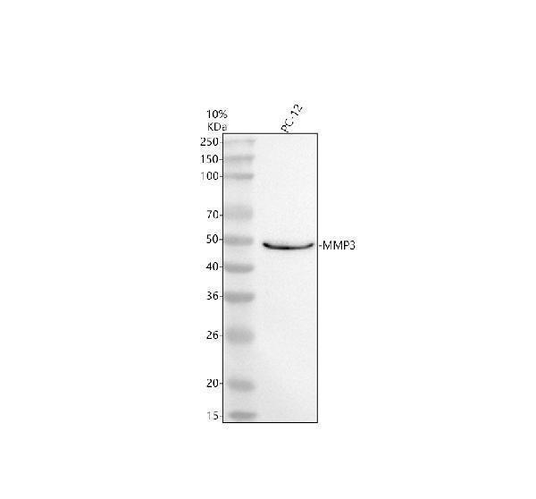
Western blot analysis of MMP3 using anti-MMP3 antibody (M00775).
Electrophoresis was performed on a 10% SDS-PAGE gel at 80V (Stacking gel) / 120V (Resolving gel) for 2 hours. The sample well of each lane was loaded with 30 ug of sample under reducing conditions.
Lane 1: rat PC-12 whole cell lysates.
After electrophoresis, proteins were transferred to a nitrocellulose membrane at 150 mA for 50-90 minutes. Blocked the membrane with 5% non-fat milk/TBS for 1.5 hour at RT. The membrane was incubated with rabbit anti-MMP3 antigen affinity purified monoclonal antibody (M00775) at 1:500 overnight at 4°C, then washed with TBS-0.1%Tween 3 times with 5 minutes each and probed with a goat anti-rabbit IgG-HRP secondary antibody at a dilution of 1:5000 for 1.5 hour at RT. The signal is developed using an ECL Plus Western Blotting Substrate (Catalog # AR1196-200) with Tanon 5200 system. A specific band was detected for MMP3 at approximately 50 kDa. The expected band size for MMP3 is at 50 kDa.

Click image to see more details
Immunofluorescent analysis of HeLa cells, using MMP3 Antibody.

Click image to see more details
Immunohistochemical analysis of paraffin-embedded human liver cancer, using MMP3 Antibody.

Click image to see more details
The role of TRADD in TNF-α-mediated cellular events. The cells were transfected with siRNA and lipofectamine 3000 for 48 h. After 48 h transfection, the medium was replaced by refresh medium and chondrocytes were treated with TNF-α (5 ng/ml) for 24 h, the protein expression of TRADD, iNOS, COX2, COL2A1, MMP13, cleaved-caspase-3, and LC3 was examined ( A – C , E , F ). After 48 h transfection with siRNA and lipofectamine 3000, the medium was replaced by refresh medium and chondrocytes were pre-treated with z-VAD for 2 h and then exposed to TNF-α for 24 h, the phosphorylation of RIPK1, RIPK3, and MLKL as well as protein expression of these three markers were detected using western blot ( G ). I , K The protein expression of iNOS, COX2, COL2A1, and MMP3 was examined in chondrocytes overexpressing TRADD after TNF-α intervention. D , H , J , L Semi-quantitative analysis of protein bands by image J and data in figures were expressed as means ± SD. * p < 0.05, ** p < 0.01. NS not significant,. NC negative control. z-VAD, Z-VAD-FMK. C-caspase-3, cleaved-caspase-3. M The graphical scheme summarized the findings of the role of TRADD in TNF-α-mediated chondrocytes events.
Index in PubMed under a CC BY license. PMID: 37002200

Click image to see more details
W-Exo-L@GelMA exhibits a strong chondrocyte-targeting effect and a pronounced action on promoting anabolism and suppressing catabolism and inflammation without causing the inhibition of chondrocyte viability. A Cell viability assessed by CCK8 assay. No obvious cytotoxicity on chondrocytes was observed when treated with W-Exo-L@GelMA loaded with 0.5, 1.0, 2.5, and 5.0 µM LRRK2-IN-1 for 48 h. Data represent mean ± SD; N = 6/group; one-way ANOVA; ns, not significant. B Immunofluorescence of Dil-labeled exosomes. The uptake of exosomes was observed in the chondrocytes when treated with Exo-L, Exo-L@GelMA or W-Exo-L@GelMA for 48 h. Dil was used for labeling exosomes (red), DAPI to label nuclei (blue), and Phalloidin to label the cytoskeleton (green). Scar bar: 200 μm. C Western blot analyses of the protein levels of anabolic, catabolic, and inflammatory factors in the IL-1β-induced chondrocytes treated with W-Exo-L@GelMA loaded with 0.5, 1.0, 2.5, and 5.0 µM LRRK2-IN-1 for 48 h. W-Exo-L@GelMA promoted COL2 and SOX9 and inhibited iNOS, COX2, MMP3, and MMP13 protein levels in a dose-dependent manner. D Quantitative analysis of the western blot results. Data represent mean ± SD; N = 3/group; *P < 0.05; **P < 0.01 by one-way ANOVA
Index in PubMed under a CC BY license. PMID: 37605203

Click image to see more details
LRRK2-IN-1 suppresses the IL-1β-induced inflammation and catabolism and induces anabolism without causing the inhibition of chondrocyte viability. A Schematic diagram of cell treatment and experimental procedures. B Cell viability assessed by CCK8 assay. No obvious inhibition of chondrocyte proliferation was observed when treated with 0.5, 1.0, 2.5, and 5.0 µM LRRK2-IN-1 for 24 h. Data represent mean ± SD; N = 6/group; one-way ANOVA; ns, not significant. C Western blot analyses of the protein levels of anabolic, catabolic, inflammatory factors in the IL-1β-induced chondrocytes treated with 0.5, 1.0, 2.5, and 5.0 µM LRRK2-IN-1 for 24 h. LRRK2-IN-1 suppressed MMP3, MMP13, iNOS, and COX2 and induced COL2 and SOX9 in a dose-dependent manner. D Quantitative analyses of the western blot results. Data represent mean ± SD; N = 3/group; *P < 0.05; **P < 0.01 by one-way ANOVA. E Immunofluorescence of iNOS, MMP13, and aggrecan expression in the IL-1β-induced chondrocytes treated with 5.0 µM LRRK2-IN-1 for 24 h. Scar bar: 400 μm
Index in PubMed under a CC BY license. PMID: 37605203

Click image to see more details
LIPUS inhibits the progression of OA. A , B Western blot and quantitative analysis of MMP13, ADAMTS5 and COX2 expression level with LIPUS application for 20 min. C , D Western blot and quantitative analysis of P62, ATG5 and LC3 expression level with LIPUS application for 20 min. E , F Western blot and quantitative analysis of ACAN, COL2A1, SOX9 and COX2 expression level with 30 mW/cm 2 LIPUS application. G Western blot and quantitative analysis of ATG5 expression level with 30 mW/cm 2 LIPUS application. H qPCR of MMP3, ADAMTS5 and MMP13 relative expression level. Data are shown as mean ± SD. *P < 0.05, **P < 0.01, ***P < 0.001
Index in PubMed under a CC BY license. PMID: 38493143

Click image to see more details
YAP exacerbated chondrocyte damage induced by IL-1β. Chondrocytes transfected with si-NC or YAP siRNA following IL-1β induction for 24 h. A qPCR of MMP3, MMP13 and ADAMTS5 relative expression level. B Western blot and quantitative analysis of COL2A1, MMP3, ADAMTS5 and MMP13 expression level. C , D Western blot and quantitative analysis of COX2, iNOS, P62 and LC3 expression level. E , F Immunofluorescence staining and fluorescence intensity analysis of COL2A1 and MMP13 relative expression level. G , H Chondrocytes were transfected with si-NC or si-YAP and then transfected with mRFP-GFP-LC3 adenovirus following IL-1β treatment for 24 h. The representative images of fluorescence and quantitative analysis of red dots and yellow dots were shown (scale bar: 10 μm). Subsequently, Chondrocytes transfected with oe-NC or oe-YAP following IL-1β induction for 24 h. I Western blot and quantitative analysis of COL2A1, MMP3, ADAMTS5 and MMP13 expression level. J , K Western blot and quantitative analysis of COX2, iNOS, Beclin-1 and P62 expression level. L – O Immunofluorescence staining and fluorescence intensity analysis of COL2A1 and MMP13 relative expression level. Data are shown as mean ± SD. *P < 0.05, **P < 0.01, ***P < 0.001
Index in PubMed under a CC BY license. PMID: 38493143

Click image to see more details
The effect of mtDNA on IRF1 and chondrocyte damage. Chondrocytes were treated with TNF-α alone or CsA alone or TNF-α combined with CsA for 12 h. (A, B) Western blots and quantitative analysis of expression level of IRF1 ( n = 3). (C, D) immunofluorescence staining and fluorescence intensity analysis of IRF1 nuclear location ( n = 3). (E, F) Western blots and quantitative analysis of expression level of iNOS, MMP13, and MMP3 ( n = 3). Data are shown as the means ± SDs. * P < 0.05, ** P < 0.01, *** P < 0.001, **** P < 0.0001
Index in PubMed under a CC BY license. PMID: 39026271

Click image to see more details
ZBP1 overexpression depends on IRF1. Chondrocytes were transfected with siNC or IRF1 siRNA for 24 h following TNF-α induction for 12 h. (A) Prediction of transcription factors using the UCSC Genome Browser database, SPP-ominer database, and Cistrome Data Browser. (B) qPCR results showing the relative mRNA expression levels of Irf1 and Zbp1 ( n = 3). (C, D) Western blots and quantitative analysis of the relative protein expression level of IRF1 ( n = 3). (E, F) Immunofluorescence staining and fluorescence intensity analysis of ZBP1 expression in chondrocytes transfected with siNC or IRF1 siRNA following TNF-α induction for 12 h ( n = 3; scale bar: 50 μm). (G-L) Western blots and quantitative analysis of the relative protein expression levels of iNOS, COX2, MMP3, MMP13, AGGRECAN, COL2A1, and SOX9 ( n = 3). (M-P) Immunofluorescence staining and fluorescence intensity analysis of COL2A1 and MMP13 expression in chondrocytes transfected with siNC or IRF1 siRNA following TNF-α induction for 12 h ( n = 3; scale bar: 50 μm). Chondrocytes were transfected with negative control siRNA(siNC) or IRF1 siRNA (si- Irf1 ) or ZBP1-plasmid (OE- Zbp1 ) or empty plasmid (OE-NC) for 24 h following TNF-α induction for 24 h. (Q-V) Western blots and quantitative analysis of the relative protein expression of AGGRECAN, COL2A1, SOX9, iNOS, COX2 MMP13, and MMP3 in chondrocytes ( n = 3). The data are shown as the means ± SDs. * P < 0.05, ** P < 0.01, *** P < 0.001, **** P < 0.0001
Index in PubMed under a CC BY license. PMID: 39026271

Click image to see more details
ZBP1 is essential for chondrocyte damage. Chondrocytes were transfected with siNC or ZBP1 siRNA for 24 h following TNF-α induction for 24 h. (A, B) Western blots and quantitative analysis of ZBP1, MMP13, and MMP3 expression levels ( n = 3). (C, D) Western blots and quantitative analysis of SOX9 expression levels. (E, F) Western blot and quantitative analysis of iNOS expression levels ( n = 3). (G) qPCR results showing the relative expression levels of Zbp1 and Inos ( n = 3). (H-K) Immunofluorescence staining and fluorescence intensity analysis of the relative expression levels of COL2A1 and MMP13 ( n = 3; scale bar: 50 μm). Chondrocytes were transfected with siNC or ZBP1 siRNA following TSZ (20 ng/ml TNF-α, 100 nM Smac mimetic, and 20 mM Z-VAD) induction for 12 h. (L, M) Western blots and quantitative analysis of ZBP1, P-RIPK3, and P-MLKL expression levels ( n = 3). The data are shown as the means ± SDs. * P < 0.05, ** P < 0.01, *** P < 0.001, **** P < 0.0001
Index in PubMed under a CC BY license. PMID: 39026271

Click image to see more details
Effects of MOIG on adhesion, migration, invasion and the expression of associated proteins of TNF-α-stimulated FLSs cells. (A) Adhesion of FLSs cells. (B) and (C) The wound healing of FLSs at 24 and 48 h; (D) and (F) The migration of FLSs at 6 h; (E) and (G) The invasion of FLSs at 24 h; ( H–M ) the expression of ICAM-1, VCAM-1, cadherin 11, MMP2 and MMP3 of FLSs. The data were expressed as mean ± SD (n = 3). # p < 0.05, ## p < 0.01, ### p < 0.001 vs. normal ctrl group; * p < 0.05, ** p < 0.01, *** p < 0.001 vs. TNF-α model group.
Index in PubMed under a CC BY license. PMID: 39444614

Click image to see more details
ICCB-19 protects against TNF-α-induced detrimental events via autophagy mechanism. After a pre-treatment of ICCB-19 for 2 h, chondrocytes were stimulated by TNF-α for 24 h. A the protein expression of ATG5 and LC3 was examined. B The semi-quantitative analysis of protein bands by image J. C Representative images of immunofluorescence staining of LC3 in chondrocytes, scale bar: 50 μm. D – F After a pre-treatment of ICCB-19 and 3-MA for 2 h, chondrocytes were stimulated by TNF-α for 24 h with or without z-VAD, the protein expression of iNOS, COX2, COL2A1, MMP13, MMP3, C-caspase-9, and C-caspase-3 were examined, and the phosphorylation of RIPK1, RIPK3, and MLKL as well as protein expression of these three markers were detected by western blot. G – H After a pre-treatment of Rapa for 2 h, chondrocytes were stimulated by TNF-α for 24 h with or without z-VAD, the protein expression of iNOS, COX2, MMP13, C-caspase-3 and the phosphorylation of RIPK1, RIPK3, and MLKL as well as protein expression of these three markers were detected by western blot. I The graphical scheme summarized the findings of protective role of ICCB-19 in TNF-α-caused detrimental events. Data are expressed as means ± SD. * p < 0.05, ** p < 0.01. z-VAD, Z-VAD-FMK. C-caspase-9, cleaved-caspase-9. C-caspase-3, cleaved-caspase-3. P-, Phosphorylated-.
Index in PubMed under a CC BY license. PMID: 37002200

Click image to see more details
LIPUS inhibits the progression of OA via YAP/RIPK1 axis. A Western blot and quantitative analysis of ACAN COL2A1, SOX9 and MMP3 expression level. B Western blot and quantitative analysis of COX2 and iNOS expression level. C Western blot of P-YAP and P-P65 expression level. D , E Immunofluorescence staining and fluorescence intensity analysis of COL2A1 and MMP13 relative expression level. F Chondrocytes were transfected with mRFP-GFP-LC3 adenovirus following IL-1β treatment for 24 h. The representative images of fluorescence and quantitative analysis of red dots and yellow dots were revealed (scale bar: 10 μm). G Western blot of P-YAP, YAP, P-P65, P65, P-IκB, IκB, P-RIPK1 and RIPK1 relative protein expression. H Images of IF staining for P65 relative expression and the distribution in chondrocytes with IL-1β induction for 15 min (scale bar: 25 μm). I Co-IP experiment of the binding between YAP and RIPK1 after LIPUS intervention. J YAP and RIPK1 colocalization in chondrocytes were detected by IF after LIPUS intervention (scale bar: 10 μm). K , L Immunohistochemistry staining to show the P-YAP-positive cell, P-RIPK1-positive cell and P-P65-positive cell in cartilage of the six groups (scale bar: 100 μm). Data are shown as mean ± SD. *P < 0.05, **P < 0.01, ***P < 0.001
Index in PubMed under a CC BY license. PMID: 38493143
Specific Publications For Anti-MMP3 Rabbit Monoclonal Antibody (M00775)
Loading publications
Recommended Resources
Here are featured tools and databases that you might find useful.
- Boster's Pathways Library
- Protein Databases
- Bioscience Research Protocol Resources
- Data Processing & Analysis Software
- Photo Editing Software
- Scientific Literature Resources
- Research Paper Management Tools
- Molecular Biology Software
- Primer Design Tools
- Bioinformatics Tools
- Phylogenetic Tree Analysis
Customer Reviews
Have you used Anti-MMP3 Rabbit Monoclonal Antibody?
Share your experimental results or join a short interview to earn up to $1,000 in product credits or other rewards.
0 Reviews For Anti-MMP3 Rabbit Monoclonal Antibody
Customer Q&As
Have a question?
Find answers in Q&As, reviews.
Can't find your answer?
Submit your question
16 Customer Q&As for Anti-MMP3 Rabbit Monoclonal Antibody
Question
Is this M00775 anti-MMP3 Rabbit Monoclonal antibody reactive to the isotypes of MMP3?
Verified Customer
Verified customer
Asked: 2020-03-23
Answer
The immunogen of M00775 anti-MMP3 Rabbit Monoclonal antibody is A synthesized peptide derived from human MMP3. Could you tell me which isotype you are interested in so I can help see if the immunogen is part of this isotype?
Boster Scientific Support
Answered: 2020-03-23
Question
We have observed staining in mouse synovium. Any tips? Is anti-MMP3 Rabbit Monoclonal antibody supposed to stain synovium positively?
Verified Customer
Verified customer
Asked: 2020-02-28
Answer
Based on literature synovium does express MMP3. Based on Uniprot.org, MMP3 is expressed in layer of synovial tissue, fibroblast, synovium, lung, among other tissues. Regarding which tissues have MMP3 expression, here are a few articles citing expression in various tissues:
Fibroblast, Pubmed ID: 3030290
Lung, Pubmed ID: 15489334
Synovium, Pubmed ID: 14702039
Boster Scientific Support
Answered: 2020-02-28
Question
I was wanting to use your anti-MMP3 Rabbit Monoclonal antibody for IHC for human layer of synovial tissue on frozen tissues, but I want to know if it has been validated for this particular application. Has this antibody been validated and is this antibody a good choice for human layer of synovial tissue identification?
Verified Customer
Verified customer
Asked: 2020-01-01
Answer
As indicated on the product datasheet, M00775 anti-MMP3 Rabbit Monoclonal antibody has been tested for IF, IHC, ICC, WB on human, mouse, rat tissues. We have an innovator award program that if you test this antibody and show it works in human layer of synovial tissue in IHC-frozen, you can get your next antibody for free.
Boster Scientific Support
Answered: 2020-01-01
Question
We appreciate helping with my inquiry over the phone. Here are the WB image, lot number and protocol we used for layer of synovial tissue using anti-MMP3 Rabbit Monoclonal antibody M00775. Let me know if you need anything else.
Verified Customer
Verified customer
Asked: 2019-11-26
Answer
We appreciate the data. You have provided everything we needed. Our lab team are working to resolve your inquiry as quickly as possible, and we appreciate your patience and understanding! Please let me know if there is anything you need in the meantime.
Boster Scientific Support
Answered: 2019-11-26
Question
I see that the anti-MMP3 Rabbit Monoclonal antibody M00775 works with IHC, what is the protocol used to produce the result images on the product page?
Verified Customer
Verified customer
Asked: 2019-08-12
Answer
You can find protocols for IHC on the "support/technical resources" section of our navigation menu. If you have any further questions, please send an email to support@bosterbio.com
Boster Scientific Support
Answered: 2019-08-12
Question
Will anti-MMP3 Rabbit Monoclonal antibody M00775 work for IHC with layer of synovial tissue?
Verified Customer
Verified customer
Asked: 2019-08-06
Answer
According to the expression profile of layer of synovial tissue, MMP3 is highly expressed in layer of synovial tissue. So, it is likely that anti-MMP3 Rabbit Monoclonal antibody M00775 will work for IHC with layer of synovial tissue.
Boster Scientific Support
Answered: 2019-08-06
Question
Is there a BSA free version of anti-MMP3 Rabbit Monoclonal antibody M00775 available?
Verified Customer
Verified customer
Asked: 2019-06-04
Answer
We appreciate your recent telephone inquiry. I can confirm that some lots of this anti-MMP3 Rabbit Monoclonal antibody M00775 are BSA free. For now, these lots are available and we can make a BSA free formula for you free of charge. It will take 3 extra days to prepare. If you require this antibody BSA free again in future, please do not hesitate to contact me and I will be pleased to check which lots we have in stock that are BSA free.
Boster Scientific Support
Answered: 2019-06-04
Question
My boss were happy with the WB result of your anti-MMP3 Rabbit Monoclonal antibody. However we have been able to see positive staining in synovium extracellular space using this antibody. Is that expected? Could you tell me where is MMP3 supposed to be expressed?
Verified Customer
Verified customer
Asked: 2019-04-30
Answer
Based on literature, synovium does express MMP3. Generally MMP3 expresses in secreted, extracellular space, extracellular. Regarding which tissues have MMP3 expression, here are a few articles citing expression in various tissues:
Fibroblast, Pubmed ID: 3030290
Lung, Pubmed ID: 15489334
Synovium, Pubmed ID: 14702039
Boster Scientific Support
Answered: 2019-04-30
Question
Does anti-MMP3 Rabbit Monoclonal antibody M00775 work on goat WB with layer of synovial tissue?
Verified Customer
Verified customer
Asked: 2019-01-15
Answer
Our lab technicians have not validated anti-MMP3 Rabbit Monoclonal antibody M00775 on goat. You can run a BLAST between goat and the immunogen sequence of anti-MMP3 Rabbit Monoclonal antibody M00775 to see if they may cross-react. If the sequence homology is close, then you can perform a pilot test. Keep in mind that since we have not validated goat samples, this use of the antibody is not covered by our guarantee. However we have an innovator award program that if you test this antibody and show it works in goat layer of synovial tissue in WB, you can get your next antibody for free.
Boster Scientific Support
Answered: 2019-01-15
Question
Is a blocking peptide available for product anti-MMP3 Rabbit Monoclonal antibody (M00775)?
Verified Customer
Verified customer
Asked: 2018-09-28
Answer
We do provide the blocking peptide for product anti-MMP3 Rabbit Monoclonal antibody (M00775). If you would like to place an order for it please contact support@bosterbio.com and make a special request.
Boster Scientific Support
Answered: 2018-09-28
Question
Would M00775 anti-MMP3 Rabbit Monoclonal antibody work on parafin embedded sections? If so, which fixation method do you recommend we use (PFA, paraformaldehyde, other)?
Verified Customer
Verified customer
Asked: 2018-06-28
Answer
As indicated on the product datasheet, M00775 anti-MMP3 Rabbit Monoclonal antibody as been validated on IHC. It is best to use PFA for fixation because it has better tissue penetration ability. PFA needs to be prepared fresh before use. Long term stored PFA turns into formalin, as the PFA molecules congregate and become formalin.
Boster Scientific Support
Answered: 2018-06-28
Question
Please see the WB image, lot number and protocol we used for layer of synovial tissue using anti-MMP3 Rabbit Monoclonal antibody M00775. Please let me know if you require anything else.
G. Huang
Verified customer
Asked: 2018-04-05
Answer
Thank you very much for the data. Our lab team are working to resolve this as quickly as possible, and we appreciate your patience and understanding! You have provided everything we needed. Please let me know if there is anything you need in the meantime.
Boster Scientific Support
Answered: 2018-04-05
Question
We are currently using anti-MMP3 Rabbit Monoclonal antibody M00775 for human tissue, and we are happy with the WB results. The species of reactivity given in the datasheet says human, mouse, rat. Is it possible that the antibody can work on goat tissues as well?
Verified Customer
Verified customer
Asked: 2018-02-28
Answer
The anti-MMP3 Rabbit Monoclonal antibody (M00775) has not been tested for cross reactivity specifically with goat tissues, though there is a good chance of cross reactivity. We have an innovator award program that if you test this antibody and show it works in goat you can get your next antibody for free. Please contact me if I can help you with anything.
Boster Scientific Support
Answered: 2018-02-28
Question
My question regarding product M00775, anti-MMP3 Rabbit Monoclonal antibody. I was wondering if it would be possible to conjugate this antibody with biotin. I would need it to be without BSA or sodium azide. I am planning on using a buffer exchange of sodium azide with PBS only. Would there be problems for me to conjugate the antibody and store it in -20 degrees in small aliquots?
C. Mangal
Verified customer
Asked: 2014-11-24
Answer
We do not recommend storing this antibody with PBS buffer only in -20 degrees. If you want to store it in -20 degrees it is best to add some cryoprotectant like glycerol. If you want carrier free M00775 anti-MMP3 Rabbit Monoclonal antibody, we can provide it to you in a special formula with trehalose and/or glycerol. These molecules will not interfere with conjugation chemistry and provide a good level of protection for the antibody from degradation. Please be sure to specify this in your purchase order.
Boster Scientific Support
Answered: 2014-11-24
Question
We ordered your anti-MMP3 Rabbit Monoclonal antibody for IF on lung a few months ago. I am using mouse, and We intend to use the antibody for IHC next. We need examining lung as well as synovium in our next experiment. Could you please give me some suggestion on which antibody would work the best for IHC?
M. Lewis
Verified customer
Asked: 2014-01-28
Answer
I have checked the website and datasheets of our anti-MMP3 Rabbit Monoclonal antibody and it appears that M00775 has been tested on mouse in both IF and IHC. Thus M00775 should work for your application. Our Boster satisfaction guarantee will cover this product for IHC in mouse even if the specific tissue type has not been validated. We do have a comprehensive range of products for IHC detection and you can check out our website bosterbio.com to find out more information about them.
Boster Scientific Support
Answered: 2014-01-28
Question
We want to test anti-MMP3 Rabbit Monoclonal antibody M00775 on human layer of synovial tissue for research purposes, then I may be interested in using anti-MMP3 Rabbit Monoclonal antibody M00775 for diagnostic purposes as well. Is the antibody suitable for diagnostic purposes?
R. Dhar
Verified customer
Asked: 2013-10-09
Answer
The products we sell, including anti-MMP3 Rabbit Monoclonal antibody M00775, are only intended for research use. They would not be suitable for use in diagnostic work. If you have the means to develop a product into diagnostic use, and are interested in collaborating with us and develop our product into an IVD product, please contact us for more discussions.
Boster Scientific Support
Answered: 2013-10-09
