Click image to see more details
Product Info Summary
| SKU: | A00276 |
|---|---|
| Size: | 80 µl |
| Reactive Species: | Mouse |
| Host: | Rabbit |
| Application: | Flow Cytometry, IHC-P, WB |
Customers Who Bought This Also Bought

Product info
Product Name
Anti-(Mouse) Epcam Antibody (C-term)
SKU/Catalog Number
A00276
Size
80 µl
Form
Liquid
Description
Boster Bio Anti-(Mouse) Epcam Antibody (C-term) (Catalog # A00276). Tested in WB, IHC-P, Flow Cytometry application(s). This antibody reacts with Mouse.
Storage & Handling
Maintain refrigerated at 2-8°C for up to 2 weeks. For long-term storage, store at -20°C in small aliquots to prevent freeze-thaw cycles.
Cite This Product
Anti-(Mouse) Epcam Antibody (C-term) (Boster Biological Technology, Pleasanton CA, USA, Catalog # A00276)
Host
Rabbit
Contents
Purified polyclonal antibody supplied in PBS with 0.09% (W/V) sodium azide.
Clonality
Polyclonal
Isotype
Rabbit IgG
Immunogen
This mouse Epcam antibody is generated from a rabbit immunized with a KLH conjugated synthetic peptide between 302-335 amino acids from the C-terminal region of mouse Epcam.
Cross-reactivity
No cross reactivity with other proteins.
Reactive Species
A00276 is reactive to Epcam in Mouse
Calculated molecular weight
35.0 kDa
Background of Epcam
May act as a physical homophilic interaction molecule between intestinal epithelial cells (IECs) and intraepithelial lymphocytes (IELs) at the mucosal epithelium for providing immunological barrier as a first line of defense against mucosal infection. Plays a role in embryonic stem cells proliferation and differentiation. Up-regulates the expression of FABP5, MYC and cyclins A and E (By similarity).
Antibody Validation
Boster validates all antibodies on WB, IHC, ICC, Immunofluorescence, and ELISA with known positive control and negative samples to ensure specificity and high affinity, including thorough antibody incubations.
Application & Images
Applications
A00276 is guaranteed for Flow Cytometry, IHC-P, WB Boster Guarantee
Recommend Dilution
WB: 1:1000
IHC-P: 1:25
FC: 1:25
Validation Images & Assay Conditions
Click image to see more details
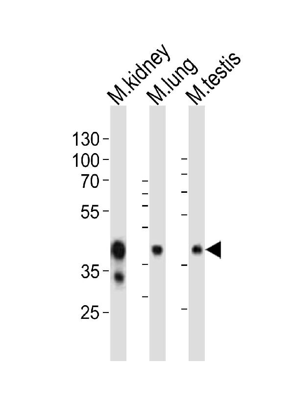
Western blot analysis of lysates from mouse kidney, mouse lung, mouse testis tissue lysate (from left to right), using Epcam Antibody (C-term). A00276 was diluted at 1:1000 at each lane. A goat anti-rabbit IgG H&L (HRP) at 1:10000 dilution was used as the secondary antibody. Lysates at 20ug per lane.

Click image to see more details
A00276 staining Epcam in Mouse colon tissue sections by Immunohistochemistry (IHC-P -paraformaldehyde-fixed, paraffin-embedded sections). Tissue was fixed with formaldehyde and blocked with 3% BSA for 0. 5 hour at room temperature; antigen retrieval was by heat mediation with a citrate buffer (pH6). Samples were incubated with primary antibody (1/25) for 1 hours at 37°C. A undiluted biotinylated goat polyvalent antibody was used as the secondary antibody.

Click image to see more details
A00276 staining Epcam in Human colorectal carcinoma tissue sections by Immunohistochemistry (IHC-P -paraformaldehyde-fixed, paraffin-embedded sections). Tissue was fixed with formaldehyde and blocked with 3% BSA for 0. 5 hour at room temperature; antigen retrieval was by heat mediation with a citrate buffer (pH6). Samples were incubated with primary antibody (1/25) for 1 hours at 37°C. A undiluted biotinylated goat polyvalent antibody was used as the secondary antibody.

Click image to see more details
Overlay histogram showing HepG2 cells stained with A00276 (red line). The cells were fixed with 2% paraformaldehyde (10 min) and then permeabilized with 90% methanol for 10 min. The cells were then icubated in 2% bovine serum albumin to block non-specific protein-protein interactions followed by the antibody (A00276, 1:25 dilution) for 60 min at 37ºC. The secondary antibody used was Alexa Fluor® 488 goat anti-rabbit lgG (H+L) at 1/400 dilution for 40 min at 37ºC. Isotype control antibody (blue line) was rabbit IgG1 (1μg/1x10^6 cells) used under the same conditions. Acquisition of >10, 000 events was performed.
Specific Publications For Anti-(Mouse) Epcam Antibody (C-term) (A00276)
Loading publications
Recommended Resources
Here are featured tools and databases that you might find useful.
- Boster's Pathways Library
- Protein Databases
- Bioscience Research Protocol Resources
- Data Processing & Analysis Software
- Photo Editing Software
- Scientific Literature Resources
- Research Paper Management Tools
- Molecular Biology Software
- Primer Design Tools
- Bioinformatics Tools
- Phylogenetic Tree Analysis
Customer Reviews
Have you used Anti-(Mouse) Epcam Antibody (C-term)?
Share your experimental results or join a short interview to earn up to $1,000 in product credits or other rewards.
0 Reviews For Anti-(Mouse) Epcam Antibody (C-term)
Customer Q&As
Have a question?
Find answers in Q&As, reviews.
Can't find your answer?
Submit your question
