Click image to see more details
Product Info Summary
| SKU: | RP1015 |
|---|---|
| Size: | 100 μg/vial |
| Reactive Species: | Mouse |
| Host: | Rabbit |
| Application: | ELISA, WB |
Customers Who Bought This Also Bought

Product info
Product Name
Anti-Interleukin-10 IL10 Antibody Picoband®
SKU/Catalog Number
RP1015
Size
100 μg/vial
Form
Lyophilized
Description
Boster Bio Anti-Interleukin-10 IL10 Antibody catalog # RP1015. Tested in ELISA, WB applications. This antibody reacts with Mouse. The brand Picoband indicates this is a premium antibody that guarantees superior quality, high affinity, and strong signals with minimal background in Western blot applications. Only our best-performing antibodies are designated as Picoband, ensuring unmatched performance.
Storage & Handling
Store at -20˚C for one year from date of receipt. After reconstitution, at 4˚C for one month. It can also be aliquotted and stored frozen at -20˚C for six months. Avoid repeated freeze-thaw cycles.
Cite This Product
Anti-Interleukin-10 IL10 Antibody Picoband® (Boster Biological Technology, Pleasanton CA, USA, Catalog # RP1015)
Host
Rabbit
Contents
Each vial contains 0.9mg NaCl, 0.2mg Na2HPO4, 0.05mg NaN3. Carrier free (No BSA) form available in stock. If you want this antibody carrier free please specify "Carrier Free" or "No BSA" in your order note.
Clonality
Polyclonal
Isotype
Rabbit IgG
Immunogen
E. coli-derived mouse IL-10 recombinant protein (Position: S19-S178).
Cross-reactivity
No cross-reactivity with other proteins
Reactive Species
RP1015 is reactive to Il10 in Mouse
Observed Molecular Weight
19 kDa
Calculated molecular weight
20.6 kDa
Background of Il10
Interleukin-10 (IL-10 or IL10), also known as human cytokine synthesis inhibitory factor (CSIF), is an anti-inflammatory cytokine. In humans IL-10 is encoded by the IL10 gene. It is capable of inhibiting synthesis of pro-inflammatory cytokines like IFN-gamma, IL-2, IL-3, TNFalpha and GM-CSF made by cells such as macrophages and regulatory T-cells.IL-10 also displays potent abilities to suppress the antigen presentation capacity of antigen presenting cells. Kim et al. (1992) showed that the mouse IL 10 gene contains 5 exons and spans about 5.2 kb of genomic DNA. Eskdale et al. (1997) mapped the IL10 gene to the junction between 1q31 and 1q32.
Antibody Validation
Boster validates all antibodies on WB, IHC, ICC, Immunofluorescence, and ELISA with known positive control and negative samples to ensure specificity and high affinity, including thorough antibody incubations.
Application & Images
Applications
RP1015 is guaranteed for ELISA, WB Boster Guarantee
Recommend Dilution
| Application | Dilution | Species |
|---|---|---|
| ELISA | 0.1-0.5μg/ml | - |
| Western blot | 0.1-0.5μg/ml | Mouse |
Validation Images & Assay Conditions
Click image to see more details
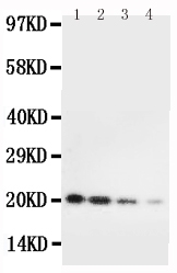
Anti-mouse IL-10 antibody, RP1015, Western blotting
Lane 1: Recombinant Mouse IL-10 Protein 10ng
Lane 2: Recombinant Mouse IL-10 Protein 5ng
Lane 3: Recombinant Mouse IL-10 Protein 2

Click image to see more details
The infiltration of MPO + neutrophils, and the cellular distribution and relative expression level detection of the TNF and IL-10 in the small intestinal and colonic mucosa at 7 days after the termination of DSS administration. (A) The MPO immunohistochemistry staining of the small intestinal mucosa: (A1) the normal group: few neutrophils were observed in the small intestinal mucosa; (A2) the DSS group: a number of accumulative MPO + neutrophils (brown) infiltrated into the mucosa epithelium; (A3) the DSS + B. subtilis- fermented milk group: only limited neutrophil infiltration could be observed in the small intestinal mucosa. (B) The MPO immunohistochemistry staining of the colonic mucosa: (B1) the normal group: few neutrophils were observed in the colonic mucosa; (B2) the DSS group: colonic epithelium and the glands disappeared, and the ulcer was locally replaced by scars and a number of accumulative MPO + neutrophils (brown) were observed in the scars; (B3) the DSS + B. subtilis -fermented milk group: only limited MPO + neutrophils observed in the colonic mucosa. (C) The TNF immunohistochemistry staining of the small intestinal mucosa: (C1) the normal group: the epithelium was integrated with faint yellow staining, suggesting low expression of TNF; (C2) the DSS group: the villus structure is not integrated, and the epithelial cells showed black brown, suggesting overexpression of TNF; (C3) the DSS + B. subtilis -fermented milk group: the villus and the glands were almost integrated, and the staining of epithelial cells was similar to that of the normal group (C1) , suggesting low expression of TNF. (D) The TNF immunohistochemistry staining of the colonic mucosa: (D1) the normal colonic mucosa: the epithelium was integrated with low TNF expression (faint yellow); ( D2 ) the DSS group: the epithelium structure and the glands were destroyed and replaced by a scar, and there were a number of TNF + inflammatory cells (black brown) in the scar; (D3) the DSS + B. subtilis -fermented milk group: the recovered epithelium showed faint yellow, suggesting low TNF expression. (E) The IL-10 immunohistochemistry staining of the small intestinal mucosa: (E1) the normal small intestinal mucosa: the IL-10 staining dispersed in the villi and the crypts with faint yellow, suggesting low-level expression of IL-10; (E2) the DSS group, the residual epithelium and the crypts were light brown, suggesting mid-level of IL-10 expression; (E3) the DSS + B. subtilis -fermented milk group: the dark brown staining of the regenerative epithelium represented high-level expression of IL-10. (F) The IL-10 immunohistochemistry staining of the colonic mucosa: (F1) the normal group: the IL-10 staining dispersed in the glands with bright yellow, suggesting low-level expression of IL-10; (F2) the DSS group: there were few IL-10 + cells in the scars; (F3) the DSS + B. subtilis -fermented milk group, the dark brown staining of the epithelial cells represented high-level expression of IL-10. (G,H) Western blotting analysis for the expression of MPO, TNF, and IL-10 in the samples containing equivalent ileum and colon. The expression level of MPO, TNF, and IL-10 in the DSS group was significantly higher than that of the normal (control) group. The expression level of MPO and TNF in the DSS + B. subtilis -fermented milk (FM) group was significantly lower than that of the DSS group, while the expression level of IL-10 in the DSS + B. subtilis -fermented milk (FM) group was significantly higher than that of the DSS group ( n = 5, * represents p < 0.05, ** represents p < 0.01).
Index in PubMed under a CC BY license. PMID: 33519783
Specific Publications For Anti-Interleukin-10 IL10 Antibody Picoband® (RP1015)
Loading publications
Recommended Resources
Here are featured tools and databases that you might find useful.
- Boster's Pathways Library
- Protein Databases
- Bioscience Research Protocol Resources
- Data Processing & Analysis Software
- Photo Editing Software
- Scientific Literature Resources
- Research Paper Management Tools
- Molecular Biology Software
- Primer Design Tools
- Bioinformatics Tools
- Phylogenetic Tree Analysis
Customer Reviews
Have you used Anti-Interleukin-10 IL10 Antibody Picoband®?
Share your experimental results or join a short interview to earn up to $1,000 in product credits or other rewards.
0 Reviews For Anti-Interleukin-10 IL10 Antibody Picoband®
Customer Q&As
Have a question?
Find answers in Q&As, reviews.
Can't find your answer?
Submit your question
15 Customer Q&As for Anti-Interleukin-10 IL10 Antibody Picoband®
Question
Does RP1015 anti-IL10 antibody work on parafin embedded sections? If so, which fixation method do you recommend we use (PFA, paraformaldehyde, other)?
Verified Customer
Verified customer
Asked: 2020-01-20
Answer
It shows on the product datasheet, RP1015 anti-IL10 antibody as been validated on ELISA. It is best to use PFA for fixation because it has better tissue penetration ability. PFA needs to be prepared fresh before use. Long term stored PFA turns into formalin, as the PFA molecules congregate and become formalin.
Boster Scientific Support
Answered: 2020-01-20
Question
My colleagues were satisfied with the WB result of your anti-IL10 antibody. However we have been able to see positive staining in t-cell secreted. using this antibody. Is that expected? Could you tell me where is IL10 supposed to be expressed?
Verified Customer
Verified customer
Asked: 2019-11-18
Answer
According to literature, t-cell does express IL10. Generally IL10 expresses in secreted. Regarding which tissues have IL10 expression, here are a few articles citing expression in various tissues:
T-cell, Pubmed ID: 1847510
Boster Scientific Support
Answered: 2019-11-18
Question
I was wanting to use your anti-IL10 antibody for ELISA for mouse esophagus on frozen tissues, but I want to know if it has been tested for this particular application. Has this antibody been tested and is this antibody a good choice for mouse esophagus identification?
C. Patel
Verified customer
Asked: 2019-11-13
Answer
It shows on the product datasheet, RP1015 anti-IL10 antibody has been validated for ELISA, WB on mouse tissues. We have an innovator award program that if you test this antibody and show it works in mouse esophagus in IHC-frozen, you can get your next antibody for free.
Boster Scientific Support
Answered: 2019-11-13
Question
Does anti-IL10 antibody RP1015 work for ELISA with esophagus?
Verified Customer
Verified customer
Asked: 2019-10-09
Answer
According to the expression profile of esophagus, IL10 is highly expressed in esophagus. So, it is likely that anti-IL10 antibody RP1015 will work for ELISA with esophagus.
Boster Scientific Support
Answered: 2019-10-09
Question
We have been able to see staining in mouse esophagus. Are there any suggestions? Is anti-IL10 antibody supposed to stain esophagus positively?
Verified Customer
Verified customer
Asked: 2019-07-08
Answer
Based on literature esophagus does express IL10. Based on Uniprot.org, IL10 is expressed in esophagus, t-cell, among other tissues. Regarding which tissues have IL10 expression, here are a few articles citing expression in various tissues:
T-cell, Pubmed ID: 1847510
Boster Scientific Support
Answered: 2019-07-08
Question
My question regards to test anti-IL10 antibody RP1015 on mouse esophagus for research purposes, then I may be interested in using anti-IL10 antibody RP1015 for diagnostic purposes as well. Is the antibody suitable for diagnostic purposes?
Verified Customer
Verified customer
Asked: 2019-07-04
Answer
The products we sell, including anti-IL10 antibody RP1015, are only intended for research use. They would not be suitable for use in diagnostic work. If you have the means to develop a product into diagnostic use, and are interested in collaborating with us and develop our product into an IVD product, please contact us for more discussions.
Boster Scientific Support
Answered: 2019-07-04
Question
Is a blocking peptide available for product anti-IL10 antibody (RP1015)?
Verified Customer
Verified customer
Asked: 2019-06-26
Answer
We do provide the blocking peptide for product anti-IL10 antibody (RP1015). If you would like to place an order for it please contact support@bosterbio.com and make a special request.
Boster Scientific Support
Answered: 2019-06-26
Question
Is this RP1015 anti-IL10 antibody reactive to the isotypes of IL10?
Verified Customer
Verified customer
Asked: 2019-06-13
Answer
The immunogen of RP1015 anti-IL10 antibody is E. coli-derived mouse IL-10 recombinant protein(Position: S19-S178). Could you tell me which isotype you are interested in so I can help see if the immunogen is part of this isotype?
Boster Scientific Support
Answered: 2019-06-13
Question
We are currently using anti-IL10 antibody RP1015 for mouse tissue, and we are satisfied with the ELISA results. The species of reactivity given in the datasheet says mouse. Is it likely that the antibody can work on primate tissues as well?
Z. Mitchell
Verified customer
Asked: 2018-10-18
Answer
The anti-IL10 antibody (RP1015) has not been validated for cross reactivity specifically with primate tissues, but there is a good chance of cross reactivity. We have an innovator award program that if you test this antibody and show it works in primate you can get your next antibody for free. Please contact me if I can help you with anything.
Boster Scientific Support
Answered: 2018-10-18
Question
We appreciate helping with my inquiry over the phone. Here are the WB image, lot number and protocol we used for esophagus using anti-IL10 antibody RP1015. Let me know if you need anything else.
Verified Customer
Verified customer
Asked: 2018-01-12
Answer
We appreciate the data. You have provided everything we needed. Our lab team are working to resolve your inquiry as quickly as possible, and we appreciate your patience and understanding! Please let me know if there is anything you need in the meantime.
Boster Scientific Support
Answered: 2018-01-12
Question
See attached the WB image, lot number and protocol we used for esophagus using anti-IL10 antibody RP1015. Please let me know if you require anything else.
A. Brown
Verified customer
Asked: 2017-05-02
Answer
Thank you very much for the data. Our lab team are working to resolve this as quickly as possible, and we appreciate your patience and understanding! You have provided everything we needed. Please let me know if there is anything you need in the meantime.
Boster Scientific Support
Answered: 2017-05-02
Question
We have tried in the past anti-IL10 antibody for ELISA on t-cell a few months ago. I am using mouse, and I plan to use the antibody for WB next. We are interested in examining t-cell as well as esophagus in our next experiment. Could give a recommendation on which antibody would work the best for WB?
J. Carter
Verified customer
Asked: 2016-03-10
Answer
I have checked the website and datasheets of our anti-IL10 antibody and it appears that RP1015 has been validated on mouse in both ELISA and WB. Thus RP1015 should work for your application. Our Boster satisfaction guarantee will cover this product for WB in mouse even if the specific tissue type has not been validated. We do have a comprehensive range of products for WB detection and you can check out our website bosterbio.com to find out more information about them.
Boster Scientific Support
Answered: 2016-03-10
Question
Is there a BSA free version of anti-IL10 antibody RP1015 available?
R. Edwards
Verified customer
Asked: 2015-03-19
Answer
We appreciate your recent telephone inquiry. I can confirm that some lots of this anti-IL10 antibody RP1015 are BSA free. For now, these lots are available and we can make a BSA free formula for you free of charge. It will take 3 extra days to prepare. If you require this antibody BSA free again in future, please do not hesitate to contact me and I will be pleased to check which lots we have in stock that are BSA free.
Boster Scientific Support
Answered: 2015-03-19
Question
I see that the anti-IL10 antibody RP1015 works with ELISA, what is the protocol used to produce the result images on the product page?
H. Parker
Verified customer
Asked: 2014-04-22
Answer
You can find protocols for ELISA on the "support/technical resources" section of our navigation menu. If you have any further questions, please send an email to support@bosterbio.com
Boster Scientific Support
Answered: 2014-04-22
Question
I have a question about product RP1015, anti-IL10 antibody. I was wondering if it would be possible to conjugate this antibody with biotin. I would need it to be without BSA or sodium azide. I am planning on using a buffer exchange of sodium azide with PBS only. Would there be problems for me to conjugate the antibody and store it in -20 degrees in small aliquots?
J. Li
Verified customer
Asked: 2013-06-24
Answer
It is not recommended storing this antibody with PBS buffer only in -20 degrees. If you want to store it in -20 degrees it is best to add some cryoprotectant like glycerol. If you want carrier free RP1015 anti-IL10 antibody, we can provide it to you in a special formula with trehalose and/or glycerol. These molecules will not interfere with conjugation chemistry and provide a good level of protection for the antibody from degradation. Please be sure to specify this in your purchase order.
Boster Scientific Support
Answered: 2013-06-24
