Click image to see more details
-

-
-
-
-
+6
Product Info Summary
| SKU: | PB9447 |
|---|---|
| Size: | 100 μg/vial |
| Reactive Species: | Human, Mouse, Rat |
| Host: | Rabbit |
| Application: | IP, IF, IHC, ICC, WB |
Customers Who Bought This Also Bought

Product info
Product Name
Anti-mtTFA/TFAM Antibody Picoband®
SKU/Catalog Number
PB9447
PB0413 is an alternative SKU for this antibody, used in previous lots.
Size
100 μg/vial
Form
Lyophilized
Description
Boster Bio Anti-mtTFA/TFAM Antibody Picoband® catalog # PB9447. Tested in IP, IF, IHC, ICC, WB applications. This antibody reacts with Human, Mouse, Rat. The brand Picoband indicates this is a premium antibody that guarantees superior quality, high affinity, and strong signals with minimal background in Western blot applications. Only our best-performing antibodies are designated as Picoband, ensuring unmatched performance.
Storage & Handling
Store at -20˚C for one year from date of receipt. After reconstitution, at 4˚C for one month. It can also be aliquotted and stored frozen at -20˚C for six months. Avoid repeated freeze-thaw cycles.
Cite This Product
Anti-mtTFA/TFAM Antibody Picoband® (Boster Biological Technology, Pleasanton CA, USA, Catalog # PB9447)
Host
Rabbit
Contents
Each vial contains 4mg Trehalose, 0.9mg NaCl and 0.2mg Na2HPO4.
Clonality
Polyclonal
Isotype
Rabbit IgG
Immunogen
A synthetic peptide corresponding to a sequence at the N-terminus of human mtTFA, different from the related mouse and rat sequences by five amino acids.
Cross-reactivity
No cross-reactivity with other proteins
Reactive Species
PB9447 is reactive to TFAM in Human, Mouse, Rat
Observed Molecular Weight
24 kDa
Calculated molecular weight
29.1 kDa
Background of TFAM
TFAM (Transcription factor A, mitochondrial), also known as TCF6 or TCF6L2, is a 162-amino acid protein that activates transcription of each mitochondrial DNA (mtDNA) strand by binding to an element of approximately 30 nucleotides present in both the light-strand and the heavy-strand promoters. By Southern blot analysis of restriction enzyme digests of human/Chinese hamster somatic cell hybrid lines, Milatovich et al. (1992) mapped TFAM sequences, which they called MTTF1, to 3 different chromosomes: chromosomes 10, 7p, and 11q. By PCR-based screening of a somatic cell hybrid panel and by fluorescence in situ hybridization, Scott (2007) stated that the sequences mapped to chromosomes 7p (TCF6L1) and 11q (MTTF1, or TCF6L3) are pseudogenes. Larsson et al. (1997) mapped the mouse mitochondrial transcription factor A gene (Tfam) to the central part of mouse chromosome 10. This region exhibits syntenic homology with human 10q21. Mitochondrial transcription factor A is a key activator of mitochondrial transcription in mammals. It also has a role in mitochondrial DNA replication, since transcription generates an RNA primer necessary for initiation of mtDNA replication.
Antibody Validation
Boster validates all antibodies on WB, IHC, ICC, Immunofluorescence, and ELISA with known positive control and negative samples to ensure specificity and high affinity, including thorough antibody incubations.
Application & Images
Applications
PB9447 is guaranteed for IP, IF, IHC, ICC, WB Boster Guarantee
Recommend Dilution
| Application | Dilution | Species |
|---|---|---|
| Western blot | 0.1-0.5μg/ml | Human, Mouse, Rat |
| Immunohistochemistry (Paraffin-embedded Section) | 2-5μg/ml | Human |
| Immunocytochemistry/Immunofluorescence | 5μg/ml | Human |
| Immunoprecipitation | 0.5-2 μg/ml | Human |
Tested application
Suggested blocking solution with 5% non-fat milk or BSA; (*)Recommended protein loading: 20-40 µg per lane
Use TE buffer pH 9.0 for antigen retrieval; (*) citrate buffer pH 6.0 is an alternative.
Validation Images & Assay Conditions
Click image to see more details
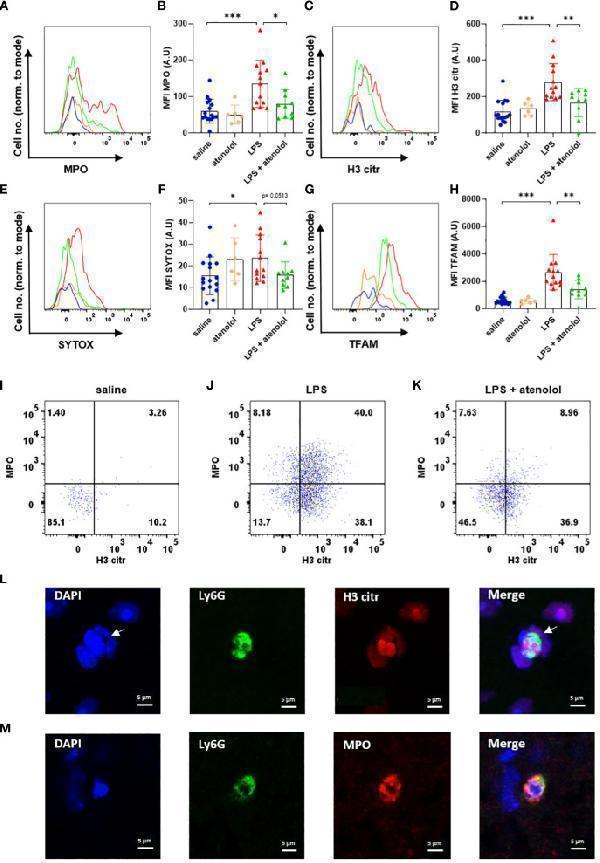
Atenolol decreased the production of NETs induced by LPS. Fluorescence intensity for a representative animal of each group for the extracellular trap markers MPO (A) , H3 citr (C) , Sytox (E) and TFAM (G) in saline (blue), atenolol (orange), LPS (red) and LPS + atenolol (green). Quantification of mean fluorescence intensities (MFIs) in live neutrophils for the extracellular trap markers MPO (B) , H3 citr (D) , Sytox (F) and TFAM (H) for saline (blue), atenolol (orange), LPS (red) and LPS + atenolol (green) groups. Representative dot plot of living neutrophils co-expressing MPO and H3 citr in saline (I) , LPS (J) and LPS + atenolol (K) groups. Representative pictures of Ly6G+ neutrophils (green) labeling with citrullinated histone H3 (H3 citr) (red). The nucleus of cells was labeled with DAPI (in blue). Arrow shows a neutrophil producing an ET (L) . Representative pictures of Ly6G+ neutrophils (green) labeling with myeloperoxidase (MPO) (red). The nucleus of cells was labeled with DAPI (in blue) (M) . Saline n=16, atenolol n=6, LPS n=13, LPS + atenolol n=10. * p<0.05, ** p<0.01, *** p<0.001.
Index in PubMed under a CC BY license. PMID: 37809074

Click image to see more details
Atenolol decreased the production of MiETs induced by LPS. Fluorescence intensity for a representative animal of each group for the extracellular trap markers MPO (A) , H3 citr (C) , Sytox (E) and TFAM (G) in saline (blue), atenolol (orange), LPS (red) and LPS + atenolol (green) groups. Quantification of mean fluorescence intensities (MFIs) in live microglia for the extracellular traps markers MPO (B) , H3 citr (D) , Sytox (F) and TFAM (H) for saline (blue), atenolol (orange), LPS (red) and LPS + atenolol (green) groups. Representative dot plot of living microglia co-expressing MPO and H3 citr in saline (I) , LPS (J) and LPS + atenolol (K) groups. Representative pictures of Iba1 + microglia (green) labeling with citrullinated histone H3 (H3 citr) (red). The nucleus of cells was labeled with DAPI (in blue) for saline (L) and LPS (M) groups. Saline n=16, atenolol n=6, LPS n=13, LPS + atenolol n=10. ** p<0.01, *** p<0.001.
Index in PubMed under a CC BY license. PMID: 37809074

Click image to see more details
Sivelestat decreased the production of ETs induced by LPS. Quantification of mean fluorescence intensities (MFIs) of extracellular trap markers MPO (A) , H3 citr (B) , Sytox (C) and TFAM (D) for saline (blue), sivelestat (turquoise), LPS (red) and LPS + sivelestat (khaki) groups in live neutrophils. Quantification of MFI of extracellular traps markers MPO (E) , H3 citr (F) , Sytox (G) and TFAM (H) for saline (blue), sivelestat (turquoise), LPS (red) and LPS + sivelestat (khaki) groups in live microglia. Representative dot plot of live neutrophils co-expressing MPO and H3 citr in saline (I) , LPS (J) and LPS + sivelestat (K) . Representative dot plot of living microglia co-expressing MPO and H3 citr in saline (L) , LPS (M) and LPS + sivelestat (N) groups. Saline n=16, sivelestat n=6, LPS n=13, LPS + sivelestat n=8. * p<0.05, ** p<0.01, ***p<0.001.
Index in PubMed under a CC BY license. PMID: 37809074

Click image to see more details
CXCR2 promotes mitochondrial dysfunction and tubular senescence in vitro . (A–J) Representative micrographs of western blot and quantitative statistical data show renal expression of (B) CXCR2 (C) active β-catenin, (D) PGC-1α, (E) COX1, (F) TFAM, (G) TOMM20, (H) p16 INK4A , (I) γ-H2AX and (J) FN in each group. * p < 0.05, ** p < 0.01, *** p < 0.001, versus the pcDNA3 group ( n = 3). (K,L) Graphical representations show the relative mRNA level of (K) CXCR2 and (L) p16 INK4A in different groups. *** p < 0.001 versus the pcDNA3 group ( n = 3). (M) Representative micrographs show SA-β-gal staining and γ-H2AX expression in different groups. Frozen sections were performed by SA-β-gal and γ-H2AX immunofluorescence staining. Arrows indicates positive staining. Scale bar = 50 or 10 μm. (N–R) Representative micrographs of western blot and quantitative statistical data show protein levels of (O) CXCR2, (P) TOMM20, (Q) p16 INK4A and (R) FN in each group. * p < 0.05 versus the control group ( n = 3). (S) Representative micrographs show SA-β-gal staining in each group. Frozen renal sections were performed by SA-β-gal staining. Arrows indicate positive staining. Scale bar, 50 μm.
Index in PubMed under a CC BY license. PMID: 35592244

Click image to see more details
O304 could promote autophagy and alleviate mitochondrial dysfunction in vitro . (A–D) Representative western blot showing the expression of p-mTOR, P62, and LC3B in different groups. HKC-8 cells were stimulated by D-Gal (10 mg/ml) for 60 h with or without O304 (50 nM). * p < 0.05 versus control (Ctrl) cells; # p < 0.05 versus D-Gal-treated group alone ( n = 3). (E) Representative micrographs show that O304 promoted acidic-pH LC3B-positive autophagolysosomes (red fluorescence). HKC-8 cells were pre-transfected with lentiviruses expressing RFP-GFP-LC3B for 12 h, and were then stimulated by D-Gal (10 mg/ml) for 60 h with or without O304 (50 nM). Natural-pH LC3B-positive autophagosomes (green fluorescence) and acidic-pH LC3B-positive autophagolysosomes (red fluorescence) were detected. (F–H) Representative western blot and quantitative data showing the expression of PGC-1α and TFAM. * p < 0.05 versus control group; # p < 0.05 versus D-Gal-treated group alone ( n = 3). (I) Representative micrographs showing the expression of mTOR, P62, TOMM20 and mitochondrial ROS production in different groups. HKC-8 cells were seeded on coverslips and stimulated by D-Gal (10 mg/ml) for 60 h with or without O304 (50 nM). The cells were immunostained with mitoSOX probe or antibodies against mTOR, P62 and TOMM20, respectively. Representative transmission electron microscopy (TEM) micrographs (middle) showing O304 protects the normal structure of mitochondria in HKC-8 cells. For mTOR, P62, TOMM20 and mitoSOX staining, arrows indicate positive staining. For TEM analyses, arrowheads indicate abnormal-shaped mitochondria.
Index in PubMed under a CC BY license. PMID: 35308246

Click image to see more details
Western blot analysis of mtTFA using anti-mtTFA antibody (PB9447).
Electrophoresis was performed on a 12% SDS-PAGE gel at 80V (Stacking gel) / 120V (Resolving gel) for 2 hours. The sample well of each lane was loaded with 30 ug of sample under reducing conditions.
Lane 1: human 293T whole cell lysates,
Lane 2: human K562 whole cell lysates,
Lane 3: human Caco-2 whole cell lysates,
Lane 4: human Raji whole cell lysates,
Lane 5: rat brain tissue lysates,
Lane 6: rat PC-12 whole cell lysates,
Lane 7: mouse brian tissue lysates,
Lane 8: mouse NIH/3T3 whole cell lysates.
After electrophoresis, proteins were transferred to a nitrocellulose membrane at 150 mA for 50-90 minutes. Blocked the membrane with 5% non-fat milk/TBS for 1.5 hour at RT. The membrane was incubated with rabbit anti-mtTFA antigen affinity purified polyclonal antibody (PB9447) at 0.5 μg/mL overnight at 4°C, then washed with TBS-0.1%Tween 3 times with 5 minutes each and probed with a goat anti-rabbit IgG-HRP secondary antibody (Catalog # BA1054) at a dilution of 1:5000 for 1.5 hour at RT. The signal is developed using an ECL Plus Western Blotting Substrate (Catalog # AR1196-200) with Tanon 5200 system. A specific band was detected for mtTFA at approximately 24 kDa. The expected band size for mtTFA is at 29 kDa.

Click image to see more details
IHC analysis of mtTFA using anti-mtTFA antibody (PB9447).
mtTFA was detected in a paraffin-embedded section of human liver cancer tissue. Heat mediated antigen retrieval was performed in EDTA buffer (pH 8.0, epitope retrieval solution). The tissue section was blocked with 10% goat serum. The tissue section was then incubated with 2 μg/ml rabbit anti-mtTFA Antibody (PB9447) overnight at 4°C. Peroxidase Conjugated Goat Anti-rabbit IgG was used as secondary antibody and incubated for 30 minutes at 37°C. The tissue section was developed using HRP Conjugated Rabbit IgG Super Vision Assay Kit (Catalog # SV0002) with DAB as the chromogen.

Click image to see more details
IHC analysis of mtTFA using anti-mtTFA antibody (PB9447).
mtTFA was detected in a paraffin-embedded section of human pancrease cancer tissue. Heat mediated antigen retrieval was performed in EDTA buffer (pH 8.0, epitope retrieval solution). The tissue section was blocked with 10% goat serum. The tissue section was then incubated with 2 μg/ml rabbit anti-mtTFA Antibody (PB9447) overnight at 4°C. Peroxidase Conjugated Goat Anti-rabbit IgG was used as secondary antibody and incubated for 30 minutes at 37°C. The tissue section was developed using HRP Conjugated Rabbit IgG Super Vision Assay Kit (Catalog # SV0002) with DAB as the chromogen.

Click image to see more details
IF analysis of mtTFA using anti-mtTFA antibody (PB9447) and anti-Tubulin Alpha antibody (M03989-3).
mtTFA was detected in immunocytochemical section of U2OS cell. Enzyme antigen retrieval was performed using IHC enzyme antigen retrieval reagent (AR0022) for 15 mins. The cells were blocked with 10% goat serum. And then incubated with 5 μg/mL rabbit anti-mtTFA Antibody (PB9447) and mouse anti-Tubulin Alpha antibody (M03989-3) overnight at 4°C. DyLight®488 Conjugated Goat Anti-Rabbit IgG (BA1127) and Cy3 Conjugated Goat Anti-Mouse IgG (BA1031) were used as secondary antibody at 1:500 dilution and incubated for 30 minutes at 37°C. The section was counterstained with DAPI. Visualize using a fluorescence microscope and filter sets appropriate for the label used.

Click image to see more details
Immunoprecipitating (IP) mtTFA in K562 whole cell lysate.
Western blot analysis of mtTFA using anti-mtTFA antibody (PB9447);
Lane 1: K562 whole cell lysates (30ug);
Lane 2: Rabbit control IgG instead of anti-mtTFA antibody in K562 whole cell lysate;
Lane 3: anti-mtTFA antibody (2μg) + K562 whole cell lysate (500μg).
After electrophoresis, proteins were transferred to a membrane. Then the membrane was incubated with rabbit anti-mtTFA antigen affinity purified polyclonal antibody (PB9447) at a dilution of 0.5 μg/mL and probed with a goat anti-rabbit IgG-HRP secondary antibody (Catalog # BA1054). The signal is developed using ECL Plus Western Blotting Substrate (Catalog # AR1196-200). A specific band was detected for mtTFA at approximately 24 kDa. The expected band size for mtTFA is at 29 kDa.
Specific Publications For Anti-mtTFA/TFAM Antibody Picoband® (PB9447)
Loading publications
Recommended Resources
Here are featured tools and databases that you might find useful.
- Boster's Pathways Library
- Protein Databases
- Bioscience Research Protocol Resources
- Data Processing & Analysis Software
- Photo Editing Software
- Scientific Literature Resources
- Research Paper Management Tools
- Molecular Biology Software
- Primer Design Tools
- Bioinformatics Tools
- Phylogenetic Tree Analysis
Customer Reviews
Have you used Anti-mtTFA/TFAM Antibody Picoband®?
Share your experimental results or join a short interview to earn up to $1,000 in product credits or other rewards.
0 Reviews For Anti-mtTFA/TFAM Antibody Picoband®
Customer Q&As
Have a question?
Find answers in Q&As, reviews.
Can't find your answer?
Submit your question
16 Customer Q&As for Anti-mtTFA/TFAM Antibody Picoband®
Question
Thank you for helping with my inquiry over the phone. Here are the WB image, lot number and protocol we used for cervix carcinoma erythroleukemia using anti-mtTFA/TFAM antibody PB9447. Let me know if you need anything else.
Verified Customer
Verified customer
Asked: 2020-05-01
Answer
Thank you for the data. You have provided everything we needed. Our lab team are working to resolve your inquiry as quickly as possible, and we appreciate your patience and understanding! Please let me know if there is anything you need in the meantime.
Boster Scientific Support
Answered: 2020-05-01
Question
I was wanting to use your anti-mtTFA/TFAM antibody for IHC-F for human cervix carcinoma erythroleukemia on frozen tissues, but I want to know if it has been validated for this particular application. Has this antibody been validated and is this antibody a good choice for human cervix carcinoma erythroleukemia identification?
Verified Customer
Verified customer
Asked: 2020-04-09
Answer
It shows on the product datasheet, PB9447 anti-mtTFA/TFAM antibody has been validated for Flow Cytometry, IF, IHC-P, IHC-F, ICC, WB on human, mouse, rat tissues. We have an innovator award program that if you test this antibody and show it works in human cervix carcinoma erythroleukemia in IHC-frozen, you can get your next antibody for free.
Boster Scientific Support
Answered: 2020-04-09
Question
Will PB9447 anti-mtTFA/TFAM antibody work on parafin embedded sections? If so, which fixation method do you recommend we use (PFA, paraformaldehyde, other)?
Verified Customer
Verified customer
Asked: 2020-04-03
Answer
It shows on the product datasheet, PB9447 anti-mtTFA/TFAM antibody as been validated on IHC-F. It is best to use PFA for fixation because it has better tissue penetration ability. PFA needs to be prepared fresh before use. Long term stored PFA turns into formalin, as the PFA molecules congregate and become formalin.
Boster Scientific Support
Answered: 2020-04-03
Question
Can you help my question with product PB9447, anti-mtTFA/TFAM antibody. I was wondering if it would be possible to conjugate this antibody with biotin. I would need it to be without BSA or sodium azide. I am planning on using a buffer exchange of sodium azide with PBS only. Would there be problems for me to conjugate the antibody and store it in -20 degrees in small aliquots?
Verified Customer
Verified customer
Asked: 2020-03-12
Answer
We do not advise storing this antibody with PBS buffer only in -20 degrees. If you want to store it in -20 degrees it is best to add some cryoprotectant like glycerol. If you want carrier free PB9447 anti-mtTFA/TFAM antibody, we can provide it to you in a special formula with trehalose and/or glycerol. These molecules will not interfere with conjugation chemistry and provide a good level of protection for the antibody from degradation. Please be sure to specify this in your purchase order.
Boster Scientific Support
Answered: 2020-03-12
Question
Is this PB9447 anti-mtTFA/TFAM antibody reactive to the isotypes of TFAM?
Verified Customer
Verified customer
Asked: 2020-02-26
Answer
The immunogen of PB9447 anti-mtTFA/TFAM antibody is A synthetic peptide corresponding to a sequence at the N-terminus of human mtTFA (214-241aa EMKSWEEQMIEVGRKDLLRRTIKKQRKY), different from the related mouse and rat sequences by five amino acids. Could you tell me which isotype you are interested in so I can help see if the immunogen is part of this isotype?
Boster Scientific Support
Answered: 2020-02-26
Question
We have been able to see staining in mouse female gonad. What should we do? Is anti-mtTFA/TFAM antibody supposed to stain female gonad positively?
Verified Customer
Verified customer
Asked: 2020-02-07
Answer
According to literature female gonad does express TFAM. According to Uniprot.org, TFAM is expressed in female gonad, hepatoma, tongue, lymphocyte, cervix carcinoma, cervix carcinoma erythroleukemia, among other tissues. Regarding which tissues have TFAM expression, here are a few articles citing expression in various tissues:
Cervix carcinoma, Pubmed ID: 18669648
Cervix carcinoma, and Erythroleukemia, Pubmed ID: 23186163
Lymphocyte, Pubmed ID: 1610904
Tongue, Pubmed ID: 14702039
Boster Scientific Support
Answered: 2020-02-07
Question
My question regards using your anti-mtTFA/TFAM antibody for mitochondrial transcription initiation studies. Has this antibody been tested with western blotting on jurkat whole cell lysates? We would like to see some validation images before ordering.
Verified Customer
Verified customer
Asked: 2019-10-07
Answer
Thanks for your inquiry. This PB9447 anti-mtTFA/TFAM antibody is tested on human 293t whole cell lysates, jurkat whole cell lysates, a431 whole cell lysates, sw620 whole cell lysates, lung cancer tissue, siha cells, rat brain tissue, mouse brain. It is guaranteed to work for Flow Cytometry, IF, IHC-P, IHC-F, ICC, WB in human, mouse, rat. Our Boster guarantee will cover your intended experiment even if the sample type has not been be directly tested.
Boster Scientific Support
Answered: 2019-10-07
Question
Is a blocking peptide available for product anti-mtTFA/TFAM antibody (PB9447)?
Verified Customer
Verified customer
Asked: 2019-09-11
Answer
We do provide the blocking peptide for product anti-mtTFA/TFAM antibody (PB9447). If you would like to place an order for it please contact support@bosterbio.com and make a special request.
Boster Scientific Support
Answered: 2019-09-11
Question
you antibody to test anti-mtTFA/TFAM antibody PB9447 on human cervix carcinoma erythroleukemia for research purposes, then I may be interested in using anti-mtTFA/TFAM antibody PB9447 for diagnostic purposes as well. Is the antibody suitable for diagnostic purposes?
Verified Customer
Verified customer
Asked: 2019-08-09
Answer
The products we sell, including anti-mtTFA/TFAM antibody PB9447, are only intended for research use. They would not be suitable for use in diagnostic work. If you have the means to develop a product into diagnostic use, and are interested in collaborating with us and develop our product into an IVD product, please contact us for more discussions.
Boster Scientific Support
Answered: 2019-08-09
Question
Here is the WB image, lot number and protocol we used for cervix carcinoma erythroleukemia using anti-mtTFA/TFAM antibody PB9447. Please let me know if you require anything else.
Verified Customer
Verified customer
Asked: 2019-05-22
Answer
Thank you very much for the data. Our lab team are working to resolve this as quickly as possible, and we appreciate your patience and understanding! You have provided everything we needed. Please let me know if there is anything you need in the meantime.
Boster Scientific Support
Answered: 2019-05-22
Question
Our team were happy with the WB result of your anti-mtTFA/TFAM antibody. However we have seen positive staining in hepatoma mitochondrion using this antibody. Is that expected? Could you tell me where is TFAM supposed to be expressed?
Verified Customer
Verified customer
Asked: 2018-04-03
Answer
From literature, hepatoma does express TFAM. Generally TFAM expresses in mitochondrion. Regarding which tissues have TFAM expression, here are a few articles citing expression in various tissues:
Cervix carcinoma, Pubmed ID: 18669648
Cervix carcinoma, and Erythroleukemia, Pubmed ID: 23186163
Lymphocyte, Pubmed ID: 1610904
Tongue, Pubmed ID: 14702039
Boster Scientific Support
Answered: 2018-04-03
Question
Do you have a BSA free version of anti-mtTFA/TFAM antibody PB9447 available?
Verified Customer
Verified customer
Asked: 2017-12-12
Answer
Thanks for your recent telephone inquiry. I can confirm that some lots of this anti-mtTFA/TFAM antibody PB9447 are BSA free. For now, these lots are available and we can make a BSA free formula for you free of charge. It will take 3 extra days to prepare. If you require this antibody BSA free again in future, please do not hesitate to contact me and I will be pleased to check which lots we have in stock that are BSA free.
Boster Scientific Support
Answered: 2017-12-12
Question
We are currently using anti-mtTFA/TFAM antibody PB9447 for rat tissue, and we are satisfied with the IHC-F results. The species of reactivity given in the datasheet says human, mouse, rat. Is it possible that the antibody can work on monkey tissues as well?
B. Krishna
Verified customer
Asked: 2017-02-23
Answer
The anti-mtTFA/TFAM antibody (PB9447) has not been validated for cross reactivity specifically with monkey tissues, though there is a good chance of cross reactivity. We have an innovator award program that if you test this antibody and show it works in monkey you can get your next antibody for free. Please contact me if I can help you with anything.
Boster Scientific Support
Answered: 2017-02-23
Question
I see that the anti-mtTFA/TFAM antibody PB9447 works with IHC-F, what is the protocol used to produce the result images on the product page?
J. Patel
Verified customer
Asked: 2016-04-22
Answer
You can find protocols for IHC-F on the "support/technical resources" section of our navigation menu. If you have any further questions, please send an email to support@bosterbio.com
Boster Scientific Support
Answered: 2016-04-22
Question
Would anti-mtTFA/TFAM antibody PB9447 work for IHC-F with cervix carcinoma erythroleukemia?
J. Collins
Verified customer
Asked: 2015-08-14
Answer
According to the expression profile of cervix carcinoma erythroleukemia, TFAM is highly expressed in cervix carcinoma erythroleukemia. So, it is likely that anti-mtTFA/TFAM antibody PB9447 will work for IHC-F with cervix carcinoma erythroleukemia.
Boster Scientific Support
Answered: 2015-08-14
Question
We have tried in the past anti-mtTFA/TFAM antibody for WB on cervix carcinoma erythroleukemia in a previous project. I am using mouse, and I plan to use the antibody for IF next. We want examining cervix carcinoma erythroleukemia as well as lymphocyte in our next experiment. Could you please give me some suggestion on which antibody would work the best for IF?
C. Jones
Verified customer
Asked: 2013-07-03
Answer
I have checked the website and datasheets of our anti-mtTFA/TFAM antibody and it seems that PB9447 has been tested on mouse in both WB and IF. Thus PB9447 should work for your application. Our Boster satisfaction guarantee will cover this product for IF in mouse even if the specific tissue type has not been validated. We do have a comprehensive range of products for IF detection and you can check out our website bosterbio.com to find out more information about them.
Boster Scientific Support
Answered: 2013-07-03
