Click image to see more details
Product Info Summary
| SKU: | A04187-2 |
|---|---|
| Size: | 100 μg/vial |
| Reactive Species: | Mouse, Rat |
| Host: | Rabbit |
| Application: | ELISA, IF, ICC, WB |
Customers Who Bought This Also Bought

Product info
Product Name
Anti-MYBBP1A Antibody Picoband®
SKU/Catalog Number
A04187-2
Size
100 μg/vial
Form
Lyophilized
Description
Boster Bio Anti-MYBBP1A Antibody Picoband® catalog # A04187-2. Tested in WB, ICC/IF, ELISA applications. This antibody reacts with Mouse, Rat.
Storage & Handling
At -20°C for one year from date of receipt. After reconstitution, at 4°C for one month. It can also be aliquotted and stored frozen at -20°C for six months. Avoid repeated freezing and thawing.
Cite This Product
Anti-MYBBP1A Antibody Picoband® (Boster Biological Technology, Pleasanton CA, USA, Catalog # A04187-2)
Host
Rabbit
Contents
Each vial contains 4 mg Trehalose, 0.9 mg NaCl, 0.2 mg Na2HPO4.
Clonality
Polyclonal
Immunogen
E.coli-derived mouse MYBBP1A recombinant protein (Position: D39-R1316). Mouse MYBBP1A shares 69.7% and 89% amino acid (aa) sequence identity with human and rat MYBBP1A, respectively.
Reactive Species
A04187-2 is reactive to MYBBP1A in Mouse, Rat
Observed Molecular Weight
150 kDa
Calculated molecular weight
152.0 kDa
Background of MYBBP1A
Enables E-box binding activity and transcription corepressor activity. Involved in circadian regulation of gene expression and negative regulation of DNA-templated transcription. Acts upstream of or within respiratory electron transport chain. Located in cytoplasm and nucleolus. Part of NLS-dependent protein nuclear import complex. Is expressed in several structures, including alimentary system; brain; genitourinary system; integumental system; and limb. Orthologous to human MYBBP1A (MYB binding protein 1a).
Antibody Validation
Boster validates all antibodies on WB, IHC, ICC, Immunofluorescence, and ELISA with known positive control and negative samples to ensure specificity and high affinity, including thorough antibody incubations.
Application & Images
Applications
A04187-2 is guaranteed for ELISA, IF, ICC, WB Boster Guarantee
Recommend Dilution
| Application | Dilution | Species |
|---|---|---|
| Western blot | 0.25-0.5 μg/ml | Mouse, Rat |
| Immunocytochemistry/Immunofluorescence | 5 μg/ml | Mouse |
| ELISA | 0.1-0.5 μg/ml |
Validation Images & Assay Conditions
Click image to see more details
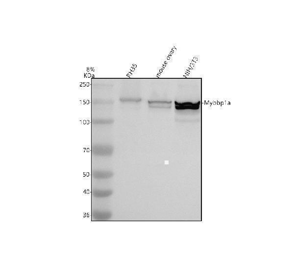
Western blot analysis of MYBBP1A using anti-MYBBP1A antibody (A04187-2).
Electrophoresis was performed on a 8% SDS-PAGE gel at 80V (Stacking gel) / 120V (Resolving gel) for 2 hours. The sample well of each lane was loaded with 30 ug of sample under reducing conditions.
Lane 1: rat RH35 whole cell lysates,
Lane 2: mouse ovary tissue lysates,
Lane 3: mouse NIH/3T3 tissue lysates.
After electrophoresis, proteins were transferred to a nitrocellulose membrane at 150 mA for 50-90 minutes. Blocked the membrane with 5% non-fat milk/TBS for 1.5 hour at RT. The membrane was incubated with rabbit anti-MYBBP1A antigen affinity purified polyclonal antibody (A04187-2) at 0.5 μg/mL overnight at 4°C, then washed with TBS-0.1%Tween 3 times with 5 minutes each and probed with a goat anti-rabbit IgG-HRP secondary antibody at a dilution of 1:5000 for 1.5 hour at RT. The signal is developed using an ECL Plus Western Blotting Substrate (Catalog # AR1196-200) with Tanon 5200 system. A specific band was detected for MYBBP1A at approximately 150 kDa. The expected band size for MYBBP1A is at 149 kDa.

Click image to see more details
IF analysis of MYBBP1A using anti-MYBBP1A antibody (A04187-2) and anti-Beta Tubulin antibody (M01857-3).
MYBBP1A was detected in an immunocytochemical section of NIH/3T3 cells. Enzyme antigen retrieval was performed using IHC enzyme antigen retrieval reagent (AR0022) for 15 mins. The cells were blocked with 10% goat serum. And then incubated with 5 μg/mL rabbit anti-MYBBP1A Antibody (A04187-2) and mouse anti-Beta Tubulin antibody (M01857-3) overnight at 4°C. DyLight®488 Conjugated Goat Anti-Rabbit IgG (BA1127) and Cy3 Conjugated Goat Anti-Mouse IgG (BA1031) were used as secondary antibody at 1:500 dilution and incubated for 30 minutes at 37°C. Visualize using a fluorescence microscope and filter sets appropriate for the label used.
Specific Publications For Anti-MYBBP1A Antibody Picoband® (A04187-2)
Loading publications
Recommended Resources
Here are featured tools and databases that you might find useful.
- Boster's Pathways Library
- Protein Databases
- Bioscience Research Protocol Resources
- Data Processing & Analysis Software
- Photo Editing Software
- Scientific Literature Resources
- Research Paper Management Tools
- Molecular Biology Software
- Primer Design Tools
- Bioinformatics Tools
- Phylogenetic Tree Analysis
Customer Reviews
Have you used Anti-MYBBP1A Antibody Picoband®?
Share your experimental results or join a short interview to earn up to $1,000 in product credits or other rewards.
0 Reviews For Anti-MYBBP1A Antibody Picoband®
Customer Q&As
Have a question?
Find answers in Q&As, reviews.
Can't find your answer?
Submit your question
