Click image to see more details
Product Info Summary
| SKU: | A06446 |
|---|---|
| Size: | 100ug |
| Reactive Species: | Mouse, Rat |
| Host: | Rabbit |
| Application: | ELISA, IP, IHC, WB |
Customers Who Bought This Also Bought

Product info
Product Name
Anti-Myosin MYL9 Antibody
SKU/Catalog Number
A06446
Size
100ug
Form
Liquid (sterile filtered)
Description
Boster Bio Anti-Myosin MYL9 Antibody (Catalog # A06446). Tested in ELISA, WB applications. This antibody reacts with Mouse, Rat.
Storage & Handling
Store vial at -20°C prior to opening. Aliquot contents and freeze at -20°C or below for extended storage. Avoid cycles of freezing and thawing. Centrifuge product if not completely clear after standing at room temperature. This product is stable for several weeks at 4°C as an undiluted liquid. Dilute only prior to immediate use. Expiration date is one (1) year from date of opening. (Ship on dry ice.)
Cite This Product
Anti-Myosin MYL9 Antibody (Boster Biological Technology, Pleasanton CA, USA, Catalog # A06446)
Host
Rabbit
Contents
0.02 M Potassium Phosphate, 0.15 M Sodium Chloride, pH 7.2, 0.01% (w/v) Sodium Azide
Clonality
Polyclonal
Isotype
IgG
Immunogen
This affinity purified antibody was prepared from whole rabbit serum produced by repeated immunizations with a synthetic peptide corresponding to amino acids aa 10-35 of human myosin light chain protein.
Reactive Species
A06446 is reactive to MYL12A in Mouse, Rat
Observed Molecular Weight
42 kDa
Background of MYL12A
Myosin is the major component of thick muscle filaments, and is a long asymmetric molecule containing a globular head and a long tail. The molecule consists of two heavy chains each ~200,000 daltons, and four light chains each ~16,000 - 21,000 daltons. Activation of smooth and cardiac muscle primarily involves pathways that increase calcium and myosin phosphorylation resulting in contraction. Myosin light chain phosphatase acts to regulate muscle contraction by dephosphorylating activated myosin light chain. The selected peptide sequence used to generate the polyclonal antibody is located near the amino terminal end of the polypeptide corresponding to the smooth/non-muscle form of myosin regulatory light chain found in cardiac myocytes in addition to smooth and non-muscle cells.
Antibody Validation
Boster validates all antibodies on WB, IHC, ICC, Immunofluorescence, and ELISA with known positive control and negative samples to ensure specificity and high affinity, including thorough antibody incubations.
Application & Images
Applications
A06446 is guaranteed for ELISA, IP, IHC, WB Boster Guarantee
Recommend Dilution
| Application | Dilution | Species |
|---|---|---|
| ELISA: 1:10 | 000 - 1:70 | 000 |
| WB: 1:500 - 1:2 | 000 | |
| This affinity-purified antibody was tested by ELISA and immunoblotting and was found to be reactive with both the unphosphorylated and mono-phosphorylated forms of the protein. Although not tested | this antibody is likely functional in immunohistochemistry and immunoprecipitation. |
Validation Images & Assay Conditions
Click image to see more details
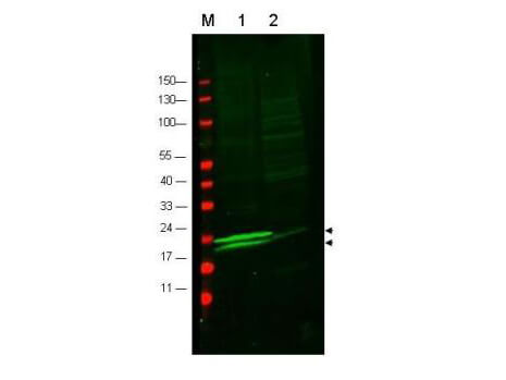
Western blot using Boster's anti-RLC of Smooth and Non-muscle Myosin antibody to detect vascular myosin (rat aorta, lane 1) but not cardiac myosin (mouse heart, lane2). Each lane was loaded with 35 µg of lysate. Arrowheads indicate the detection of both mono-phosphorylated (upper) and unphosphorylated (lower) forms of the protein. After blocking with 5% Normal goat serum and 0.5% BLOTTO in PBS, the membrane was probed with the primary antibody diluted in blocking buffer to 1:600 for 2 h at room temperature. The membrane was washed and reacted with a 1:10,000 dilution of IRDye800™ conjugated Gt-a-Rabbit IgG [H&L] MX (611-132-122) for 45 min at room temperature (800 nm channel, green). Molecular weight estimation was made by comparison to prestained MW markers in lane M (700 nm channel, red). IRDye™800 fluorescence image was captured using the Odyssey® Infrared Imaging System developed by LI-COR. IRDye is a trademark of LI-COR, Inc. Other detection systems will yield similar results.
Specific Publications For Anti-Myosin MYL9 Antibody (A06446)
Loading publications
Recommended Resources
Here are featured tools and databases that you might find useful.
- Boster's Pathways Library
- Protein Databases
- Bioscience Research Protocol Resources
- Data Processing & Analysis Software
- Photo Editing Software
- Scientific Literature Resources
- Research Paper Management Tools
- Molecular Biology Software
- Primer Design Tools
- Bioinformatics Tools
- Phylogenetic Tree Analysis
Customer Reviews
Have you used Anti-Myosin MYL9 Antibody?
Share your experimental results or join a short interview to earn up to $1,000 in product credits or other rewards.
0 Reviews For Anti-Myosin MYL9 Antibody
Customer Q&As
Have a question?
Find answers in Q&As, reviews.
Can't find your answer?
Submit your question
1 Customer Q&As for Anti-Myosin MYL9 Antibody
Question
We are currently using anti-Myosin antibody A06446 for human tissue, and we are satisfied with the WB results. The species of reactivity given in the datasheet says human. Is it likely that the antibody can work on dog tissues as well?
Verified Customer
Verified customer
Asked: 2019-12-24
Answer
The anti-Myosin antibody (A06446) has not been tested for cross reactivity specifically with dog tissues, but there is a good chance of cross reactivity. We have an innovator award program that if you test this antibody and show it works in dog you can get your next antibody for free. Please contact me if I can help you with anything.
Boster Scientific Support
Answered: 2019-12-24
