Click image to see more details
Product Info Summary
| SKU: | MA1063 |
|---|---|
| Size: | 100 μg/vial |
| Reactive Species: | Human, Mouse, Rabbit, Rat |
| Host: | Mouse |
| Application: | IHC, WB |
Customers Who Bought This Also Bought

Product info
Product Name
Anti-Myosin(Skeletal, Fast) MYH2 Antibody (Monoclonal, MY-32)
SKU/Catalog Number
MA1063
BM0096 is an alternative SKU for this antibody, used in previous lots.
Size
100 μg/vial
Form
Lyophilized
Description
Boster Bio Anti-Myosin (Skeletal, Fast) MYH2 Antibody (Monoclonal, MY-32) catalog # MA1063. Tested in IHC, WB applications. This antibody reacts with Human, Mouse, Rabbit, Rat.
Storage & Handling
Store at -20˚C for one year from date of receipt. After reconstitution, at 4˚C for one month. It can also be aliquotted and stored frozen at -20˚C for six months. Avoid repeated freeze-thaw cycles.
Cite This Product
Anti-Myosin(Skeletal, Fast) MYH2 Antibody (Monoclonal, MY-32) (Boster Biological Technology, Pleasanton CA, USA, Catalog # MA1063)
Host
Mouse
Contents
Mouse IgG in stabilizing components, 1.2% sodium acetate and 0.01mg NaN3.
Clonality
Monoclonal
Clone Number
MY-32
Isotype
Mouse IgG1
Immunogen
Rabbit muscle myosin.
Cross-reactivity
No cross-reactivity with other proteins
Reactive Species
MA1063 is reactive to MYH2 in Human, Mouse, Rabbit, Rat
Observed Molecular Weight
200 kDa
Background of MYH2
Myosin is composed of 2 heavy chains of about 200,000 daltons each and 4 light chains of about 20,000 daltons each. The light chains are of 2 distinct types: the phosphorylatable, regulatory, or MLC2 type, and the nonphosphorylatable, alkali, or MLC1 and MLC3 types (MYL1 according to the HGM symbols). Skeletal Myosin (Fast),, also known as MYL1, mapped to the region 2q32.1-qter.
Antibody Validation
Boster validates all antibodies on WB, IHC, ICC, Immunofluorescence, and ELISA with known positive control and negative samples to ensure specificity and high affinity, including thorough antibody incubations.
Application & Images
Applications
MA1063 is guaranteed for IHC, WB Boster Guarantee
Recommend Dilution
| Application | Dilution | Species |
|---|---|---|
| Immunohistochemistry (Paraffin-embedded Section) | 1.5-2μg/ml | Human, mouse, rabbit, rat |
| Western blot | 1-2μg/ml | Human, mouse, rabbit, rat |
Tested application
Suggested blocking solution with 5% non-fat milk or BSA; (*)Recommended protein loading: 20-40 µg per lane
Validation Images & Assay Conditions
Click image to see more details
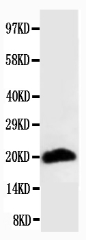
Anti-Myosin(Skeletal, Fast) antibody (monoclonal), MA1063, Western blotting
WB: Rat Skeletal Muscle Tissue Lysate

Click image to see more details
miR-411-5p activate p38MAPK pathway by targeting SPRY4-induced terminal differentiation of RMS. ( a ) Western blot analysis of p38MAPK activation (P-p38MAPK: p-38) in RMS cells treated with HA-tagged MKK6EE protein-expressing pcDNA3, SPRY4 siRNA, or both. ( b ) Western blot analysis of p38MAPK activation (P-p38MAPK:p-38) in RMS cells treated with miR-411-5p-M, miR-411-5p-I, and MKK6EE protein-expressing pcDNA3 plus miR-411-5p-M. ( c ) Terminal differentiation of RD cells after treatment with SPRY4 siRNA or miR-411-5p-M for 6 and 48 h. Cells stained with apoptosis marker (caspase-3); cell cycle was analyzed using propidium iodide (PI) and showed changed morphology (MHC and myosin) after activation of p38MAPK caused by SPRY4 siRNA or miR-411-5p-M. Scale bars: panel (Caspase-3)=100 μ m, panel (MHC and Myosin)=20 μ m. ( d ) Quantitation of terminal differentiation of RD cells after treatment with SPRY4 siRNA or miR-411-5p-M for 6 and 48 h. ( e ) Graphical representation of cell cycle phase proportions in RD cells after treatment with miR-411-5p-M for 6 and 48 h. ( f ) Proliferation of RD cells was evaluated by [ 3 H] thymidine incorporation assay after treatment with SPRY4 siRNA or miR-411-5p-M for 4 days. The amount of [ 3 H] thymidine incorporated was measured with a liquid scintillation counter. The OD at 557 nm was determined using a microplate reader. ( g ) Western blot analysis of cleaved caspase-3 in RMS cells treated with SPRY4 siRNA or miR-411-5p-M for 24 and 72 h. Each assay ( a – g ) was conducted at least three times independently. Error bars indicate S.D. * P <0.05; ** P <0.01; *** P <0.005
Index in PubMed under a CC BY license. PMID: 26291313
Specific Publications For Anti-Myosin(Skeletal, Fast) MYH2 Antibody (Monoclonal, MY-32) (MA1063)
Loading publications
Recommended Resources
Here are featured tools and databases that you might find useful.
- Boster's Pathways Library
- Protein Databases
- Bioscience Research Protocol Resources
- Data Processing & Analysis Software
- Photo Editing Software
- Scientific Literature Resources
- Research Paper Management Tools
- Molecular Biology Software
- Primer Design Tools
- Bioinformatics Tools
- Phylogenetic Tree Analysis
Customer Reviews
Have you used Anti-Myosin(Skeletal, Fast) MYH2 Antibody (Monoclonal, MY-32)?
Share your experimental results or join a short interview to earn up to $1,000 in product credits or other rewards.
0 Reviews For Anti-Myosin(Skeletal, Fast) MYH2 Antibody (Monoclonal, MY-32)
Customer Q&As
Have a question?
Find answers in Q&As, reviews.
Can't find your answer?
Submit your question
