Click image to see more details
-

-
-
-
-
+1
Product Info Summary
| SKU: | PA1929 |
|---|---|
| Size: | 100 μg/vial |
| Reactive Species: | Human, Mouse, Rat |
| Host: | Rabbit |
| Application: | IHC, WB |
Customers Who Bought This Also Bought

Product info
Product Name
Anti-NADPH oxidase 4/NOX4 Antibody Picoband®
SKU/Catalog Number
PA1929
BA2813 is an alternative SKU for this antibody, used in previous lots.
Size
100 μg/vial
Form
Lyophilized
Description
Boster Bio Anti-NADPH oxidase 4/NOX4 Antibody catalog # PA1929. Tested in IHC, WB applications. This antibody reacts with Human, Mouse, Rat. The brand Picoband indicates this is a premium antibody that guarantees superior quality, high affinity, and strong signals with minimal background in Western blot applications. Only our best-performing antibodies are designated as Picoband, ensuring unmatched performance.
Storage & Handling
Store at -20˚C for one year from date of receipt. After reconstitution, at 4˚C for one month. It can also be aliquotted and stored frozen at -20˚C for six months. Avoid repeated freeze-thaw cycles.
Cite This Product
Anti-NADPH oxidase 4/NOX4 Antibody Picoband® (Boster Biological Technology, Pleasanton CA, USA, Catalog # PA1929)
Host
Rabbit
Contents
Each vial contains 4 mg Trehalose, 0.9 mg NaCl and 0.2 mg Na2HPO4.
Clonality
Polyclonal
Isotype
Rabbit IgG
Immunogen
A synthetic peptide corresponding to a sequence at the C-terminus of mouse NADPH oxidase 4, identical to the related rat sequence and different from the related human sequence by two amino acids.
Cross-reactivity
No cross-reactivity with other proteins
Reactive Species
PA1929 is reactive to Nox4 in Human, Mouse, Rat
Observed Molecular Weight
67 kDa
Calculated molecular weight
66.5 kDa
Background of Nox4
NADPH oxidase 4 is an enzyme that in humans is encoded by the NOX4 gene, and is a member of the NOX family of NADPH oxidases. This gene encodes a member of the NOX-family of enzymes that functions as the catalytic subunit the NADPH oxidase complex. The encoded protein is localized to non-phagocytic cells where it acts as an oxygen sensor and catalyzes the reduction of molecular oxygen to various reactive oxygen species (ROS). The ROS generated by this protein have been implicated in numerous biological functions including signal transduction, cell differentiation and tumor cell growth. A pseudogene has been identified on the other arm of chromosome 11. Alternative splicing results in multiple transcript variants.
Antibody Validation
Boster validates all antibodies on WB, IHC, ICC, Immunofluorescence, and ELISA with known positive control and negative samples to ensure specificity and high affinity, including thorough antibody incubations.
Application & Images
Applications
PA1929 is guaranteed for IHC, WB Boster Guarantee
Assay Dilutions Recommendation
The recommendations below provide a starting point for assay optimization. The actual working concentration varies and should be decided by the user.
Western blot, 0.1-0.5μg/ml, Human, Mouse, Rat
Immunohistochemistry (Paraffin-embedded Section), 2-5μg/ml, Human
Validation Images & Assay Conditions
Click image to see more details
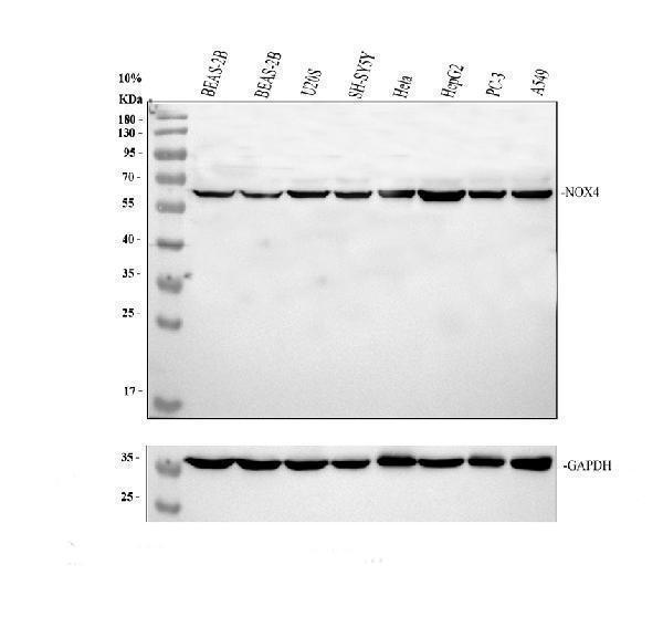
Western blot analysis of NOX4 using anti-NOX4 antibody (PA1929).
Electrophoresis was performed on a 10% SDS-PAGE gel at 80V (Stacking gel) / 120V (Resolving gel) for 2 hours. The sample well of each lane was loaded with 30 ug of sample under reducing conditions.
Lane 1: human BEAS-2B whole cell lysates,
Lane 2: human BEAS-2B whole cell lysates,
Lane 3: human U2OS whole cell lysates,
Lane 4: human SH-SY5Y whole cell lysates,
Lane 5: human Hela whole cell lysates,
Lane 6: human HepG2 whole cell lysates,
Lane 7: human PC-3 whole cell lysates,
Lane 8: human A549 whole cell lysates.
After electrophoresis, proteins were transferred to a nitrocellulose membrane at 150 mA for 50-90 minutes. Blocked the membrane with 5% non-fat milk/TBS for 1.5 hour at RT. The membrane was incubated with rabbit anti-NOX4 antigen affinity purified polyclonal antibody (PA1929) at 0.5 μg/mL overnight at 4°C, then washed with TBS-0.1%Tween 3 times with 5 minutes each and probed with a goat anti-rabbit IgG-HRP secondary antibody (Catalog # BA1054) at a dilution of 1:5000 for 1.5 hour at RT. The signal is developed using an ECL Plus Western Blotting Substrate (Catalog # AR1196-200) with Tanon 5200 system. A specific band was detected for NOX4 at approximately 67 kDa. The expected band size for NOX4 is at 67 kDa.

Click image to see more details
Western blot analysis of NOX4 using anti-NOX4 antibody (PA1929).
Electrophoresis was performed on a 10% SDS-PAGE gel at 80V (Stacking gel) / 120V (Resolving gel) for 2 hours. The sample well of each lane was loaded with 30 ug of sample under reducing conditions.
Lane 1: rat heart tissue lysates,
Lane 2: mouse NIH/3T3 whole cell lysates.
After electrophoresis, proteins were transferred to a nitrocellulose membrane at 150 mA for 50-90 minutes. Blocked the membrane with 5% non-fat milk/TBS for 1.5 hour at RT. The membrane was incubated with rabbit anti-NOX4 antigen affinity purified polyclonal antibody (PA1929) at 0.5 μg/mL overnight at 4°C, then washed with TBS-0.1%Tween 3 times with 5 minutes each and probed with a goat anti-rabbit IgG-HRP secondary antibody (Catalog # BA1054) at a dilution of 1:5000 for 1.5 hour at RT. The signal is developed using an ECL Plus Western Blotting Substrate (Catalog # AR1196-200) with Tanon 5200 system. A specific band was detected for NOX4 at approximately 67 kDa. The expected band size for NOX4 is at 67 kDa.

Click image to see more details
IHC analysis of NOX4 using anti-NOX4 antibody (PA1929).
NOX4 was detected in a paraffin-embedded section of human kidney tissue. Heat mediated antigen retrieval was performed in EDTA buffer (pH 8.0, epitope retrieval solution). The tissue section was blocked with 10% goat serum. The tissue section was then incubated with 2 μg/ml rabbit anti-NOX4 Antibody (PA1929) overnight at 4°C. Peroxidase Conjugated Goat Anti-rabbit IgG was used as secondary antibody and incubated for 30 minutes at 37°C. The tissue section was developed using HRP Conjugated Rabbit IgG Super Vision Assay Kit (Catalog # SV0002) with DAB as the chromogen.

Click image to see more details
Patients with IMN exhibited increased serum levels of EVs, and oxidative stress enhanced EVs production in vitro . (A) TEM images displayed the morphology of EVs isolated from the serum of IMN patients. Scale bar = 100 nm. Patients with MCD served as controls. NTA images showed the distribution of EVs. (B) Representative images of immune colloidal gold electron microscopy demonstrated the presence of PLA2R on the surface of EVs, indicated by black dots. Scale bar = 200 nm. (C, D) Images from nano-flow cytometry and a corresponding statistical bar chart illustrated the proportion of PLA2R + EVs in the serum of IMN patients compared to those with MCD. (E) A positive correlation was observed between the proportion of PLA2R + EVs and serum aPLA2Rab levels. (F) Representative Western blot analyses showed the PLA2R expression levels of EVs isolated from the lung tissue of smokers and non-smokers. (G) The levels of oxidative stress, PLA2R expression, and EVs production in Beas-2B cells were examined after stimulation with LPS, with or without GW4869 intervention. Representative Western blot (G) and quantitative data (H–L) are presented. The expression levels of NOX4 (H) , NOX2 (I) , TSG101 (J) , CD63 (K) , and PLA2R (L) were significantly upregulated by LPS, while GSH reversed this upregulation. (* p < 0.05 versus control; # p < 0.05 versus LPS). (M–R) The levels of oxidative stress, PLA2R expression, and EVs production in Beas-2B cells stimulated by RM8785 were analyzed, with or without GW4869 intervention. Representative Western blot (M) and quantitative data (N–R) are presented. The expression levels of NOX4 (N) , NOX2 (O) , TSG101 (P) , CD63 (Q) , and PLA2R (R) were significantly upregulated by RM8785, while GW4869 effectively reversed this upregulation. (* p < 0.05 versus control; # p < 0.05 versus PM2.5).
Index in PubMed under a CC BY license. PMID: 39744137

Click image to see more details
PM2.5 induces oxidative stress and PLA2R overexpression in bronchial epithelium. (A–F) Levels of oxidative stress, PLA2R expression, and EVs production in Beas-2B cells stimulated by LPS, with or without GSH intervention. Western blot (A) and quantitative data (B–F) are presented. (B, C) NOX2 and NOX4 are oxidative stress markers from the NADPH oxidase family. (D) PLA2R expression levels. (E, F) TSG101 and CD63 are markers for EVs. All are significantly upregulated by LPS, while GSH effectively reverses this upregulation. (* p < 0.05 versus control; #P < 0.05 versus LPS). (G) Representative micrographs show immunofluorescence staining of PLA2R in Beas-2B cells treated with LPS, with or without GSH. Scale bar = 100 μm. (H–M) Levels of oxidative stress, PLA2R expression, and EVs production in Beas-2B cells stimulated by RM8785, with or without GSH intervention. Representative Western blot (H) and quantitative data (I–M) are provided. The expression levels of NOX4 (I) , NOX2 (J) , PLA2R (K) , CD63 (L) , and TSG101 (M) are all significantly upregulated by RM8785, while GSH effectively reverses this upregulation. (* p < 0.05 versus control; #P < 0.05 versus PM2.5).
Index in PubMed under a CC BY license. PMID: 39744137
Specific Publications For Anti-NADPH oxidase 4/NOX4 Antibody Picoband® (PA1929)
Loading publications
Recommended Resources
Here are featured tools and databases that you might find useful.
- Boster's Pathways Library
- Protein Databases
- Bioscience Research Protocol Resources
- Data Processing & Analysis Software
- Photo Editing Software
- Scientific Literature Resources
- Research Paper Management Tools
- Molecular Biology Software
- Primer Design Tools
- Bioinformatics Tools
- Phylogenetic Tree Analysis
Customer Reviews
Have you used Anti-NADPH oxidase 4/NOX4 Antibody Picoband®?
Share your experimental results or join a short interview to earn up to $1,000 in product credits or other rewards.
0 Reviews For Anti-NADPH oxidase 4/NOX4 Antibody Picoband®
Customer Q&As
Have a question?
Find answers in Q&As, reviews.
Can't find your answer?
Submit your question
5 Customer Q&As for Anti-NADPH oxidase 4/NOX4 Antibody Picoband®
Question
We bought anti-NADPH oxidase 4/NOX4 antibody for IHC-P on nephron tubule last year. I am using rat, and We want to use the antibody for Flow Cytometry next. I am interested in examining nephron tubule as well as lung in our next experiment. Could you please give me some suggestion on which antibody would work the best for Flow Cytometry?
Verified Customer
Verified customer
Asked: 2020-01-16
Answer
I have checked the website and datasheets of our anti-NADPH oxidase 4/NOX4 antibody and it appears that PA1929 has been tested on rat in both IHC-P and Flow Cytometry. Thus PA1929 should work for your application. Our Boster satisfaction guarantee will cover this product for Flow Cytometry in rat even if the specific tissue type has not been validated. We do have a comprehensive range of products for Flow Cytometry detection and you can check out our website bosterbio.com to find out more information about them.
Boster Scientific Support
Answered: 2020-01-16
Question
We have seen staining in human nephron tubule. Do you have any suggestions? Is anti-NADPH oxidase 4/NOX4 antibody supposed to stain nephron tubule positively?
Verified Customer
Verified customer
Asked: 2019-12-13
Answer
Based on literature nephron tubule does express NOX4. Based on Uniprot.org, NOX4 is expressed in nephron tubule, kidney, fetal kidney, lung, pulmonary artery, ovary, among other tissues. Regarding which tissues have NOX4 expression, here are a few articles citing expression in various tissues:
Fetal kidney, Pubmed ID: 11376945
Kidney, Pubmed ID: 10869423, 11032835
Lung, Pubmed ID: 15721269
Ovary, Pubmed ID: 15489334
Pulmonary artery, Pubmed ID: 14702039
Boster Scientific Support
Answered: 2019-12-13
Question
We are interested in using your anti-NADPH oxidase 4/NOX4 antibody for defense response studies. Has this antibody been tested with western blotting on hepg2 whole cell lysates? We would like to see some validation images before ordering.
K. Li
Verified customer
Asked: 2018-02-23
Answer
I appreciate your inquiry. This PA1929 anti-NADPH oxidase 4/NOX4 antibody is validated on human 293t whole cell lysates, hepg2 whole cell lysates, sw620 whole cell lysates, rat kidney tissue, tissue lysate, u20s cells. It is guaranteed to work for Flow Cytometry, IHC-P, IHC-F, ICC, WB in human, mouse, rat. Our Boster guarantee will cover your intended experiment even if the sample type has not been be directly tested.
Boster Scientific Support
Answered: 2018-02-23
Question
We are currently using anti-NADPH oxidase 4/NOX4 antibody PA1929 for human tissue, and we are happy with the ICC results. The species of reactivity given in the datasheet says human, mouse, rat. Is it true that the antibody can work on feline tissues as well?
S. Jackson
Verified customer
Asked: 2015-03-12
Answer
The anti-NADPH oxidase 4/NOX4 antibody (PA1929) has not been validated for cross reactivity specifically with feline tissues, though there is a good chance of cross reactivity. We have an innovator award program that if you test this antibody and show it works in feline you can get your next antibody for free. Please contact me if I can help you with anything.
Boster Scientific Support
Answered: 2015-03-12
Question
Our team were content with the WB result of your anti-NADPH oxidase 4/NOX4 antibody. However we have observed positive staining in nephron tubule endoplasmic reticulum membrane using this antibody. Is that expected? Could you tell me where is NOX4 supposed to be expressed?
A. Krishna
Verified customer
Asked: 2014-06-25
Answer
According to literature, nephron tubule does express NOX4. Generally NOX4 expresses in endoplasmic reticulum membrane. Regarding which tissues have NOX4 expression, here are a few articles citing expression in various tissues:
Fetal kidney, Pubmed ID: 11376945
Kidney, Pubmed ID: 10869423, 11032835
Lung, Pubmed ID: 15721269
Ovary, Pubmed ID: 15489334
Pulmonary artery, Pubmed ID: 14702039
Boster Scientific Support
Answered: 2014-06-25
