Click image to see more details
Product Info Summary
| SKU: | A08224 |
|---|---|
| Size: | 100 μg/vial |
| Reactive Species: | Human, Mouse, Rat |
| Host: | Rabbit |
| Application: | ELISA, IHC, WB |
Customers Who Bought This Also Bought

Product info
Product Name
Anti-NDUFA1 Antibody Picoband®
SKU/Catalog Number
A08224
Size
100 μg/vial
Form
Lyophilized
Description
Boster Bio Anti-NDUFA1 Antibody Picoband® catalog # A08224. Tested in WB, IHC, ELISA applications. This antibody reacts with Human, Mouse, Rat. The brand Picoband indicates this is a premium antibody that guarantees superior quality, high affinity, and strong signals with minimal background in Western blot applications. Only our best-performing antibodies are designated as Picoband, ensuring unmatched performance.
Storage & Handling
At -20°C for one year from date of receipt. After reconstitution, at 4°C for one month. It can also be aliquotted and stored frozen at -20°C for six months. Avoid repeated freezing and thawing.
Cite This Product
Anti-NDUFA1 Antibody Picoband® (Boster Biological Technology, Pleasanton CA, USA, Catalog # A08224)
Host
Rabbit
Contents
Each vial contains 4 mg Trehalose, 0.9 mg NaCl, 0.2 mg Na2HPO4.
Clonality
Polyclonal
Immunogen
E.coli-derived human NDUFA1 recombinant protein (Position: M1-D70). Human NDUFA1 shares 82.9% amino acid (aa) sequence identity with mouse NDUFA1.
Reactive Species
A08224 is reactive to NDUFA1 in Human, Mouse, Rat
Observed Molecular Weight
10 kDa
Calculated molecular weight
8.1 kDa
Background of NDUFA1
NDUFA1(NADH-UBIQUINONE OXIDOREDUCTASE 1 ALPHA SUBCOMPLEX, 1), also called MWFE, B. TAURUS, HOMOLOG OF, encodes a subunit of mitochondrial NADH: ubiquinone oxidoreductase, also known as mitochondrial complex I. The NDUFA1 gene is mapped to chromosome Xq24. The deduced polypeptide sequence of NDUFA1 was found to have an N-terminal hydrophobic domain, likely to be a transmembrane domain, and a C-terminal hydrophilic domain. And the NDUFA1 gene contains 3 exons and spans about 5.0 kb of genomic DNA. Complementation with hamster Ndufa1 cDNA restored the rotenone-sensitive complex I activity of these mutant cells to approximately 100% of the parent cell activity. The findings established that the MWFE polypeptide is absolutely essential for an active complex I in mammals. The NDUFA1 peptide is one of about 31 components of the "hydrophobic protein"(HP) fraction of complex I which is involved in proton translocation. Thus the NDUFA1 peptide may also participate in that function.
Antibody Validation
Boster validates all antibodies on WB, IHC, ICC, Immunofluorescence, and ELISA with known positive control and negative samples to ensure specificity and high affinity, including thorough antibody incubations.
Application & Images
Applications
A08224 is guaranteed for ELISA, IHC, WB Boster Guarantee
Recommend Dilution
| Application | Dilution | Species |
|---|---|---|
| Western blot | 0.1-0.25 μg/ml | Human, Mouse, Rat |
| Immunohistochemistry(Paraffin-embedded Section) | 2-5 μg/ml | Human |
| ELISA | 0.1-0.5 μg/ml | - |
Tested application
Suggested blocking solution with 5% non-fat milk or BSA; (*)Recommended protein loading: 20-40 µg per lane
Use TE buffer pH 9.0 for antigen retrieval; (*) citrate buffer pH 6.0 is an alternative.
Validation Images & Assay Conditions
Click image to see more details
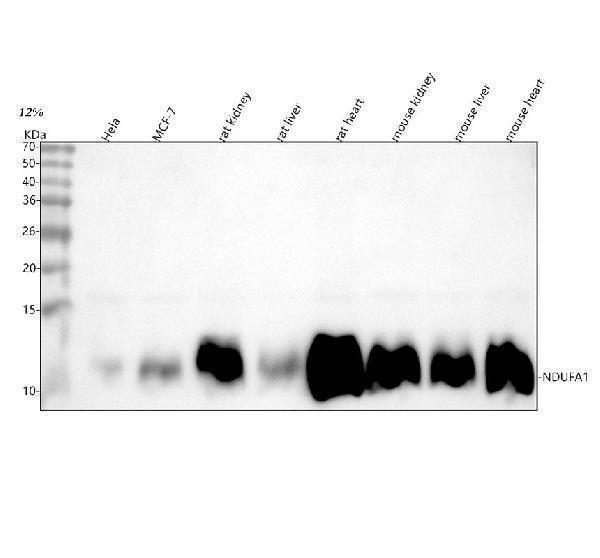
Western blot analysis of NDUFA1 using anti-NDUFA1 antibody (A08224).
Electrophoresis was performed on a 5-20% SDS-PAGE gel at 70V (Stacking gel) / 90V (Resolving gel) for 2-3 hours. The sample well of each lane was loaded with 30 ug of sample under reducing conditions.
Lane 1: human Hela whole cell lysates,
Lane 2: human MCF-7 whole cell lysates,
Lane 3: rat kidney tissue lysates,
Lane 4: rat liver tissue lysates,
Lane 5: rat heart tissue lysates,
Lane 6: mouse kidney tissue lysates,
Lane 7: mouse liver tissue lysates,
Lane 8: mouse heart tissue lysates.
After electrophoresis, proteins were transferred to a nitrocellulose membrane at 150 mA for 50-90 minutes. Blocked the membrane with 5% non-fat milk/TBS for 1.5 hour at RT. The membrane was incubated with rabbit anti-NDUFA1 antigen affinity purified polyclonal antibody (Catalog # A08224) at 0.25 μg/mL overnight at 4°C, then washed with TBS-0.1%Tween 3 times with 5 minutes each and probed with a goat anti-rabbit IgG-HRP secondary antibody at a dilution of 1:5000 for 1.5 hour at RT. The signal is developed using an Enhanced Chemiluminescent detection (ECL) kit (Catalog # EK1002) with Tanon 5200 system. A specific band was detected for NDUFA1 at approximately 10 kDa. The expected band size for NDUFA1 is at 8 kDa.

Click image to see more details
Western blot analysis of NDUFA1 using anti-NDUFA1 antibody (A08224).
Electrophoresis was performed on a 5-20% SDS-PAGE gel at 70V (Stacking gel) / 90V (Resolving gel) for 2-3 hours. The sample well of each lane was loaded with 30 ug of sample under reducing conditions.
Lane 1: human THP-1 whole cell lysates.
After electrophoresis, proteins were transferred to a nitrocellulose membrane at 150 mA for 50-90 minutes. Blocked the membrane with 5% non-fat milk/TBS for 1.5 hour at RT. The membrane was incubated with rabbit anti-NDUFA1 antigen affinity purified polyclonal antibody (A08224) at 0.5 μg/mL overnight at 4°C, then washed with TBS-0.1%Tween 3 times with 5 minutes each and probed with a goat anti-rabbit IgG-HRP secondary antibody at a dilution of 1:5000 for 1.5 hour at RT. The signal is developed using an Enhanced Chemiluminescent detection (ECL) kit (Catalog # EK1002) with Tanon 5200 system. A specific band was detected for NDUFA1 at approximately 10 kDa. The expected band size for NDUFA1 is at 8 kDa.

Click image to see more details
IHC analysis of NDUFA1 using anti-NDUFA1 antibody (A08224).
NDUFA1 was detected in a paraffin-embedded section of human prostate cancer tissue. Heat mediated antigen retrieval was performed in EDTA buffer (pH 8.0, epitope retrieval solution). The tissue section was blocked with 10% goat serum. The tissue section was then incubated with 2 μg/ml rabbit anti-NDUFA1 Antibody (A08224) overnight at 4°C. Peroxidase Conjugated Goat Anti-rabbit IgG was used as secondary antibody and incubated for 30 minutes at 37°C. The tissue section was developed using HRP Conjugated Rabbit IgG Super Vision Assay Kit (Catalog # SV0002) with DAB as the chromogen.
Specific Publications For Anti-NDUFA1 Antibody Picoband® (A08224)
Loading publications
Recommended Resources
Here are featured tools and databases that you might find useful.
- Boster's Pathways Library
- Protein Databases
- Bioscience Research Protocol Resources
- Data Processing & Analysis Software
- Photo Editing Software
- Scientific Literature Resources
- Research Paper Management Tools
- Molecular Biology Software
- Primer Design Tools
- Bioinformatics Tools
- Phylogenetic Tree Analysis
Customer Reviews
Have you used Anti-NDUFA1 Antibody Picoband®?
Share your experimental results or join a short interview to earn up to $1,000 in product credits or other rewards.
0 Reviews For Anti-NDUFA1 Antibody Picoband®
Customer Q&As
Have a question?
Find answers in Q&As, reviews.
Can't find your answer?
Submit your question
