Click image to see more details
Product Info Summary
| SKU: | A00969 |
|---|---|
| Size: | 100 μg/vial |
| Reactive Species: | Human |
| Host: | Rabbit |
| Application: | ELISA, Flow Cytometry, IF, ICC, WB |
Customers Who Bought This Also Bought

Product info
Product Name
Anti-NFAT1/NFATC2 Antibody Picoband®
SKU/Catalog Number
A00969
Size
100 μg/vial
Form
Lyophilized
Description
Boster Bio Anti-NFAT1/NFATC2 Antibody Picoband® catalog # A00969. Tested in ELISA, Flow Cytometry, IF, ICC, WB applications. This antibody reacts with Human. The brand Picoband indicates this is a premium antibody that guarantees superior quality, high affinity, and strong signals with minimal background in Western blot applications. Only our best-performing antibodies are designated as Picoband, ensuring unmatched performance.
Storage & Handling
Store at -20˚C for one year from date of receipt. After reconstitution, at 4˚C for one month. It can also be aliquotted and stored frozen at -20˚C for six months. Avoid repeated freeze-thaw cycles.
Cite This Product
Anti-NFAT1/NFATC2 Antibody Picoband® (Boster Biological Technology, Pleasanton CA, USA, Catalog # A00969)
Host
Rabbit
Contents
Each vial contains 4 mg Trehalose, 0.9 mg NaCl and 0.2 mg Na2HPO4.
Clonality
Polyclonal
Isotype
Rabbit IgG
Immunogen
E. coli-derived human NFAT1 recombinant protein (Position: Q594-H676).
Cross-reactivity
No cross-reactivity with other proteins.
Reactive Species
A00969 is reactive to NFATC2 in Human
Observed Molecular Weight
135 kDa
Calculated molecular weight
100.1 kDa
Background of NFATC2
NFATC2 (Nuclear factor of activated T-cells, cytoplasmic 2), also known as NFATP or the 'preexisting component' of NFAT, is present in the cytosolic fraction of unstimulated T cells, which is also a member of the nuclear factor of activated T cells (NFAT) family. The NFATC2 gene is mapped on 20q13.2. NFATC2 is highly homologous to NFATC1 over a limited domain which shows similarity to the Dorsal/Rel family but has a wider tissue distribution. Ectopic expression of NFATC2 inhibited the basal activity of the human CDK4 promoter. Additionally, both Calna-/- and Nfatc2 -/- mice had elevated protein levels of Cdk4, confirming a negative regulatory role for the calcineurin/NFAT pathway. NFATC2 controls myoblast fusion at a specific stage of myogenesis after the initial formation of a myotube and is necessary for further cell growth. Overexpression of NFATC2 promoted differentiation of osteoclast precursor cells into tartrate-resistant acid phosphatase-positive (TRAP-positive) multinucleated osteoclast-like cells even in the absence of RANKL.
Antibody Validation
Boster validates all antibodies on WB, IHC, ICC, Immunofluorescence, and ELISA with known positive control and negative samples to ensure specificity and high affinity, including thorough antibody incubations.
Application & Images
Applications
A00969 is guaranteed for ELISA, Flow Cytometry, IF, ICC, WB Boster Guarantee
Recommend Dilution
| Application | Dilution | Species |
|---|---|---|
| Western blot | 0.1-0.5μg/ml | Human |
| Immunocytochemistry/Immunofluorescence | 5 μg/ml | Human |
| Flow Cytometry(Fixed) | 1-3 μg/1x106 cells | Human |
| ELISA | 0.1-0.5μg/ml | - |
Tested application
Suggested blocking solution with 5% non-fat milk or BSA; (*)Recommended protein loading: 20-40 µg per lane
Validation Images & Assay Conditions
Click image to see more details
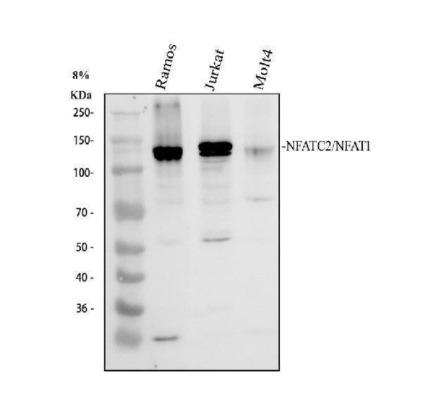
Western blot analysis of NFAT1/NFATC2 using anti-NFAT1/NFATC2 antibody (A00969).
Electrophoresis was performed on a 8% SDS-PAGE gel at 80V (Stacking gel) / 120V (Resolving gel) for 2 hours. The sample well of each lane was loaded with 30 ug of sample under reducing conditions.
Lane 1: human Ramos whole cell lysates,
Lane 2: human Jurkat whole cell lysates,
Lane 3: human MOLT-4 whole cell lysates.
After electrophoresis, proteins were transferred to a nitrocellulose membrane at 150 mA for 50-90 minutes. Blocked the membrane with 5% non-fat milk/TBS for 1.5 hour at RT. The membrane was incubated with rabbit anti-NFAT1/NFATC2 antigen affinity purified polyclonal antibody (A00969) at 0.25 μg/mL overnight at 4°C, then washed with TBS-0.1%Tween 3 times with 5 minutes each and probed with a goat anti-rabbit IgG-HRP secondary antibody (Catalog # BA1054) at a dilution of 1:5000 for 1.5 hour at RT. The signal is developed using an ECL Plus Western Blotting Substrate (Catalog # AR1196-200) with Tanon 5200 system. A specific band was detected for NFAT1/NFATC2 at approximately 135 kDa. The expected band size for NFAT1/NFATC2 is at 100 kDa.

Click image to see more details
IF analysis of NFAT1/NFATC2 using anti-NFAT1/NFATC2 antibody (A06945-1) and anti-Tubulin Alpha antibody (M03989-3).
NFAT1/NFATC2 was detected in immunocytochemical section of Hela cell. Enzyme antigen retrieval was performed using IHC enzyme antigen retrieval reagent (AR0022) for 15 mins. The cells were blocked with 10% goat serum. And then incubated with 5 μg/mL rabbit anti-NFAT1/NFATC2 Antibody (A06945-1) and mouse anti-Tubulin Alpha antibody (M03989-3) overnight at 4°C. DyLight®488 Conjugated Goat Anti-Rabbit IgG (BA1127) and Cy3 Conjugated Goat Anti-Mouse IgG (BA1031) were used as secondary antibody at 1:500 dilution and incubated for 30 minutes at 37°C. Visualize using a fluorescence microscope and filter sets appropriate for the label used.

Click image to see more details
Flow Cytometry analysis of Jurkat cells using anti-NFAT1/NFATC2 antibody (A00969).
Overlay histogram showing Jurkat cells stained with A00969 (Blue line). To facilitate intracellular staining, cells were fixed with 4% paraformaldehyde and permeabilized with permeabilization buffer. The cells were blocked with 10% normal goat serum. And then incubated with rabbit anti-NFAT1/NFATC2 Antibody (A00969, 1 μg/1x106 cells) for 30 min at 20°C. DyLight®488 conjugated goat anti-rabbit IgG (BA1127, 5-10 μg/1x106 cells) was used as secondary antibody for 30 minutes at 20°C. Isotype control antibody (Green line) was rabbit IgG (1 μg/1x106) used under the same conditions. Unlabelled sample without incubation with primary antibody and secondary antibody (Red line) was used as a blank control.
Specific Publications For Anti-NFAT1/NFATC2 Antibody Picoband® (A00969)
Loading publications
Recommended Resources
Here are featured tools and databases that you might find useful.
- Boster's Pathways Library
- Protein Databases
- Bioscience Research Protocol Resources
- Data Processing & Analysis Software
- Photo Editing Software
- Scientific Literature Resources
- Research Paper Management Tools
- Molecular Biology Software
- Primer Design Tools
- Bioinformatics Tools
- Phylogenetic Tree Analysis
Customer Reviews
Have you used Anti-NFAT1/NFATC2 Antibody Picoband®?
Share your experimental results or join a short interview to earn up to $1,000 in product credits or other rewards.
0 Reviews For Anti-NFAT1/NFATC2 Antibody Picoband®
Customer Q&As
Have a question?
Find answers in Q&As, reviews.
Can't find your answer?
Submit your question
1 Customer Q&As for Anti-NFAT1/NFATC2 Antibody Picoband®
Question
We are currently using anti-NFAT1/NFATC2 antibody A00969 for rat tissue, and we are well pleased with the ELISA results. The species of reactivity given in the datasheet says human, mouse, rat. Is it likely that the antibody can work on pig tissues as well?
Verified Customer
Verified customer
Asked: 2018-01-01
Answer
The anti-NFAT1/NFATC2 antibody (A00969) has not been tested for cross reactivity specifically with pig tissues, but there is a good chance of cross reactivity. We have an innovator award program that if you test this antibody and show it works in pig you can get your next antibody for free. Please contact me if I can help you with anything.
Boster Scientific Support
Answered: 2018-01-01
