Click image to see more details
Product Info Summary
| SKU: | A01070 |
|---|---|
| Size: | 100 μg/vial |
| Reactive Species: | Human, Mouse, Rat |
| Host: | Rabbit |
| Application: | WB |
Customers Who Bought This Also Bought

Product info
Product Name
Anti-nNOS (neuronal)/NOS1 Antibody Picoband®
SKU/Catalog Number
A01070
Size
100 μg/vial
Form
Lyophilized
Description
Boster Bio Anti-nNOS (neuronal)/NOS1 Antibody Picoband® catalog # A01070. Tested in WB applications. This antibody reacts with Human, Mouse, Rat. The brand Picoband indicates this is a premium antibody that guarantees superior quality, high affinity, and strong signals with minimal background in Western blot applications. Only our best-performing antibodies are designated as Picoband, ensuring unmatched performance.
Storage & Handling
Store at -20˚C for one year from date of receipt. After reconstitution, at 4˚C for one month. It can also be aliquotted and stored frozen at -20˚C for six months. Avoid repeated freeze-thaw cycles.
Cite This Product
Anti-nNOS (neuronal)/NOS1 Antibody Picoband® (Boster Biological Technology, Pleasanton CA, USA, Catalog # A01070)
Host
Rabbit
Contents
Each vial contains antibody formulated with stabilizing components, 0.9 mg NaCl, 0.2 mg Na2HPO4, and 0.05 mg NaN3.
*This antibody is supplied in a stabilized formulation.
Compatibility with conjugation reactions depends on the chemistry of the conjugation method used.
For conjugation methods that are not compatible with the stabilizing components present in this formulation, a carrier-free antibody format is required.
Clonality
Polyclonal
Isotype
Rabbit IgG
Immunogen
A synthetic peptide corresponding to a sequence at the C-terminus of human nNOS(neuronal), identical to the related mouse sequence, and different from the related rat sequence by one amino acid.
Cross-reactivity
No cross-reactivity with other proteins.
Reactive Species
A01070 is reactive to NOS1 in Human, Mouse, Rat
Observed Molecular Weight
160 kDa
Calculated molecular weight
161.0 kDa
Background of NOS1
Nitric oxide synthase 1 (neuronal), also known as NOS1, is an enzyme that in humans is encoded by the NOS1 gene. The protein encoded by this gene belongs to the family of nitric oxide synthases, which synthesize nitric oxide from L-arginine. Nitric oxide is a reactive free radical, which acts as a biologic mediator in several processes, including neurotransmission, and antimicrobial and antitumoral activities. In the brain and peripheral nervous system, nitric oxide displays many properties of a neurotransmitter, and has been implicated in neurotoxicity associated with stroke and neurodegenerative diseases, neural regulation of smooth muscle, including peristalsis, and penile erection. This protein is ubiquitously expressed, with high level of expression in skeletal muscle. Multiple transcript variants that differ in the 5' UTR have been described for this gene but the full-length nature of these transcripts is not known. Additionally, alternatively spliced transcript variants encoding different isoforms (some testis-specific) have been found for this gene.
Antibody Validation
Boster validates all antibodies on WB, IHC, ICC, Immunofluorescence, and ELISA with known positive control and negative samples to ensure specificity and high affinity, including thorough antibody incubations.
Application & Images
Applications
A01070 is guaranteed for WB Boster Guarantee
Recommend Dilution
| Application | Dilution | Species |
|---|---|---|
| Western blot | 0.1-0.5μg/ml | Mouse, Rat, Human |
Tested application
Suggested blocking solution with 5% non-fat milk or BSA; (*)Recommended protein loading: 20-40 µg per lane
Validation Images & Assay Conditions
Click image to see more details
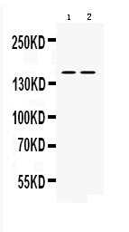
Western blot analysis of nNOS using anti-nNOS antibody (A01070).
Electrophoresis was performed on a 5-20% SDS-PAGE gel at 70V (Stacking gel) / 90V (Resolving gel) for 2-3 hours. The sample well of each lane was loaded with 50ug of sample under reducing conditions.
lane 1: rat brain tissue lysates,
lane 2: mouse brain tissue lysates.
After Electrophoresis, proteins were transferred to a Nitrocellulose membrane at 150mA for 50-90 minutes. Blocked the membrane with 5% Non-fat Milk/ TBS for 1.5 hour at RT. The membrane was incubated with rabbit anti-nNOS antigen affinity purified polyclonal antibody (Catalog # A01070) at 0.5 μg/mL overnight at 4°C, then washed with TBS-0.1%Tween 3 times with 5 minutes each and probed with a goat anti-rabbit IgG-HRP secondary antibody at a dilution of 1:10000 for 1.5 hour at RT. The signal is developed using an Enhanced Chemiluminescent detection (ECL) kit (Catalog # EK1002) with Tanon 5200 system. A specific band was detected for nNOS at approximately 160KD. The expected band size for nNOS is at 160KD.
Specific Publications For Anti-nNOS (neuronal)/NOS1 Antibody Picoband® (A01070)
Loading publications
Recommended Resources
Here are featured tools and databases that you might find useful.
- Boster's Pathways Library
- Protein Databases
- Bioscience Research Protocol Resources
- Data Processing & Analysis Software
- Photo Editing Software
- Scientific Literature Resources
- Research Paper Management Tools
- Molecular Biology Software
- Primer Design Tools
- Bioinformatics Tools
- Phylogenetic Tree Analysis
Customer Reviews
Have you used Anti-nNOS (neuronal)/NOS1 Antibody Picoband®?
Share your experimental results or join a short interview to earn up to $1,000 in product credits or other rewards.
0 Reviews For Anti-nNOS (neuronal)/NOS1 Antibody Picoband®
Customer Q&As
Have a question?
Find answers in Q&As, reviews.
Can't find your answer?
Submit your question
4 Customer Q&As for Anti-nNOS (neuronal)/NOS1 Antibody Picoband®
Question
My question regards using your anti-nNOS (neuronal)/NOS1 antibody for negative regulation of calcium ion transport studies. Has this antibody been tested with western blotting on mouse brain? We would like to see some validation images before ordering.
Verified Customer
Verified customer
Asked: 2019-12-31
Answer
We appreciate your inquiry. This A01070 anti-nNOS (neuronal)/NOS1 antibody is tested on mouse brain. It is guaranteed to work for WB in human, mouse, rat. Our Boster guarantee will cover your intended experiment even if the sample type has not been be directly tested.
Boster Scientific Support
Answered: 2019-12-31
Question
Our lab were content with the WB result of your anti-nNOS (neuronal)/NOS1 antibody. However we have observed positive staining in skeletal muscle cell membrane using this antibody. Is that expected? Could you tell me where is NOS1 supposed to be expressed?
Verified Customer
Verified customer
Asked: 2018-04-27
Answer
From what I have seen in literature, skeletal muscle does express NOS1. Generally NOS1 expresses in cell membrane, sarcolemma. Regarding which tissues have NOS1 expression, here are a few articles citing expression in various tissues:
Brain, Pubmed ID: 7678401
Cerebellum, Pubmed ID: 7515942
Retina, Pubmed ID: 8879752
Skeletal muscle, Pubmed ID: 9791007
Testis, Pubmed ID: 9111048
Boster Scientific Support
Answered: 2018-04-27
Question
We are currently using anti-nNOS (neuronal)/NOS1 antibody A01070 for rat tissue, and we are well pleased with the WB results. The species of reactivity given in the datasheet says human, mouse, rat. Is it true that the antibody can work on bovine tissues as well?
Verified Customer
Verified customer
Asked: 2018-03-19
Answer
The anti-nNOS (neuronal)/NOS1 antibody (A01070) has not been validated for cross reactivity specifically with bovine tissues, though there is a good chance of cross reactivity. We have an innovator award program that if you test this antibody and show it works in bovine you can get your next antibody for free. Please contact me if I can help you with anything.
Boster Scientific Support
Answered: 2018-03-19
Question
We have seen staining in mouse vastus lateralis. Are there any suggestions? Is anti-nNOS (neuronal)/NOS1 antibody supposed to stain vastus lateralis positively?
D. Krishna
Verified customer
Asked: 2016-06-03
Answer
From literature vastus lateralis does express NOS1. From Uniprot.org, NOS1 is expressed in vastus lateralis, cerebellum, brain, retina, testis, skeletal muscle, among other tissues. Regarding which tissues have NOS1 expression, here are a few articles citing expression in various tissues:
Brain, Pubmed ID: 7678401
Cerebellum, Pubmed ID: 7515942
Retina, Pubmed ID: 8879752
Skeletal muscle, Pubmed ID: 9791007
Testis, Pubmed ID: 9111048
Boster Scientific Support
Answered: 2016-06-03
