Click image to see more details
-

-
-
-
-
+6
Product Info Summary
| SKU: | A00176-2 |
|---|---|
| Size: | 100 μg/vial |
| Reactive Species: | Human, Mouse, Rat |
| Host: | Rabbit |
| Application: | Flow Cytometry, IP, IF, IHC, ICC, WB |
Customers Who Bought This Also Bought

Product info
Product Name
Anti-p38 alpha/MAPK14 Antibody Picoband®
SKU/Catalog Number
A00176-2
Size
100 μg/vial
Form
Lyophilized
Description
Boster Bio Anti-p38 alpha/MAPK14 Antibody Picoband® catalog # A00176-2. Tested in Flow Cytometry, IP, ICC/IF, IHC, WB applications. This antibody reacts with Human, Mouse, Rat. The brand Picoband indicates this is a premium antibody that guarantees superior quality, high affinity, and strong signals with minimal background in Western blot applications. Only our best-performing antibodies are designated as Picoband, ensuring unmatched performance.
Storage & Handling
At -20°C for one year from date of receipt. After reconstitution, at 4°C for one month. It can also be aliquotted and stored frozen at -20°C for six months. Avoid repeated freezing and thawing.
Cite This Product
Anti-p38 alpha/MAPK14 Antibody Picoband® (Boster Biological Technology, Pleasanton CA, USA, Catalog # A00176-2)
Host
Rabbit
Contents
Each vial contains 4 mg Trehalose, 0.9 mg NaCl, 0.2 mg Na2HPO4.
Clonality
Polyclonal
Isotype
Rabbit IgG
Immunogen
A synthetic peptide corresponding to a sequence at the C-terminus of human p38 alpha/MAPK14, identical to the related mouse and rat sequences.
Cross-reactivity
No cross-reactivity with other proteins.
Reactive Species
A00176-2 is reactive to MAPK14 in Human, Mouse, Rat
Observed Molecular Weight
38 kDa
Calculated molecular weight
41.3 kDa
Background of MAPK14
Mitogen-activated protein kinase 14, also called p38-α, is an enzyme that in humans is encoded by the MAPK14 gene. The protein encoded by this gene is a member of the MAP kinase family. MAP kinases act as an integration point for multiple biochemical signals, and are involved in a wide variety of cellular processes such as proliferation, differentiation, transcription regulation and development. This kinase is activated by various environmental stresses and proinflammatory cytokines. The activation requires its phosphorylation by MAP kinase kinases (MKKs), or its autophosphorylation triggered by the interaction of MAP3K7IP1/TAB1 protein with this kinase. The substrates of this kinase include transcription regulator ATF2, MEF2C, and MAX, cell cycle regulator CDC25B, and tumor suppressor p53, which suggest the roles of this kinase in stress related transcription and cell cycle regulation, as well as in genotoxic stress response. Four alternatively spliced transcript variants of this gene encoding distinct isoforms have been reported.
Antibody Validation
Boster validates all antibodies on WB, IHC, ICC, Immunofluorescence, and ELISA with known positive control and negative samples to ensure specificity and high affinity, including thorough antibody incubations.
Application & Images
Applications
A00176-2 is guaranteed for Flow Cytometry, IP, IF, IHC, ICC, WB Boster Guarantee
Recommend Dilution
| Application | Dilution | Species |
|---|---|---|
| Western blot | 0.25-0.5 μg/ml | Human, Mouse, Rat |
| Immunohistochemistry(Paraffin-embedded Section) | 2-5 μg/ml | Human, Mouse, Rat |
| Immunocytochemistry/Immunofluorescence | 5 μg/ml | Human |
| Immunoprecipitation | 0.5-2 μg/ml | Human |
| Flow Cytometry (Fixed) | 1-3 μg/1x106 cells | Human |
Tested application
Suggested blocking solution with 5% non-fat milk or BSA; (*)Recommended protein loading: 20-40 µg per lane
Use TE buffer pH 9.0 for antigen retrieval; (*) citrate buffer pH 6.0 is an alternative.
Validation Images & Assay Conditions
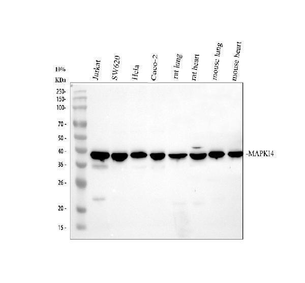
Click image to see more details
Western blot analysis of p38 alpha/MAPK14 using anti-p38 alpha/MAPK14 antibody (A00176-2).
Electrophoresis was performed on a 10% SDS-PAGE gel at 80V (Stacking gel) / 120V (Resolving gel) for 2 hours. The sample well of each lane was loaded with 30 ug of sample under reducing conditions.
Lane 1: human Jurkat whole cell lysates,
Lane 2: human SW620 whole cell lysates,
Lane 3: human Hela whole cell lysates,
Lane 4: human CACO-2 whole cell lysates,
Lane 5: rat lung tissue lysates,
Lane 6: rat heart tissue lysates,
Lane 7: mouse lung tissue lysates,
Lane 8: mouse heart tissue lysates.
After electrophoresis, proteins were transferred to a nitrocellulose membrane at 150 mA for 50-90 minutes. Blocked the membrane with 5% non-fat milk/TBS for 1.5 hour at RT. The membrane was incubated with rabbit anti-p38 alpha/MAPK14 antigen affinity purified polyclonal antibody (A00176-2) at 0.5 μg/mL overnight at 4°C, then washed with TBS-0.1%Tween 3 times with 5 minutes each and probed with a goat anti-rabbit IgG-HRP secondary antibody (Catalog # BA1054) at a dilution of 1:5000 for 1.5 hour at RT. The signal is developed using an ECL Plus Western Blotting Substrate (Catalog # AR1196-200) with Tanon 5200 system. A specific band was detected for p38 alpha/MAPK14 at approximately 38 kDa. The expected band size for p38 alpha/MAPK14 is at 41 kDa.

Click image to see more details
Western blot analysis of p38 alpha using anti-p38 alpha antibody (A00176-2).
Electrophoresis was performed on a 8% SDS-PAGE gel at 80V (Stacking gel) / 120V (Resolving gel) for 2 hours. The sample well of each lane was loaded with 30 μg of sample under reducing conditions.
Lane 1: mouse spleen tissue lysates,
Lane 2: mouse RAW264.7 whole cell lysates.
After electrophoresis, proteins were transferred to a nitrocellulose membrane at 150 mA for 50-90 minutes. Blocked the membrane with 5% non-fat milk/TBS for 1.5 hour at RT. The membrane was incubated with rabbit anti-p38 alpha antigen affinity purified polyclonal antibody (A00176-2) at 0.5 μg/mL overnight at 4°C, then washed with TBS-0.1%Tween-20 3 times with 5 minutes each and probed with a goat anti-rabbit IgG-HRP secondary antibody (Catalog # BA1054) at a dilution of 1:5000 for 1.5 hour at RT. The signal is developed using an ECL Plus Western Blotting Substrate (Catalog # AR1196-200) with Tanon 5200 system. A specific band was detected for p38 alpha at approximately 41 kDa. The expected band size for p38 alpha is at 41 kDa.

Click image to see more details
Western blot analysis of p38 MAPK/MAPK14 using anti-p38 MAPK/MAPK14 antibody (A00176-2).
Electrophoresis was performed on a 10% SDS-PAGE gel at 80V (Stacking gel) / 120V (Resolving gel) for 2 hours. The sample well of each lane was loaded with 30 ug of sample under reducing conditions.
Lane 1: human Hela- WT whole cell lysates,
Lane 2: human Hela-MAPK14 KO whole cell lysates.
After electrophoresis, proteins were transferred to a nitrocellulose membrane at 150 mA for 50-90 minutes. Blocked the membrane with 5% non-fat milk/TBS for 1.5 hour at RT. The membrane was incubated with rabbit anti-p38 MAPK/MAPK14 antigen affinity purified polyclonal antibody (A00176-2) at 0.5 μg/mL overnight at 4°C, then washed with TBS-0.1%Tween 3 times with 5 minutes each and probed with a goat anti-rabbit IgG-HRP secondary antibody at a dilution of 1:5000 for 1.5 hour at RT. The signal is developed using an ECL Plus Western Blotting Substrate (Catalog # AR1196-200) with Tanon 5200 system. A specific band was detected for p38 MAPK/MAPK14 at approximately 38 kDa. The expected band size for p38 MAPK/MAPK14 is at 41 kDa.

Click image to see more details
IHC analysis of p38 alpha/MAPK14 using anti-p38 alpha/MAPK14 antibody (A00176-2).
p38 alpha/MAPK14 was detected in a paraffin-embedded section of human breast cancer tissue. Heat mediated antigen retrieval was performed in EDTA buffer (pH 8.0, epitope retrieval solution). The tissue section was blocked with 10% goat serum. The tissue section was then incubated with 2 μg/ml rabbit anti-p38 alpha/MAPK14 Antibody (A00176-2) overnight at 4°C. Peroxidase Conjugated Goat Anti-rabbit IgG was used as secondary antibody and incubated for 30 minutes at 37°C. The tissue section was developed using HRP Conjugated Rabbit IgG Super Vision Assay Kit (Catalog # SV0002) with DAB as the chromogen.

Click image to see more details
IHC analysis of p38 alpha/MAPK14 using anti-p38 alpha/MAPK14 antibody (A00176-2).
p38 alpha/MAPK14 was detected in a paraffin-embedded section of mouse brain tissue. Heat mediated antigen retrieval was performed in EDTA buffer (pH 8.0, epitope retrieval solution). The tissue section was blocked with 10% goat serum. The tissue section was then incubated with 2 μg/ml rabbit anti-p38 alpha/MAPK14 Antibody (A00176-2) overnight at 4°C. Peroxidase Conjugated Goat Anti-rabbit IgG was used as secondary antibody and incubated for 30 minutes at 37°C. The tissue section was developed using HRP Conjugated Rabbit IgG Super Vision Assay Kit (Catalog # SV0002) with DAB as the chromogen.

Click image to see more details
IHC analysis of p38 alpha/MAPK14 using anti-p38 alpha/MAPK14 antibody (A00176-2).
p38 alpha/MAPK14 was detected in a paraffin-embedded section of rat brain tissue. Heat mediated antigen retrieval was performed in EDTA buffer (pH 8.0, epitope retrieval solution). The tissue section was blocked with 10% goat serum. The tissue section was then incubated with 2 μg/ml rabbit anti-p38 alpha/MAPK14 Antibody (A00176-2) overnight at 4°C. Peroxidase Conjugated Goat Anti-rabbit IgG was used as secondary antibody and incubated for 30 minutes at 37°C. The tissue section was developed using HRP Conjugated Rabbit IgG Super Vision Assay Kit (Catalog # SV0002) with DAB as the chromogen.

Click image to see more details
IF analysis of P38 Alpha/MAPK14 using anti-P38 Alpha/MAPK14 antibody (A00176-2) and anti-Alpha Tubulin antibody (M03989-3).
P38 Alpha/MAPK14 was detected in an immunocytochemical section of Hela cells. Enzyme antigen retrieval was performed using IHC enzyme antigen retrieval reagent (AR0022) for 15 mins. The cells were blocked with 10% goat serum. And then incubated with 5 μg/mL rabbit anti-P38 Alpha/MAPK14 Antibody (A00176-2) and mouse anti-Alpha Tubulin antibody (M03989-3) overnight at 4°C. Fluoro488 Conjugated Goat Anti-Rabbit IgG (BA1127) and Cy3 Conjugated Goat Anti-Mouse IgG (BA1031) were used as secondary antibody at 1:500 dilution and incubated for 30 minutes at 37°C. Visualize using a fluorescence microscope and filter sets appropriate for the label used.

Click image to see more details
Immunoprecipitating p38 alpha/MAPK14 in Jurkat whole cell lysate.
Western blot analysis of p38 alpha/MAPK14 using anti-p38 alpha/MAPK14 antibody (A00176-2);
Lane 1: Jurkat whole cell lysates (30ug);
Lane 2: Rabbit control IgG instead of anti-p38 alpha/MAPK14 antibody in Jurkat whole cell lysate;
Lane 3: anti-p38 alpha/MAPK14 antibody (2μg) + Jurkat whole cell lysate (500μg).
After electrophoresis, proteins were transferred to a membrane. Then the membrane was incubated with rabbit anti-p38 alpha/MAPK14 antigen affinity purified polyclonal antibody (A00176-2) at a dilution of 0.5 μg/mL and probed with a goat anti-rabbit IgG-HRP secondary antibody (Catalog # BA1054). The signal is developed using ECL Plus Western Blotting Substrate (Catalog # AR1196-200). A specific band was detected for p38 alpha/MAPK14 at approximately 38 kDa. The expected band size for p38 alpha/MAPK14 is at 41 kDa.

Click image to see more details
Flow Cytometry analysis of CACO-2 cells using anti-P38 Alpha/MAPK14 antibody (A00176-2).
Overlay histogram showing CACO-2 cells stained with A00176-2 (Blue line). To facilitate intracellular staining, cells were fixed with 4% paraformaldehyde and permeabilized with permeabilization buffer. The cells were blocked with 10% normal goat serum. And then incubated with rabbit anti-P38 Alpha/MAPK14 Antibody (A00176-2, 1 μg/1x106 cells) for 30 min at 20°C. DyLight®488 conjugated goat anti-rabbit IgG (BA1127, 5-10 μg/1x106 cells) was used as secondary antibody for 30 minutes at 20°C. Isotype control antibody (Green line) was rabbit IgG (1 μg/1x106) used under the same conditions. Unlabelled sample without incubation with primary antibody and secondary antibody (Red line) was used as a blank control.

Click image to see more details
(A) Protein expression levels of LC3-I, LC3-II, p38 MAPK, and p-p38 MAPK in SMMC-7721 cells measured by Western blot after treatment with 5 μM PL, 10 μM 3-MA autophagy inhibitor, 10 μM rapamycin autophagy agonist, 10 μM SB202190, and 10 μM SB203580 p38 MAPK inhibitor. (B) LC3-I, LC3-II, p38 MAPK, and p-p38 MAPK were used as internal reference total proteins for gray value analysis. * p < 0.05, ** p < 0.01 *** p < 0.001, compared with the PL group. # p < 0.05, ## p < 0.01 ### p < 0.001 vs. the control group. (C) Protein expression levels of LC3-I, LC3-II, p38 MAPK, and p-p38 MAPK in BEL-7404 cells measured by Western blot after treatment with 5 μM PL, 10 μM 3-MA autophagy inhibitor, 10 μM rapamycin autophagy agonist, 10 μM SB202190, and 10 μM SB203580 p38 MAPK inhibitor. (D) LC3-I, LC3-II, p38 MAPK, and p-p38 MAPK were used as internal reference total proteins for gray value analysis. * p < 0.05, ** p < 0.01 *** p < 0.001, compared with the PL group. # p < 0.05, ## p < 0.01 ### p < 0.001 vs. the control group.
Index in PubMed under a CC BY license. PMID: 33912033
Specific Publications For Anti-p38 alpha/MAPK14 Antibody Picoband® (A00176-2)
Loading publications
Recommended Resources
Here are featured tools and databases that you might find useful.
- Boster's Pathways Library
- Protein Databases
- Bioscience Research Protocol Resources
- Data Processing & Analysis Software
- Photo Editing Software
- Scientific Literature Resources
- Research Paper Management Tools
- Molecular Biology Software
- Primer Design Tools
- Bioinformatics Tools
- Phylogenetic Tree Analysis
Customer Reviews
Have you used Anti-p38 alpha/MAPK14 Antibody Picoband®?
Share your experimental results or join a short interview to earn up to $1,000 in product credits or other rewards.
0 Reviews For Anti-p38 alpha/MAPK14 Antibody Picoband®
Customer Q&As
Have a question?
Find answers in Q&As, reviews.
Can't find your answer?
Submit your question
