Click image to see more details
-

-
-
-
-
+2
Product Info Summary
| SKU: | A01419-2 |
|---|---|
| Size: | 0.1 mg |
| Reactive Species: | Human, Mouse, Rat |
| Host: | Rabbit |
| Application: | ELISA, IF, IHC-P, WB |
Customers Who Bought This Also Bought

Product info
Product Name
Anti-PAK2 Antibody
SKU/Catalog Number
A01419-2
Size
0.1 mg
Form
Liquid
Description
Boster Bio Anti-PAK2 Antibody (Catalog # A01419-2). Tested in ELISA, WB, IHC-P, IF applications. This antibody reacts with Human, Mouse, Rat.
Storage & Handling
PAK2 antibody can be stored at 4°C for three months and -20°C, stable for up to one year. Avoid repeated freeze-thaw cycles. Antibodies should not be exposed to prolonged high temperatures.
Cite This Product
Anti-PAK2 Antibody (Boster Biological Technology, Pleasanton CA, USA, Catalog # A01419-2)
Host
Rabbit
Contents
PAK2 Antibody is supplied in PBS containing 0.02% sodium azide.
Clonality
Polyclonal
Isotype
IgG
Immunogen
Anti-PAK2 antibody was raised against a peptide corresponding to 14 amino acids near the carboxy terminus of human PAK2. The immunogen is located within amino acids 440-490 of PAK2.
Reactive Species
A01419-2 is reactive to PAK2 in Human, Mouse, Rat
Observed Molecular Weight
68 kDa
Calculated molecular weight
58.0 kDa
Background of PAK2
The p21-activated kinases (PAKs) are serine-threonine kinases that bind to the active forms of Cdc42 and Rac. They are divided into two groups, the first of which include PAK1, 2 and 3, and can be activated by Cdc42/Rac binding. Group 1 PAKs contain an autoinhibitory domain whose activity is regulated by Cdc42/Rac binding. The group 1 PAKs are known to be involved in cellular processes such as gene transcription, apoptosis, and cell morphology and motility. Much less is known about the second group, which includes PAK4, 5 and 6, and are not activated by Cdc42/Rac binding. Of the six PAK proteins, only PAK2 is ubiquitously expressed and cleaved by caspase-3. This cleavage removes the amino-terminal regulatory domain and generates a constitutively active kinase fragment. Recent experiments have shown that following cleavage, the active fragment is myristoylated and directed to the plasma membrane and membrane ruffles where it promotes cell death via increased signaling through the c-Jun N-terminal kinase pathway, but without compromising mitochondrial integrity.
Antibody Validation
Boster validates all antibodies on WB, IHC, ICC, Immunofluorescence, and ELISA with known positive control and negative samples to ensure specificity and high affinity, including thorough antibody incubations.
Application & Images
Applications
A01419-2 is guaranteed for ELISA, IF, IHC-P, WB Boster Guarantee
Recommend Dilution
| Application | Dilution | Species |
|---|---|---|
| Antibody validated: Western Blot in human | mouse and rat samples; Immunohistochemistry and Immunofluorescence in mouse samples. All other applications and species not yet tested. Optimal dilutions for each application should be determined by the researcher. |
Validation Images & Assay Conditions
Click image to see more details
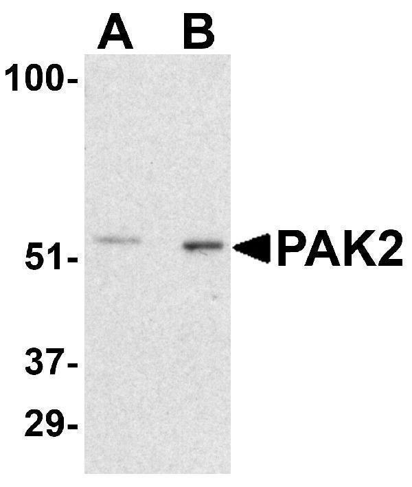
Western Blot Validation in Rat Bladder Tissue Lysate
Loading: 15 μg of lysates per lane.
Antibodies: PAK2 A01419-2 (A: 0.5 μg/mL, B: 1 μg/mL), 1h incubation at RT in 5% NFDM/TBST.
Secondary: Goat anti-rabbit IgG HRP conjugate at 1:10000 dilution.

Click image to see more details
Independent Antibody Validation (IAV) via Protein Expression Profile in Cell Lines
Loading: 15 μg of lysates per lane.
Antibodies: PAK2 A01419-2 (1 μg/mL), PAK2 3887 (1 μg/mL) and beta-actin (1 μg/mL), 1h incubation at RT in 5% NFDM/TBST.
Secondary: Goat anti-rabbit IgG HRP conjugate at 1:10000 dilution.

Click image to see more details
Western Blot Validation in Human, Mouse and Rat Cell Lines
Loading: 15 μg of lysates per lane.
Antibodies: PAK2 A01419-2 (1 μg/mL per lane), 1h incubation at RT in 5% NFDM/TBST.
Secondary: Goat anti-rabbit IgG HRP conjugate at 1:10000 dilution.

Click image to see more details
Western Blot Validation in Mouse Tissues
Loading: 15 μg of lysates per lane.
Antibodies: PAK2 A01419-2 (1 μg/mL per lane), 1h incubation at RT in 5% NFDM/TBST.
Secondary: Goat anti-rabbit IgG HRP conjugate at 1:10000 dilution.

Click image to see more details
Immunohistochemistry Validation of PAK2 in Mouse Spleen Tissue
Immunohistochemical analysis of paraffin-embedded mouse kidney tissue using anti-PAK2 antibody (A01419-2) at 10 μg/ml. Tissue was fixed with formaldehyde and blocked with 10% serum for 1 h at RT; antigen retrieval was by heat mediation with a citrate buffer (pH6). Samples were incubated with primary antibody overnight at 4˚C. A goat anti-rabbit IgG H&L (HRP) at 1/250 was used as secondary. Counter stained with Hematoxylin.

Click image to see more details
Immunofluorescence Validation of PAK2 in Mouse Spleen Cells
Immunofluorescent analysis of 4% paraformaldehyde-fixed mouse spleen labeling PAK2 with A01419-2 at 20 μg/mL, followed by goat anti-rabbit IgG secondary antibody at 1/500 dilution (red).
Specific Publications For Anti-PAK2 Antibody (A01419-2)
Loading publications
Recommended Resources
Here are featured tools and databases that you might find useful.
- Boster's Pathways Library
- Protein Databases
- Bioscience Research Protocol Resources
- Data Processing & Analysis Software
- Photo Editing Software
- Scientific Literature Resources
- Research Paper Management Tools
- Molecular Biology Software
- Primer Design Tools
- Bioinformatics Tools
- Phylogenetic Tree Analysis
Customer Reviews
Have you used Anti-PAK2 Antibody?
Share your experimental results or join a short interview to earn up to $1,000 in product credits or other rewards.
0 Reviews For Anti-PAK2 Antibody
Customer Q&As
Have a question?
Find answers in Q&As, reviews.
Can't find your answer?
Submit your question
