Click image to see more details
-

-
-
-
-
+3
Product Info Summary
| SKU: | M00669 |
|---|---|
| Size: | 100 μl |
| Reactive Species: | Human, Mouse |
| Host: | Rabbit |
| Application: | Flow Cytometry, IF, IHC, ICC, WB |
Customers Who Bought This Also Bought

Product info
Product Name
Anti-PAX5/Bsap Rabbit Monoclonal Antibody
SKU/Catalog Number
M00669
BM4806 is an alternative SKU for this antibody, used in previous lots.
Size
100 μl
Form
Liquid
Description
Boster Bio Anti-PAX5/Bsap Rabbit Monoclonal Antibody catalog # M00669. Tested in WB, IHC, ICC/IF, Flow Cytometry applications. This antibody reacts with Human, Mouse.
Storage & Handling
Store at -20°C for one year. For short term storage and frequent use, store at 4°C for up to one month. Avoid repeated freeze-thaw cycles.
Cite This Product
Anti-PAX5/Bsap Rabbit Monoclonal Antibody (Boster Biological Technology, Pleasanton CA, USA, Catalog # M00669)
Host
Rabbit
Contents
Rabbit IgG in stabilizing components, phosphate buffered saline, pH 7.4, 150mM NaCl, 0.02% sodium azide and 50% glycerol.
*This antibody is supplied in a stabilized formulation.
Compatibility with conjugation reactions depends on the chemistry of the conjugation method used.
For conjugation methods that are not compatible with the stabilizing components present in this formulation, a carrier-free antibody format is required.
Clonality
Monoclonal
Clone Number
ICD-16
Isotype
Rabbit IgG
Immunogen
A synthesized peptide derived from human PAX5
Reactive Species
M00669 is reactive to PAX5 in Human, Mouse
Observed Molecular Weight
45 kDa
Calculated molecular weight
42.1 kDa
Antibody Validation
Boster validates all antibodies on WB, IHC, ICC, Immunofluorescence, and ELISA with known positive control and negative samples to ensure specificity and high affinity, including thorough antibody incubations.
Application & Images
Applications
M00669 is guaranteed for Flow Cytometry, IF, IHC, ICC, WB Boster Guarantee
Recommend Dilution
WB 1:500-2000
IHC 1:50-200
ICC/IF 1:50-200
FC 1:30
Tested application
Suggested blocking solution with 5% non-fat milk or BSA; (*)Recommended protein loading: 20-40 µg per lane
Use TE buffer pH 9.0 for antigen retrieval; (*) citrate buffer pH 6.0 is an alternative.
Validation Images & Assay Conditions
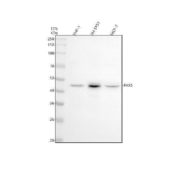
Click image to see more details
Western blot analysis of BSAP/PAX5 using anti-BSAP/PAX5 antibody (M00669).
Electrophoresis was performed on a 10% SDS-PAGE gel at 80V (Stacking gel) / 120V (Resolving gel) for 2 hours. The sample well of each lane was loaded with 30 ug of sample under reducing conditions.
Lane 1: human THP-1 whole cell lysates,
Lane 2: human SH-SY5Y whole cell lysates,
Lane 3: human MCF7 whole cell lysates.
After electrophoresis, proteins were transferred to a nitrocellulose membrane at 150 mA for 50-90 minutes. Blocked the membrane with 5% non-fat milk/TBS for 1.5 hour at RT. The membrane was incubated with rabbit anti-BSAP/PAX5 antigen affinity purified monoclonal antibody (M00669) at 1: 1000 overnight at 4°C, then washed with TBS-0.1%Tween 3 times with 5 minutes each and probed with a goat anti-rabbit IgG-HRP secondary antibody at a dilution of 1:5000 for 1.5 hour at RT. The signal is developed using an ECL Plus Western Blotting Substrate (Catalog # AR1196-200) with Tanon 5200 system. A specific band was detected for BSAP/PAX5 at approximately 45 kDa. The expected band size for BSAP/PAX5 is at 42 kDa.

Click image to see more details
IHC analysis of BSAP/PAX5 using anti-BSAP/PAX5 antibody (M00669).
BSAP/PAX5 was detected in a paraffin-embedded section of human spleen tissue. Heat mediated antigen retrieval was performed in EDTA buffer (pH 8.0, epitope retrieval solution). The tissue section was blocked with 10% goat serum. The tissue section was then incubated with 1: 50 rabbit anti-BSAP/PAX5 Antibody (M00669) overnight at 4°C. Peroxidase Conjugated Goat Anti-rabbit IgG was used as secondary antibody and incubated for 30 minutes at 37°C. The tissue section was developed using HRP Conjugated Rabbit IgG Super Vision Assay Kit (Catalog # SV0002) with DAB as the chromogen.

Click image to see more details
IHC analysis of BSAP/PAX5 using anti-BSAP/PAX5 antibody (M00669).
BSAP/PAX5 was detected in a paraffin-embedded section of human tonsil tissue. Heat mediated antigen retrieval was performed in EDTA buffer (pH 8.0, epitope retrieval solution). The tissue section was blocked with 10% goat serum. The tissue section was then incubated with 1: 50 rabbit anti-BSAP/PAX5 Antibody (M00669) overnight at 4°C. Peroxidase Conjugated Goat Anti-rabbit IgG was used as secondary antibody and incubated for 30 minutes at 37°C. The tissue section was developed using HRP Conjugated Rabbit IgG Super Vision Assay Kit (Catalog # SV0002) with DAB as the chromogen.

Click image to see more details
IHC analysis of BSAP/PAX5 using anti-BSAP/PAX5 antibody (M00669).
BSAP/PAX5 was detected in a paraffin-embedded section of mouse spleen tissue. Heat mediated antigen retrieval was performed in EDTA buffer (pH 8.0, epitope retrieval solution). The tissue section was blocked with 10% goat serum. The tissue section was then incubated with 1: 50 rabbit anti-BSAP/PAX5 Antibody (M00669) overnight at 4°C. Peroxidase Conjugated Goat Anti-rabbit IgG was used as secondary antibody and incubated for 30 minutes at 37°C. The tissue section was developed using HRP Conjugated Rabbit IgG Super Vision Assay Kit (Catalog # SV0002) with DAB as the chromogen.

Click image to see more details
Immunofluorescent analysis of Ramos cells, using PAX5 Antibody.

Click image to see more details
Immunofluorescent analysis using the Antibody at 1:50 dilution.
Specific Publications For Anti-PAX5/Bsap Rabbit Monoclonal Antibody (M00669)
Loading publications
Recommended Resources
Here are featured tools and databases that you might find useful.
- Boster's Pathways Library
- Protein Databases
- Bioscience Research Protocol Resources
- Data Processing & Analysis Software
- Photo Editing Software
- Scientific Literature Resources
- Research Paper Management Tools
- Molecular Biology Software
- Primer Design Tools
- Bioinformatics Tools
- Phylogenetic Tree Analysis
Customer Reviews
Have you used Anti-PAX5/Bsap Rabbit Monoclonal Antibody?
Share your experimental results or join a short interview to earn up to $1,000 in product credits or other rewards.
0 Reviews For Anti-PAX5/Bsap Rabbit Monoclonal Antibody
Customer Q&As
Have a question?
Find answers in Q&As, reviews.
Can't find your answer?
Submit your question
4 Customer Q&As for Anti-PAX5/Bsap Rabbit Monoclonal Antibody
Question
My question regarding product M00669, anti-PAX5/Bsap Rabbit Monoclonal antibody. I was wondering if it would be possible to conjugate this antibody with biotin. I would need it to be without BSA or sodium azide. I am planning on using a buffer exchange of sodium azide with PBS only. Would there be problems for me to conjugate the antibody and store it in -20 degrees in small aliquots?
Verified Customer
Verified customer
Asked: 2020-04-15
Answer
It is not recommended storing this antibody with PBS buffer only in -20 degrees. If you want to store it in -20 degrees it is best to add some cryoprotectant like glycerol. If you want carrier free M00669 anti-PAX5/Bsap Rabbit Monoclonal antibody, we can provide it to you in a special formula with trehalose and/or glycerol. These molecules will not interfere with conjugation chemistry and provide a good level of protection for the antibody from degradation. Please be sure to specify this in your purchase order.
Boster Scientific Support
Answered: 2020-04-15
Question
I see that the anti-PAX5/Bsap Rabbit Monoclonal antibody M00669 works with WB, what is the protocol used to produce the result images on the product page?
Verified Customer
Verified customer
Asked: 2019-06-24
Answer
You can find protocols for WB on the "support/technical resources" section of our navigation menu. If you have any further questions, please send an email to support@bosterbio.com
Boster Scientific Support
Answered: 2019-06-24
Question
I was wanting to use your anti-PAX5/Bsap Rabbit Monoclonal antibody for WB for mouse buccal mucosa cell on frozen tissues, but I want to know if it has been validated for this particular application. Has this antibody been validated and is this antibody a good choice for mouse buccal mucosa cell identification?
R. Lewis
Verified customer
Asked: 2016-08-24
Answer
It shows on the product datasheet, M00669 anti-PAX5/Bsap Rabbit Monoclonal antibody has been tested for IF, WB on human, mouse tissues. We have an innovator award program that if you test this antibody and show it works in mouse buccal mucosa cell in IHC-frozen, you can get your next antibody for free.
Boster Scientific Support
Answered: 2016-08-24
Question
We appreciate helping with my inquiry over the phone. Here are the WB image, lot number and protocol we used for buccal mucosa cell using anti-PAX5/Bsap Rabbit Monoclonal antibody M00669. Let me know if you need anything else.
B. Krishna
Verified customer
Asked: 2014-11-05
Answer
Thank you for the data. You have provided everything we needed. Our lab team are working to resolve your inquiry as quickly as possible, and we appreciate your patience and understanding! Please let me know if there is anything you need in the meantime.
Boster Scientific Support
Answered: 2014-11-05
