Click image to see more details
Product Info Summary
| SKU: | A06996-3 |
|---|---|
| Size: | 100 μg/vial |
| Reactive Species: | Human, Mouse, Rat |
| Host: | Rabbit |
| Application: | IF, ICC, WB |
Customers Who Bought This Also Bought

Product info
Product Name
Anti-Periphilin 1/PPHLN1 Antibody Picoband®
SKU/Catalog Number
A06996-3
Size
100 μg/vial
Form
Lyophilized
Description
Boster Bio Anti-Periphilin 1/PPHLN1 Antibody Picoband® catalog # A06996-3. Tested in WB, ICC, IF applications. This antibody reacts with Human, Mouse, Rat. The brand Picoband indicates this is a premium antibody that guarantees superior quality, high affinity, and strong signals with minimal background in Western blot applications. Only our best-performing antibodies are designated as Picoband, ensuring unmatched performance.
Storage & Handling
At -20°C for one year from date of receipt. After reconstitution, at 4°C for one month. It can also be aliquotted and stored frozen at -20°C for six months. Avoid repeated freezing and thawing.
Cite This Product
Anti-Periphilin 1/PPHLN1 Antibody Picoband® (Boster Biological Technology, Pleasanton CA, USA, Catalog # A06996-3)
Host
Rabbit
Contents
Each vial contains 4 mg Trehalose, 0.9 mg NaCl, 0.2 mg Na2HPO4.
Clonality
Polyclonal
Immunogen
A synthetic peptide corresponding to a sequence in the middle region of human Periphilin 1/PPHLN1.
Reactive Species
A06996-3 is reactive to PPHLN1 in Human, Mouse, Rat
Observed Molecular Weight
60 kDa
Calculated molecular weight
52.7 kDa
Background of PPHLN1
The protein encoded by this gene is one of the several proteins that become sequentially incorporated into the cornified cell envelope during the terminal differentiation of keratinocyte at the outer layers of epidermis. This protein interacts with periplakin, which is known as a precursor of the cornified cell envelope. The cellular localization pattern and insolubility of this protein suggest that it may play a role in epithelial differentiation and contribute to epidermal integrity and barrier formation. Multiple alternatively spliced transcript variants encoding distinct isoforms have been observed.
Antibody Validation
Boster validates all antibodies on WB, IHC, ICC, Immunofluorescence, and ELISA with known positive control and negative samples to ensure specificity and high affinity, including thorough antibody incubations.
Application & Images
Applications
A06996-3 is guaranteed for IF, ICC, WB Boster Guarantee
Recommend Dilution
| Application | Dilution | Species |
|---|---|---|
| Western blot | 0.25-0.5 μg/ml | Human, Mouse, Rat |
| Immunocytochemistry/Immunofluorescence | 5 μg/ml | Human |
| Flow Cytometry (Fixed) | 1-3 μg/1x106 cells | Human |
Validation Images & Assay Conditions
Click image to see more details
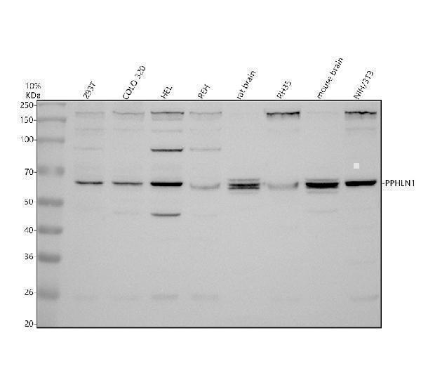
Western blot analysis of PPHLN1 using anti-PPHLN1 antibody (A06996-3).
Electrophoresis was performed on a 10% SDS-PAGE gel at 80V (Stacking gel) / 120V (Resolving gel) for 2 hours. The sample well of each lane was loaded with 30 ug of sample under reducing conditions.
Lane 1: human 293T whole cell lysates,
Lane 2: human COLO-320 whole cell lysates,
Lane 3: human HEL whole cell lysates,
Lane 4: human REH whole cell lysates,
Lane 5: rat brain tissue lysates,
Lane 6: rat RH35 whole cell lysates,
Lane 7: mouse brain tissue lysates,
Lane 8: mouse NIH/3T3 whole cell lysates.
After electrophoresis, proteins were transferred to a nitrocellulose membrane at 150 mA for 50-90 minutes. Blocked the membrane with 5% non-fat milk/TBS for 1.5 hour at RT. The membrane was incubated with rabbit anti-PPHLN1 antigen affinity purified polyclonal antibody (A06996-3) at 0.5 μg/mL overnight at 4°C, then washed with TBS-0.1%Tween 3 times with 5 minutes each and probed with a goat anti-rabbit IgG-HRP secondary antibody at a dilution of 1:5000 for 1.5 hour at RT. The signal is developed using an ECL Plus Western Blotting Substrate (Catalog # AR1196-200) with Tanon 5200 system. A specific band was detected for PPHLN1 at approximately 60 kDa. The expected band size for PPHLN1 is at 53 kDa.

Click image to see more details
IF analysis of PPHLN1 using anti-PPHLN1 antibody (A06996-3) and anti-Alpha Tubulin antibody (M03989-3).
PPHLN1 was detected in an immunocytochemical section of U2OS cells. Enzyme antigen retrieval was performed using IHC enzyme antigen retrieval reagent (AR0022) for 15 mins. The cells were blocked with 10% goat serum. And then incubated with 5 μg/mL rabbit anti-PPHLN1 Antibody (A06996-3) and mouse anti-Alpha Tubulin antibody (M03989-3) overnight at 4°C. Fluoro488 Conjugated Goat Anti-Rabbit IgG (BA1127) and Cy3 Conjugated Goat Anti-Mouse IgG (BA1031) were used as secondary antibody at 1:500 dilution and incubated for 30 minutes at 37°C. Visualize using a fluorescence microscope and filter sets appropriate for the label used.
Specific Publications For Anti-Periphilin 1/PPHLN1 Antibody Picoband® (A06996-3)
Loading publications
Recommended Resources
Here are featured tools and databases that you might find useful.
- Boster's Pathways Library
- Protein Databases
- Bioscience Research Protocol Resources
- Data Processing & Analysis Software
- Photo Editing Software
- Scientific Literature Resources
- Research Paper Management Tools
- Molecular Biology Software
- Primer Design Tools
- Bioinformatics Tools
- Phylogenetic Tree Analysis
Customer Reviews
Have you used Anti-Periphilin 1/PPHLN1 Antibody Picoband®?
Share your experimental results or join a short interview to earn up to $1,000 in product credits or other rewards.
0 Reviews For Anti-Periphilin 1/PPHLN1 Antibody Picoband®
Customer Q&As
Have a question?
Find answers in Q&As, reviews.
Can't find your answer?
Submit your question
