Click image to see more details
-

-
-
-
-
+3
Product Info Summary
| SKU: | PB9349 |
|---|---|
| Size: | 100 μg/vial |
| Reactive Species: | Human, Mouse, Rat |
| Host: | Rabbit |
| Application: | Flow Cytometry, IF, IHC, ICC, WB |
Customers Who Bought This Also Bought

Product info
Product Name
Anti-Peroxiredoxin 3/PRDX3 Antibody Picoband®
SKU/Catalog Number
PB9349
Size
100 μg/vial
Form
Lyophilized
Description
Boster Bio Anti-Peroxiredoxin 3/PRDX3 Antibody Picoband® catalog # PB9349. Tested in Flow Cytometry, IF, IHC, ICC, WB applications. This antibody reacts with Human, Mouse, Rat. The brand Picoband indicates this is a premium antibody that guarantees superior quality, high affinity, and strong signals with minimal background in Western blot applications. Only our best-performing antibodies are designated as Picoband, ensuring unmatched performance.
Storage & Handling
Store at -20˚C for one year from date of receipt. After reconstitution, at 4˚C for one month. It can also be aliquotted and stored frozen at -20˚C for six months. Avoid repeated freeze-thaw cycles.
Cite This Product
Anti-Peroxiredoxin 3/PRDX3 Antibody Picoband® (Boster Biological Technology, Pleasanton CA, USA, Catalog # PB9349)
Host
Rabbit
Contents
Each vial contains antibody formulated with stabilizing components, 0.9 mg NaCl, 0.2 mg Na2HPO4, and 0.05 mg NaN3.
*This antibody is supplied in a stabilized formulation.
Compatibility with conjugation reactions depends on the chemistry of the conjugation method used.
For conjugation methods that are not compatible with the stabilizing components present in this formulation, a carrier-free antibody format is required.
Clonality
Polyclonal
Isotype
Rabbit IgG
Immunogen
E.coli-derived human Peroxiredoxin 3 recombinant protein (Position: T110-Q256). Human Peroxiredoxin 3 shares 93% amino acid (aa) sequence identity with both mouse and rat Peroxiredoxin 3.
Cross-reactivity
No cross-reactivity with other proteins.
Reactive Species
PB9349 is reactive to PRDX3 in Human, Mouse, Rat
Observed Molecular Weight
25 kDa
Calculated molecular weight
27.7 kDa
Background of PRDX3
PRDX3 (peroxiredoxin 3) also known as AOP-1, MER5, SP-22 or PRX3, is localized exclusively in mitochondria. The deduced 256-amino acid human AOP1 protein shares 86% amino acid sequence similarity with mouse Aop1, and significant similarity with both the human proliferation-associated gene A product and the mouse stress-induced peritoneal macrophage protein Msp23. The PRDX3 gene is mapped on 10q26.11. Expression of PRDX3 is induced by MYC and is reduced in c-myc -/- cells. Chromatin immunoprecipitation analysis spanning the entire PRDX3 genomic sequence revealed that MYC binds preferentially to a 930-bp region surrounding exon 1. Results using mitochondria-specific fluorescent probes demonstrated that PRDX3 is essential for maintaining mitochondrial mass and membrane potential in transformed rat and human cells. These data provided evidence that PRDX3 is a MYC target gene that is required to maintain normal mitochondrial function.
Antibody Validation
Boster validates all antibodies on WB, IHC, ICC, Immunofluorescence, and ELISA with known positive control and negative samples to ensure specificity and high affinity, including thorough antibody incubations.
Application & Images
Applications
PB9349 is guaranteed for Flow Cytometry, IF, IHC, ICC, WB Boster Guarantee
Recommend Dilution
| Application | Dilution | Species |
|---|---|---|
| Western blot | 0.1-0.5μg/ml | |
| Immunohistochemistry (Paraffin-embedded Section) | 0.5-1μg/ml | |
| Immunocytochemistry/Immunofluorescence | 2μg/ml | |
| Flow Cytometry (Fixed) | 1-3μg/1x106 cells |
Tested application
Suggested blocking solution with 5% non-fat milk or BSA; (*)Recommended protein loading: 20-40 µg per lane
Use TE buffer pH 9.0 for antigen retrieval; (*) citrate buffer pH 6.0 is an alternative.
Validation Images & Assay Conditions
Click image to see more details
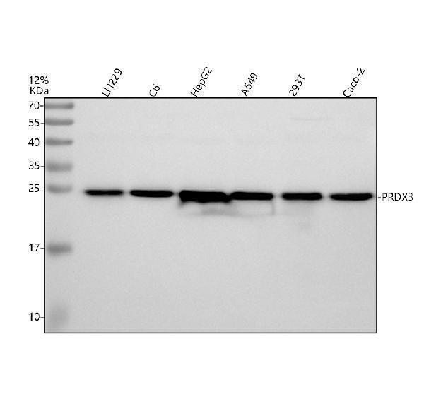
Western blot analysis of PRDX3 using anti-PRDX3 antibody (PB9349).
Electrophoresis was performed on a 12% SDS-PAGE gel at 80V (Stacking gel) / 120V (Resolving gel) for 2 hours. The sample well of each lane was loaded with 30 ug of sample under reducing conditions.
Lane 1: human LN229 whole cell lysates,
Lane 2: rat C6 whole cell lysates,
Lane 3: human HepG2 whole cell lysates,
Lane 4: human A549 whole cell lysates,
Lane 5: human 293T whole cell lysates,
Lane 6: human CACO-2 whole cell lysates.
After electrophoresis, proteins were transferred to a nitrocellulose membrane at 150 mA for 50-90 minutes. Blocked the membrane with 5% non-fat milk/TBS for 1.5 hour at RT. The membrane was incubated with rabbit anti-PRDX3 antigen affinity purified polyclonal antibody (PB9349) at 0.5 μg/mL overnight at 4°C, then washed with TBS-0.1%Tween 3 times with 5 minutes each and probed with a goat anti-rabbit IgG-HRP secondary antibody (Catalog # BA1054) at a dilution of 1:5000 for 1.5 hour at RT. The signal is developed using an ECL Plus Western Blotting Substrate (Catalog # AR1196-200) with Tanon 5200 system. A specific band was detected for PRDX3 at approximately 25 kDa. The expected band size for PRDX3 is at 28 kDa.

Click image to see more details
IHC analysis of Peroxiredoxin 3 using anti-Peroxiredoxin 3 antibody (PB9349).
Peroxiredoxin 3 was detected in paraffin-embedded section of Human Intestinal Cancer Tissue. Heat mediated antigen retrieval was performed in citrate buffer (pH6, epitope retrieval solution) for 20 mins. The tissue section was blocked with 10% goat serum. The tissue section was then incubated with 1μg/ml rabbit anti-Peroxiredoxin 3 Antibody (PB9349) overnight at 4°C. Biotinylated goat anti-rabbit IgG was used as secondary antibody and incubated for 30 minutes at 37°C. The tissue section was developed using Strepavidin-Biotin-Complex (SABC)(Catalog # SA1022) with DAB as the chromogen.

Click image to see more details
IHC analysis of Peroxiredoxin 3 using anti-Peroxiredoxin 3 antibody (PB9349).
Peroxiredoxin 3 was detected in immunocytochemical section of Hela cell. Enzyme antigen retrieval was performed using IHC enzyme antigen retrieval reagent (AR0022) for 15 mins. The cells were blocked with 10% goat serum. And then incubated with 1μg/ml rabbit anti-Peroxiredoxin 3 Antibody (PB9349) overnight at 4°C. Biotinylated goat anti-rabbit IgG was used as secondary antibody and incubated for 30 minutes at 37°C. The section was developed using Strepavidin-Biotin-Complex (SABC)(Catalog # SA1022) with DAB as the chromogen.

Click image to see more details
IHC analysis of Peroxiredoxin 3 using anti-Peroxiredoxin 3 antibody (PB9349).
Peroxiredoxin 3 was detected in immunocytochemical section of MCF-7 cell. Enzyme antigen retrieval was performed using IHC enzyme antigen retrieval reagent (AR0022) for 15 mins. The cells were blocked with 10% goat serum. And then incubated with 1μg/ml rabbit anti-Peroxiredoxin 3 Antibody (PB9349) overnight at 4°C. Biotinylated goat anti-rabbit IgG was used as secondary antibody and incubated for 30 minutes at 37°C. The section was developed using Strepavidin-Biotin-Complex (SABC)(Catalog # SA1022) with DAB as the chromogen.

Click image to see more details
IHC analysis of Peroxiredoxin 3 using anti-Peroxiredoxin 3 antibody (PB9349).
Peroxiredoxin 3 was detected in immunocytochemical section of SMMC-7721 cell. Enzyme antigen retrieval was performed using IHC enzyme antigen retrieval reagent (AR0022) for 15 mins. The cells were blocked with 10% goat serum. And then incubated with 1μg/ml rabbit anti-Peroxiredoxin 3 Antibody (PB9349) overnight at 4°C. Biotinylated goat anti-rabbit IgG was used as secondary antibody and incubated for 30 minutes at 37°C. The section was developed using Strepavidin-Biotin-Complex (SABC)(Catalog # SA1022) with DAB as the chromogen.

Click image to see more details
IF analysis of Peroxiredoxin 3 using anti-Peroxiredoxin 3 antibody (PB9349).
Peroxiredoxin 3 was detected in immunocytochemical section of U20S cells. Enzyme antigen retrieval was performed using IHC enzyme antigen retrieval reagent (AR0022) for 15 mins. The cells were blocked with 10% goat serum. And then incubated with 2μg/mL rabbit anti-Peroxiredoxin 3 Antibody (PB9349) overnight at 4°C. DyLight®594 Conjugated Goat Anti-Rabbit IgG (BA1142) was used as secondary antibody at 1:100 dilution and incubated for 30 minutes at 37°C. The section was counterstained with DAPI. Visualize using a fluorescence microscope and filter sets appropriate for the label used.

Click image to see more details
Flow Cytometry analysis of U937 cells using anti-Peroxiredoxin 3 antibody (PB9349).
Overlay histogram showing U937 cells stained with PB9349 (Blue line). To facilitate intracellular staining, cells were fixed with 4% paraformaldehyde and permeabilized with permeabilization buffer. The cells were blocked with 10% normal goat serum. And then incubated with rabbit anti-Peroxiredoxin 3 Antibody (PB9349,1μg/1x106 cells) for 30 min at 20°C. DyLight®488 conjugated goat anti-rabbit IgG (BA1127, 5-10μg/1x106 cells) was used as secondary antibody for 30 minutes at 20°C. Isotype control antibody (Green line) was rabbit IgG (1μg/1x106) used under the same conditions. Unlabelled sample without incubation with primary antibody and secondary antibody (Red line) was used as a blank control.
Specific Publications For Anti-Peroxiredoxin 3/PRDX3 Antibody Picoband® (PB9349)
Loading publications
Recommended Resources
Here are featured tools and databases that you might find useful.
- Boster's Pathways Library
- Protein Databases
- Bioscience Research Protocol Resources
- Data Processing & Analysis Software
- Photo Editing Software
- Scientific Literature Resources
- Research Paper Management Tools
- Molecular Biology Software
- Primer Design Tools
- Bioinformatics Tools
- Phylogenetic Tree Analysis
Customer Reviews
Have you used Anti-Peroxiredoxin 3/PRDX3 Antibody Picoband®?
Share your experimental results or join a short interview to earn up to $1,000 in product credits or other rewards.
0 Reviews For Anti-Peroxiredoxin 3/PRDX3 Antibody Picoband®
Customer Q&As
Have a question?
Find answers in Q&As, reviews.
Can't find your answer?
Submit your question
1 Customer Q&As for Anti-Peroxiredoxin 3/PRDX3 Antibody Picoband®
Question
We are currently using anti-Peroxiredoxin 3/PRDX3 antibody PB9349 for human tissue, and we are content with the IHC results. The species of reactivity given in the datasheet says human, mouse, rat. Is it possible that the antibody can work on canine tissues as well?
Verified Customer
Verified customer
Asked: 2019-06-03
Answer
The anti-Peroxiredoxin 3/PRDX3 antibody (PB9349) has not been tested for cross reactivity specifically with canine tissues, though there is a good chance of cross reactivity. We have an innovator award program that if you test this antibody and show it works in canine you can get your next antibody for free. Please contact me if I can help you with anything.
Boster Scientific Support
Answered: 2019-06-03
