Click image to see more details
Product Info Summary
| SKU: | A01784-1 |
|---|---|
| Size: | 100 μg/vial |
| Reactive Species: | Human |
| Host: | Rabbit |
| Application: | IF, IHC, ICC, WB |
Customers Who Bought This Also Bought

Product info
Product Name
Anti-Superoxide Dismutase 3/SOD3 Antibody Picoband®
SKU/Catalog Number
A01784-1
Size
100 μg/vial
Form
Lyophilized
Description
Boster Bio Anti-Superoxide Dismutase 3/SOD3 Antibody Picoband® catalog # A01784-1. Tested in IF, IHC, ICC, WB applications. This antibody reacts with Human. The brand Picoband indicates this is a premium antibody that guarantees superior quality, high affinity, and strong signals with minimal background in Western blot applications. Only our best-performing antibodies are designated as Picoband, ensuring unmatched performance.
Storage & Handling
Store at -20˚C for one year from date of receipt. After reconstitution, at 4˚C for one month. It can also be aliquotted and stored frozen at -20˚C for six months. Avoid repeated freeze-thaw cycles.
Cite This Product
Anti-Superoxide Dismutase 3/SOD3 Antibody Picoband® (Boster Biological Technology, Pleasanton CA, USA, Catalog # A01784-1)
Host
Rabbit
Contents
Each vial contains 4 mg Trehalose, 0.9 mg NaCl and 0.2 mg Na2HPO4.
Clonality
Polyclonal
Isotype
Rabbit IgG
Immunogen
A synthetic peptide corresponding to a sequence at the N-terminus of human SOD3.
Cross-reactivity
No cross-reactivity with other proteins.
Reactive Species
A01784-1 is reactive to SOD3 in Human
Observed Molecular Weight
33 kDa
Calculated molecular weight
25.9 kDa
Background of SOD3
SOD3 (SUPEROXIDE DISMUTASE 3), also called SUPEROXIDE DISMUTASE, EXTRACELLULAR, EC-SOD, and Cu-Zn, is an enzyme that in humans is encoded by the SOD3 gene. This gene encodes a member of the superoxide dismutase (SOD) protein family. SODs are antioxidant enzymes that catalyze the dismutation of two superoxide radicals into hydrogen peroxide and oxygen. Hendrickson et al. (1990) mapped the SOD3 gene to 4pter-q21 by a study of somatic cell hybrids. Stern et al. (2003) narrowed the assignment to 4p15.3-p15.1 by somatic cell and radiation hybrid analysis, linkage mapping, and FISH. The product of this gene is thought to protect the brain, lungs, and other tissues from oxidative stress. The protein is secreted into the extracellular space and forms a glycosylated homotetramer that is anchored to the extracellular matrix (ECM) and cell surfaces through an interaction with heparan sulfate proteoglycan and collagen. A fraction of the protein is cleaved near the C-terminus before secretion to generate circulating tetramers that do not interact with the ECM.
Antibody Validation
Boster validates all antibodies on WB, IHC, ICC, Immunofluorescence, and ELISA with known positive control and negative samples to ensure specificity and high affinity, including thorough antibody incubations.
Application & Images
Applications
A01784-1 is guaranteed for IF, IHC, ICC, WB Boster Guarantee
Recommend Dilution
| Application | Dilution | Species |
|---|---|---|
| Western blot | 0.25-0.5 μg/ml | |
| Immunohistochemistry(Paraffin-embedded Section) | 0.5-1 μg/ml | |
| Immunocytochemistry/Immunofluorescence | 5 μg/ml |
Tested application
Suggested blocking solution with 5% non-fat milk or BSA; (*)Recommended protein loading: 20-40 µg per lane
Use TE buffer pH 9.0 for antigen retrieval; (*) citrate buffer pH 6.0 is an alternative.
Validation Images & Assay Conditions
Click image to see more details
IHC analysis of SOD3 using anti-SOD3 antibody (A01784-1).
SOD3 was detected in paraffin-embedded section of human lung cancer tissue. Heat mediated antigen retrieval was performed in citrate buffer (pH6, epitope retrieval solution) for 20 mins. The tissue section was blocked with 10% goat serum. The tissue section was then incubated with 1μg/ml rabbit anti-SOD3 Antibody (A01784-1) overnight at 4°C. Biotinylated goat anti-rabbit IgG was used as secondary antibody and incubated for 30 minutes at 37°C. The tissue section was developed using Strepavidin-Biotin-Complex (SABC)(Catalog # SA1022) with DAB as the chromogen.

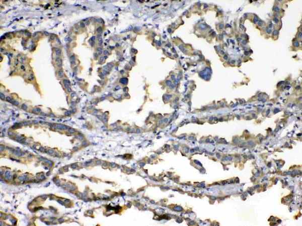
Click image to see more details
IHC analysis of SOD3 using anti-SOD3 antibody (A01784-1). SOD3 was detected in paraffin-embedded section of human mammary cancer tissue. Heat mediated antigen retrieval was performed in citrate buffer (pH6, epitope retrieval solution) for 20 mins. The tissue section was blocked with 10% goat serum. The tissue section was then incubated with 1μg/ml rabbit anti-SOD3 Antibody (A01784-1) overnight at 4°C. Biotinylated goat anti-rabbit IgG was used as secondary antibody and incubated for 30 minutes at 37°C. The tissue section was developed using Strepavidin-Biotin-Complex (SABC)(Catalog # SA1022) with DAB as the chromogen.

Click image to see more details
Western blot analysis of SOD3 using anti-SOD3 antibody (A01784-1).
Electrophoresis was performed on a 5-20% SDS-PAGE gel at 70V (Stacking gel) / 90V (Resolving gel) for 2-3 hours. The sample well of each lane was loaded with 30 ug of sample under reducing conditions.
Lane 1: human PC-3 whole cell lysates.
After electrophoresis, proteins were transferred to a nitrocellulose membrane at 150 mA for 50-90 minutes. Blocked the membrane with 5% non-fat milk/TBS for 1.5 hour at RT. The membrane was incubated with rabbit anti-SOD3 antigen affinity purified polyclonal antibody (Catalog # A01784-1) at 0.5 μg/mL overnight at 4°C, then washed with TBS-0.1%Tween 3 times with 5 minutes each and probed with a goat anti-rabbit IgG-HRP secondary antibody at a dilution of 1:5000 for 1.5 hour at RT. The signal is developed using an Enhanced Chemiluminescent detection (ECL) kit (Catalog # EK1002) with Tanon 5200 system. A specific band was detected for SOD3 at approximately 33 kDa. The expected band size for SOD3 is at 27 kDa.

Click image to see more details
IF analysis of SOD3 using anti-SOD3 antibody (A01784-1).
SOD3 was detected in an immunocytochemical section of PC-3 cells. Enzyme antigen retrieval was performed using IHC enzyme antigen retrieval reagent (AR0022) for 15 mins. The cells were blocked with 10% goat serum. And then incubated with 5 μg/mL rabbit anti-SOD3 Antibody (A01784-1) overnight at 4°C. DyLight®488 Conjugated Goat Anti-Rabbit IgG (BA1127) was used as secondary antibody at 1:100 dilution and incubated for 30 minutes at 37°C. The section was counterstained with DAPI. Visualize using a fluorescence microscope and filter sets appropriate for the label used.
Specific Publications For Anti-Superoxide Dismutase 3/SOD3 Antibody Picoband® (A01784-1)
Loading publications
Recommended Resources
Here are featured tools and databases that you might find useful.
- Boster's Pathways Library
- Protein Databases
- Bioscience Research Protocol Resources
- Data Processing & Analysis Software
- Photo Editing Software
- Scientific Literature Resources
- Research Paper Management Tools
- Molecular Biology Software
- Primer Design Tools
- Bioinformatics Tools
- Phylogenetic Tree Analysis
Customer Reviews
Have you used Anti-Superoxide Dismutase 3/SOD3 Antibody Picoband®?
Share your experimental results or join a short interview to earn up to $1,000 in product credits or other rewards.
0 Reviews For Anti-Superoxide Dismutase 3/SOD3 Antibody Picoband®
Customer Q&As
Have a question?
Find answers in Q&As, reviews.
Can't find your answer?
Submit your question
