Click image to see more details
Product Info Summary
| SKU: | A00467 |
|---|---|
| Size: | 100ug |
| Reactive Species: | Human |
| Host: | Rabbit |
| Application: | ELISA, IP, IHC, WB |
Customers Who Bought This Also Bought

Product info
Product Name
Anti-PIN1 Antibody
SKU/Catalog Number
A00467
Size
100ug
Form
Liquid (sterile filtered)
Description
Boster Bio Anti-PIN1 Antibody (Catalog # A00467). Tested in ELISA, IHC, WB applications. This antibody reacts with Human.
Storage & Handling
Store vial at -20°C prior to opening. Aliquot contents and freeze at -20°C or below for extended storage. Avoid cycles of freezing and thawing. Centrifuge product if not completely clear after standing at room temperature. This product is stable for several weeks at 4°C as an undiluted liquid. Dilute only prior to immediate use. Expiration date is one (1) year from date of opening. (Ship on dry ice.)
Cite This Product
Anti-PIN1 Antibody (Boster Biological Technology, Pleasanton CA, USA, Catalog # A00467)
Host
Rabbit
Contents
0.02 M Potassium Phosphate, 0.15 M Sodium Chloride, pH 7.2, 0.01% (w/v) Sodium Azide
Clonality
Polyclonal
Isotype
IgG
Immunogen
This affinity purified antibody was prepared from whole rabbit serum produced by repeated immunizations with a synthetic peptide corresponding to an internal sequence of human Pin1.
Cross-reactivity
No cross reactivity with other proteins.
Reactive Species
A00467 is reactive to PIN1 in Human
Observed Molecular Weight
42 kDa
Calculated molecular weight
18.2 kDa
Background of PIN1
Pin1 (peptidylprolyl cis/trans isomerase NIMA-interacting 1 protein) is an essential peptidylprolyl isomerase that regulates mitosis, presumably by interacting with NIMA and attenuating its mitosis-promoting activity. Pin1 displays a preference for an acidic residue N-terminal to the isomerized proline bond and also catalyzes pSer/Thr-Pro cis/trans isomerizations. Pin1 shows a nuclear localization.
Antibody Validation
Boster validates all antibodies on WB, IHC, ICC, Immunofluorescence, and ELISA with known positive control and negative samples to ensure specificity and high affinity, including thorough antibody incubations.
Application & Images
Applications
A00467 is guaranteed for ELISA, IP, IHC, WB Boster Guarantee
Recommend Dilution
| Application | Dilution | Species |
|---|---|---|
| ELISA: 1:2 | 500 - 1:10 | 000 |
| WB: 1:500 - 1:3 | 000 | |
| This affinity purified antibody has been tested for use in ELISA | Immunohistochemistry | and western blotting. Specific conditions for reactivity should be optimized by the end user. Expect a band approximately 18 kDa in size corresponding to Pin1 by western blotting in the appropriate cell lysate or extract. Lysates from 3T3, Jurkat, 293 or HeLa cells, as well as HeLa nuclear extract, are recommended for use as positive controls. |
Validation Images & Assay Conditions
Click image to see more details
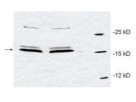
Western blot using Boster's affinity purified anti-Pin1 antibody to detect endogenous Pin1 in HeLa whole cell lysates. The sample was run in duplicate. A band representing Pin1 is indicated by the arrowhead. Cell lysates were electrophoresed using a straight 15% polyacrylamide gel, followed by transfer to nitrocellulose. The membrane was probed with the primary antibody at a 1:700 dilution. A 1:5,000 dilution of HRP Gt-a-Rabbit IgG (611-103-122) was used with a 15 sec exposure time. Personal Communication, L. D'agostino and A. Giordano, SHRO, Philadelphia, PA.

Click image to see more details
Western Blot of Rabbit anti-PIN1 antibody. Lane 1: T98G cells treated with scrambled (scr). Lane 2: T98G cells treated with PIN1 kd1 shRNA. Lane 3: T98G cells treated with PIN1-overexpressing plasmid HAP1. Lane 4: T98G cells treated with PIN1-overexpressing plasmid HAP1S67E. Lane 5: T98G cells treated with PIN1 kd2 shRNA. Load: 25 µg per lane. Primary antibody: PIN 1 antibody at 1:400 for overnight at 4°C. Secondary antibody: IRDye800™ rabbit secondary antibody at 1:10,000 for 45 min at RT. Block: 5% BLOTTO overnight at 4°C. Predicted size: 18 kDa for PIN-1. Other band(s): normalized with a-tubulin (α-tub) antibody.

Click image to see more details
Immunoprecipitation of Rabbit anti-PIN1 antibody. Lane 1: T98G cells incubated with GST-Pin1. Lane 2: T98G cells incubated with GST-CDK4/cyclinD1. Lane 3: T98G cells incubated with GST-Pin1 and GST-CDK4/cyclinD1. Immunoprecipitated with pRb antibody. Load: 25 µg per lane. Primary antibody: anti-GST 1:400 for overnight at 4°C. Secondary antibody: IRDye800™ secondary antibody at 1:10,000 for 45 min at RT. Block: 5% BLOTTO overnight at 4°C.

Click image to see more details
Immunohistochemistry of rabbit anti-PIN1 antibody. Tissue: testis. Fixation: formalin fixed paraffin embedded. Antigen retrieval: not required. Primary antibody: Anti-PIN1 at 5 µg/mL for 1 h at RT. Secondary antibody: Peroxidase rabbit secondary antibody at 1:10,000 for 45 min at RT. Staining: PIN-1 as precipitated red signal with hematoxylin purple nuclear counterstain.
Specific Publications For Anti-PIN1 Antibody (A00467)
Loading publications
Recommended Resources
Here are featured tools and databases that you might find useful.
- Boster's Pathways Library
- Protein Databases
- Bioscience Research Protocol Resources
- Data Processing & Analysis Software
- Photo Editing Software
- Scientific Literature Resources
- Research Paper Management Tools
- Molecular Biology Software
- Primer Design Tools
- Bioinformatics Tools
- Phylogenetic Tree Analysis
Customer Reviews
Have you used Anti-PIN1 Antibody?
Share your experimental results or join a short interview to earn up to $1,000 in product credits or other rewards.
0 Reviews For Anti-PIN1 Antibody
Customer Q&As
Have a question?
Find answers in Q&As, reviews.
Can't find your answer?
Submit your question
