Click image to see more details
-

-
-
-
-
+4
Product Info Summary
| SKU: | A00600 |
|---|---|
| Size: | 100ug |
| Reactive Species: | Human, Mouse |
| Host: | Rabbit |
| Application: | ELISA, Flow Cytometry, IF, IHC, WB |
Customers Who Bought This Also Bought

Product info
Product Name
Anti-PPAR alpha Antibody
SKU/Catalog Number
A00600
Size
100ug
Form
Liquid (sterile filtered)
Description
Boster Bio Anti-PPAR alpha Antibody (Catalog # A00600). Tested in ELISA, IF, IHC, WB applications. This antibody reacts with Human, Mouse.
Storage & Handling
Store vial at -20°C prior to opening. Aliquot contents and freeze at -20°C or below for extended storage. Avoid cycles of freezing and thawing. Centrifuge product if not completely clear after standing at room temperature. This product is stable for several weeks at 4°C as an undiluted liquid. Dilute only prior to immediate use. Expiration date is one (1) year from date of opening. (Ship on dry ice.)
Cite This Product
Anti-PPAR alpha Antibody (Boster Biological Technology, Pleasanton CA, USA, Catalog # A00600)
Host
Rabbit
Contents
0.02 M Potassium Phosphate, 0.15 M Sodium Chloride, pH 7.2, 0.01% (w/v) Sodium Azide
Clonality
Polyclonal
Isotype
IgG
Immunogen
PPAR alpha Antibody was prepared from whole rabbit serum produced by repeated immunizations with a synthetic peptide corresponding to a N-Terminal region near amino acids 1-25 of mouse PPAR alpha.
Cross-reactivity
No cross reactivity with other proteins.
Reactive Species
A00600 is reactive to PPARA in Human, Mouse
Calculated molecular weight
52.3 kDa
Background of PPARA
Since their discovery in the early 1990's, the peroxisome proliferator activated receptors (PPARs) have attracted significant attention. This is primarily because PPARs serve as receptors for two very important classes of drugs: the hypolipidemic fibrates and the insulin sensitizing thiazolidinediones. Peroxisome proliferators are non-genotoxic carcinogens that are purported to exert their effect on cells through their interaction with members of the nuclear hormone receptor family termed PPARs. Nuclear hormone receptors are ligand-dependent intracellular proteins that stimulate transcription of specific genes by binding to specific DNA sequences following activation by the appropriate ligand. Upon binding fatty acids or hypolipidemic drugs, PPARs form heterodimers with retinoid X receptors (RXRs) and these heterodimers regulate the expression of target genes. There are 3 known subtypes of PPARs: PPAR-alpha, PPAR-delta and PPAR-gamma. Mostly target genes are involved in the catabolism of fatty acids. Conversely, PPAR-gamma is activated by peroxisome proliferators such as prostaglandins, leukotrienes and Anti diabetic thiazolidinediones and affects the expression of genes involved in the storage of the fatty acids. PPAR-gamma may also be involved in adipocyte differentiation. It has also been shown that PPARs can induce transcription of acyl coenzyme A oxidase and cytochrome P450 through interaction with specific response elements. Anti-Ppar Antibody is useful for research interested in transcription and metabolic pathways.
Antibody Validation
Boster validates all antibodies on WB, IHC, ICC, Immunofluorescence, and ELISA with known positive control and negative samples to ensure specificity and high affinity, including thorough antibody incubations.
Application & Images
Applications
A00600 is guaranteed for ELISA, Flow Cytometry, IF, IHC, WB Boster Guarantee
Recommend Dilution
| Application | Dilution | Species |
|---|---|---|
| ELISA: 1:75 | 000 - 1:125 | 000 |
| WB: 1:500 - 1:2 | 000 | |
| Anti-PPAR alpha Antibody has been tested in ELISA | Western Blot | Immunohistochemistry, and Immunofluorescence. Expect a single band approximately 52 kDa in size corresponding to PPAR alpha by western blot in the appropriate tissue or cell lysate. A 1:200 dilution is suggested for Immunohistochemistry. Specific conditions for reactivity should be optimized by the end user. |
Validation Images & Assay Conditions
Click image to see more details
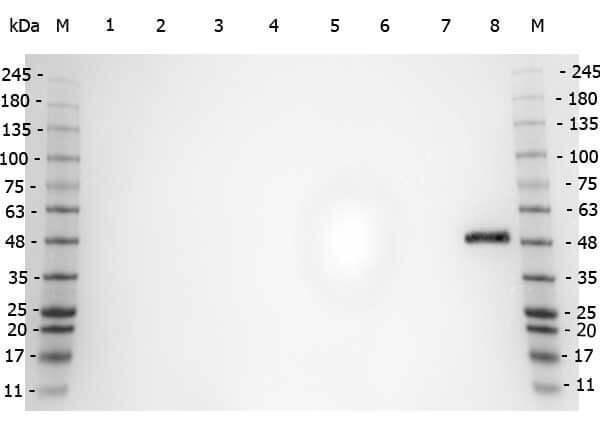
Western Blot of Rabbit anti-PPAR Alpha (N-terminal specific) antibody. Marker: Opal Pre-stained ladder . Lane 1: HEK293 lysate . Lane 2: HeLa Lysate . Lane 3: MCF-7 Lysate . Lane 4: Jurkat Lysate . Lane 5: A431 Lysate . Lane 6: LNCaP Lysate . Lane 7: A-172 Lysate . Lane 8: NIH/3T3 Lysate . Load: 35 µg per lane. Primary antibody: PPAR Alpha (N-terminal specific) antibody at 1ug/mL overnight at 4°C. Secondary antibody: Peroxidase rabbit secondary antibody at 1:30,000 for 60 min at RT. Blocking Buffer: 1% Casein-TTBS for 30 min at RT. Predicted/Observed size: 52 kDa for PPAR Alpha.

Click image to see more details
Western Blot of Rabbit anti-PPAR Alpha (N-terminal Specific) antibody. Lane M: Prestained Molecular Weight Markers. Lane 1: NIH/3T3 . Load: 10 µg per lane. Primary antibody: PPAR Alpha (N-terminal specific) antibody at 1:1,000 for overnight at 4°C. Secondary antibody: Peroxidase rabbit secondary antibody at 1:40,000 for 30 min at RT. Block: Blocking Buffer for Fluorescent Western Blotting at RT for 30 min. Predicted/Observed size: ~50 kDa for PPAR Alpha.

Click image to see more details
Immunohistochemistry (Formalin/PFA-fixed paraffin-embedded sections) showing Boster's PPAR alpha antibody staining of PPAR alpha protein in mouse liver tissue section (Formalin/PFA-fixed paraffin-embedded sections). Tissue underwent formaldehyde fixation before enzymatic antigen retrieval with 0.05% protease in PBS for 5 minutes. Sample was then blocked with 5% serum for 20 minutes at 20°C. The primary antibody was diluted 1:50 and incubated with sample in Tris plus 5% normal goat serum for 1 hour at 20°C. A Biotin conjugated goat polyclonal to rabbit IgG was used at dilution at 1:500 as secondary antibody. Images show nuclear staining in hepatocytes (perfusion-fixed mouse, 10 and 40x microscope magnification).

Click image to see more details
Affinity Purified Anti-PPAR alpha (N -terminal specific) (Rabbit) is shown to detect a 52 kDa band corresponding to PPAR alpha present in a 3T3 whole cell lysate. Approximately 20 µg of lysate was loaded per lane for SDS-PAGE. Detection occurred after using a 1:500 (lane 1) or 1:1000 (lane 2) dilution of antibody followed by 1:2000 dilution of HRP Goat-a-Rabbit IgG for visualization.

Click image to see more details
Immunohistochemistry using Boster's anti-PPAR antibody, showing staining of PPAR alpha in rat brain sections, highlighting cytoplasmic staining in ependymal cells and neurons in frontal cortex. Bottom image shows subventricular zone (svz) of lateral ventrical (exit point of progenitor olfactory neurones); top image shows frontal cortex in the same section. Cytoplasmic staining is also observed in the corpus callosum (bottom image) and in dendritic fields of the cortex. Formalin/PFA-fixed paraffin-embedded sections of rat brain tissue were incubated with the primary antibody at 1:200 for 1 hour. Antigen retrieval was performed by heat induction in citrate buffer pH 6.0.

Click image to see more details
Immunofluorescence Microscopy of Rabbit anti-PPAR alpha antibody. Tissue: HepG2 cells. Fixation: 4% formaldehyde fixed (10 min). Antigen retrieval: not required. Primary antibody: PPAR alpha antibody at 1 µg/mL overnight at 4°C. Secondary antibody: Alexa Fluor® 488 goat anti-rabbit IgG (H+L) (green) used at a 1:1000, Alexa Fluor® 594 WGA was used to label plasma membranes (red) at a 1:200 dilution for 1h for 45 min at RT. Localization: PPAR alpha is nuclear and occasionally cytoplasmic. Staining: PPAR alpha as green fluorescent signal with DAPI (blue) nuclear counterstain.

Click image to see more details
ELISA results of purified Rabbit anti-PPAR Alpha (N-terminal specific) Antibody tested against BSA-conjugated peptide of immunizing peptide. Each well was coated in duplicate with 0.1µg of conjugate. The starting dilution of antibody was 5μg/ml and the X-axis represents the Log10 of a 3-fold dilution. This titration is a 4-parameter curve fit where the IC50 is defined as the titer of the antibody. Assay performed using 3% fish gel, Goat anti-Rabbit IgG Antibody Peroxidase Conjugated (Min X Bv Ch Gt GP Ham Hs Hu Ms Rt & Sh Serum Proteins) and TMB ELISA Peroxidase Substrate .

Click image to see more details
Immunofluorescence microscopy of Rabbit Anti-PPAR alpha (N-terminal specific) antibody using (A) Mouse NIH/3T3 or (B) Human HEK293 cells fixed with MeOH. (C) Secondary antibody only with NIH/3T3 cells. Anti-PPAR alpha antibody was used at 10 µg/mL, 1h at RT⁰. Secondary antibody: Anti-RABBIT IgG DyLight™ 488 Conjugated Preadsorbed at 5 ug/ml for 1 h at RT. Staining: PPAR as green fluorescent signal with DAPI (blue) nuclear counterstain.
Specific Publications For Anti-PPAR alpha Antibody (A00600)
Loading publications
Recommended Resources
Here are featured tools and databases that you might find useful.
- Boster's Pathways Library
- Protein Databases
- Bioscience Research Protocol Resources
- Data Processing & Analysis Software
- Photo Editing Software
- Scientific Literature Resources
- Research Paper Management Tools
- Molecular Biology Software
- Primer Design Tools
- Bioinformatics Tools
- Phylogenetic Tree Analysis
Customer Reviews
Have you used Anti-PPAR alpha Antibody?
Share your experimental results or join a short interview to earn up to $1,000 in product credits or other rewards.
0 Reviews For Anti-PPAR alpha Antibody
Customer Q&As
Have a question?
Find answers in Q&As, reviews.
Can't find your answer?
Submit your question
