Click image to see more details
-

-
-
-
-
+1
Product Info Summary
| SKU: | M00449-2 |
|---|---|
| Size: | 100 µl/vial |
| Reactive Species: | Human |
| Host: | Rabbit |
| Application: | Flow Cytometry, IF, ICC, WB |
Customers Who Bought This Also Bought

Product info
Product Name
Anti-PPAR gamma Rabbit Monoclonal Antibody
SKU/Catalog Number
M00449-2
Size
100 µl/vial
Form
Liquid
Description
Boster Bio Anti-PPAR gamma Rabbit Monoclonal Antibody catalog # M00449-2. Tested in WB, ICC/IF, Flow Cytometry applications. This antibody reacts with Human.
Storage & Handling
Store at -20°C for one year. For short term storage and frequent use, store at 4°C for up to one month. Avoid repeated freeze-thaw cycles.
Cite This Product
Anti-PPAR gamma Rabbit Monoclonal Antibody (Boster Biological Technology, Pleasanton CA, USA, Catalog # M00449-2)
Host
Rabbit
Contents
Rabbit IgG in stabilizing components, phosphate buffered saline, pH 7.4, 150mM NaCl, 0.02% sodium azide and 50% glycerol.
*This antibody is supplied in a stabilized formulation.
Compatibility with conjugation reactions depends on the chemistry of the conjugation method used.
For conjugation methods that are not compatible with the stabilizing components present in this formulation, a carrier-free antibody format is required.
Clonality
Monoclonal
Clone Number
31P97
Isotype
IgG
Immunogen
A synthesized peptide derived from human PPAR gamma
Reactive Species
M00449-2 is reactive to PPARG in Human
Observed Molecular Weight
58 kDa
Calculated molecular weight
57.6 kDa
Antibody Validation
Boster validates all antibodies on WB, IHC, ICC, Immunofluorescence, and ELISA with known positive control and negative samples to ensure specificity and high affinity, including thorough antibody incubations.
Application & Images
Applications
M00449-2 is guaranteed for Flow Cytometry, IF, ICC, WB Boster Guarantee
Recommend Dilution
WB 1:500-2000
ICC/IF 1:50-200
FC 1:50
Validation Images & Assay Conditions
Click image to see more details
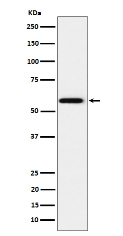
Western blot analysis of PPAR gamma expression in HeLa cell lysate.

Click image to see more details
H19 and PPARγ are upregulated in the femoral head and BMSCs of patients with SONFH. (A, B) X-ray photo and pathological structure of the femoral head from an ARCO stage V SONFH patient. The images show alterations in the morphology of the femoral head, characterized by collapse and flattening, as well as radiographic signs indicative of hip osteoarthritis. (C) Morphology of BMSCs from a patient with SONFH. (D, E) Expression levels of H19 and PPARγ in the femoral head and BMSCs from a patient with SONFH. All experimental procedures were performed in triplicate with internal normalization to GAPDH expression levels. The relative expression levels of each gene were analyzed using the 2 −△△Ct method (n = 8, all data are shown as the mean ± SD of three independent experiments, *p < 0.05, **p < 0.01).
Index in PubMed under a CC BY license. PMID: 40259926

Click image to see more details
H19 participates in increased adipogenesis of BMSCs and positively regulates PPARγ expression in SONFH. (A) Expression level of H19 in BMSCs after transfection with sh-H19. (B) Oil Red O staining (200×) of BMSCs after transfection with sh-H19. (C, D) Quantification of Oil Red O staining (200×) of BMSCs after transfection with sh-H19. (E, F) The expression of fatty acid-binding protein 4 (FABP4) was detected by qRT-PCR and Western blot after knocking-down of H19. (G) The expression levels of H19 and PPARγ 1 week following the knockdown of H19. (n = 3, all data are shown as the mean ± SD of three independent experiments, *p < 0.05, **p < 0.01).
Index in PubMed under a CC BY license. PMID: 40259926

Click image to see more details
H19 modulates PPARγ expression through miR-130b-3p. (A) Three databases, StarBase, DIANA tools, and miRWalk, were used to predict candidate micro (mi)RNAs as shown in the Venn diagram. (B) Expression levels of three candidate miRNAs (miR-301b-3p, miR-130b-3p, and miR-130a-3p) after knockdown of H19. (C) The binding sites of miR-130b-3p with H19 and PPARγ. (D) Effect of miR-130b-3p on the luciferase activity of wild-type (WT)-H19 and mutant (MT)-H19 reporter systems. (E) Effect of miR-130b-3p on the luciferase activity of WT-FABP4 and MT-FABP4 reporter systems was detected via luciferase reporter assay. (F, G) The expression of PPARγ is significantly decreased upon knocking down H19. This reduction could be reversed through co-transfection with a miR-130b-3p inhibitor. (H, I) The expression of PPARγ was significantly augmented when H19 was upregulated. This elevation in PPARγ expression could be counteracted by co-transfecting with a miR-130b-3p mimic. (n = 3, all data are shown as the mean ± SD of three independent experiments, **p < 0.01, ***p < 0.001).
Index in PubMed under a CC BY license. PMID: 40259926

Click image to see more details
All lanes use the Antibody at 1:3K dilution for 1 hour at room temperature.
Specific Publications For Anti-PPAR gamma Rabbit Monoclonal Antibody (M00449-2)
Loading publications
Recommended Resources
Here are featured tools and databases that you might find useful.
- Boster's Pathways Library
- Protein Databases
- Bioscience Research Protocol Resources
- Data Processing & Analysis Software
- Photo Editing Software
- Scientific Literature Resources
- Research Paper Management Tools
- Molecular Biology Software
- Primer Design Tools
- Bioinformatics Tools
- Phylogenetic Tree Analysis
Customer Reviews
Have you used Anti-PPAR gamma Rabbit Monoclonal Antibody?
Share your experimental results or join a short interview to earn up to $1,000 in product credits or other rewards.
0 Reviews For Anti-PPAR gamma Rabbit Monoclonal Antibody
Customer Q&As
Have a question?
Find answers in Q&As, reviews.
Can't find your answer?
Submit your question
