Click image to see more details
Product Info Summary
| SKU: | A01178-1 |
|---|---|
| Size: | 100 μg/vial |
| Reactive Species: | Human, Mouse, Rat |
| Host: | Rabbit |
| Application: | ELISA, IF, IHC, ICC, WB |
Customers Who Bought This Also Bought

Product info
Product Name
Anti-Prohibitin/PHB Antibody Picoband®
SKU/Catalog Number
A01178-1
Size
100 μg/vial
Form
Lyophilized
Description
Boster Bio Anti-Prohibitin/PHB Antibody Picoband® catalog # A01178-1. Tested in ELISA, IF, IHC, ICC, WB applications. This antibody reacts with Human, Mouse, Rat. The brand Picoband indicates this is a premium antibody that guarantees superior quality, high affinity, and strong signals with minimal background in Western blot applications. Only our best-performing antibodies are designated as Picoband, ensuring unmatched performance.
Storage & Handling
Store at -20˚C for one year from date of receipt. After reconstitution, at 4˚C for one month. It can also be aliquotted and stored frozen at -20˚C for six months. Avoid repeated freeze-thaw cycles.
Cite This Product
Anti-Prohibitin/PHB Antibody Picoband® (Boster Biological Technology, Pleasanton CA, USA, Catalog # A01178-1)
Host
Rabbit
Contents
Each vial contains 4mg Trehalose, 0.9mg NaCl and 0.2mg Na2HPO4.
Clonality
Polyclonal
Isotype
Rabbit IgG
Immunogen
E.coli-derived human Prohibitin/PHB recombinant protein (Position: M1-I257).
Cross-reactivity
No cross-reactivity with other proteins.
Reactive Species
A01178-1 is reactive to PHB in Human, Mouse, Rat
Observed Molecular Weight
30 kDa
Calculated molecular weight
29.8 kDa
Background of PHB
PHB (Prohibitin), also known as PHB1, is a protein that in humans is encoded by the PHB gene. White et al. (1991) mapped the PHB gene to chromosome 17 by analysis of human-mouse somatic cell hybrid cell lines using a genomic fragment of human prohibitin DNA isolated from a library using the rat prohibitin cDNA clone. By in situ hybridization, they localized the gene to 17q21. Sato et al. (1992) isolated the human homolog of the rat prohibitin gene and mapped it to 17q12-q21 by in situ hybridization. Proliferation of tumor cells depends on new blood vessel formation (angiogenesis) that accompanies malignant progression. Anticancer therapies using angiogenesis inhibitors or cytotoxic agents targeted to the vasculature of tumors have been evaluated in clinical trials. Although white fat is a nonmalignant tissue, it has the capability to quickly proliferate and expand. Furthermore, it is highly vascularized. Rupnick et al. (2002) showed that nonspecific angiogenesis inhibitors can prevent the development of obesity of mice.
Antibody Validation
Boster validates all antibodies on WB, IHC, ICC, Immunofluorescence, and ELISA with known positive control and negative samples to ensure specificity and high affinity, including thorough antibody incubations.
Application & Images
Applications
A01178-1 is guaranteed for ELISA, IF, IHC, ICC, WB Boster Guarantee
Recommend Dilution
| Application | Dilution | Species |
|---|---|---|
| Western blot | 0.25-0.5μg/ml | Human, Mouse, Rat |
| Immunohistochemistry (Paraffin-embedded Section) | 2-5μg/ml | Human |
| Immunocytochemistry/Immunofluorescence | 5 μg/ml | Human |
| ELISA | 0.1-0.5μg/ml | - |
Tested application
Suggested blocking solution with 5% non-fat milk or BSA; (*)Recommended protein loading: 20-40 µg per lane
Use TE buffer pH 9.0 for antigen retrieval; (*) citrate buffer pH 6.0 is an alternative.
Validation Images & Assay Conditions
Click image to see more details
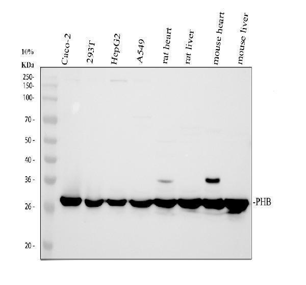
Western blot analysis of Prohibitin/PHB using anti-Prohibitin/PHB antibody (A01178-1).
Electrophoresis was performed on a 10% SDS-PAGE gel at 80V (Stacking gel) / 120V (Resolving gel) for 2 hours. The sample well of each lane was loaded with 30 ug of sample under reducing conditions.
Lane 1: human CACO-2 whole cell lysates,
Lane 2: human 293T whole cell lysates,
Lane 3: human HepG2 whole cell lysates,
Lane 4: human A549 whole cell lysates,
Lane 5: rat heart tissue lysates,
Lane 6: rat liver tissue lysates,
Lane 7: mouse heart tissue lysates,
Lane 8: mouse liver tissue lysates.
After electrophoresis, proteins were transferred to a nitrocellulose membrane at 150 mA for 50-90 minutes. Blocked the membrane with 5% non-fat milk/TBS for 1.5 hour at RT. The membrane was incubated with rabbit anti-Prohibitin/PHB antigen affinity purified polyclonal antibody (A01178-1) at 0.5 μg/mL overnight at 4°C, then washed with TBS-0.1%Tween 3 times with 5 minutes each and probed with a goat anti-rabbit IgG-HRP secondary antibody (Catalog # BA1054) at a dilution of 1:5000 for 1.5 hour at RT. The signal is developed using an ECL Plus Western Blotting Substrate (Catalog # AR1196-200) with Tanon 5200 system. A specific band was detected for Prohibitin/PHB at approximately 30 kDa. The expected band size for Prohibitin/PHB is at 30 kDa.

Click image to see more details
IHC analysis of Prohibitin/PHB using anti-Prohibitin/PHB antibody (A01178-1).
Prohibitin/PHB was detected in a paraffin-embedded section of human colon cancer tissue. Heat mediated antigen retrieval was performed in EDTA buffer (pH 8.0, epitope retrieval solution). The tissue section was blocked with 10% goat serum. The tissue section was then incubated with 2 μg/ml rabbit anti-Prohibitin/PHB Antibody (A01178-1) overnight at 4°C. Peroxidase Conjugated Goat Anti-rabbit IgG was used as secondary antibody and incubated for 30 minutes at 37°C. The tissue section was developed using HRP Conjugated Rabbit IgG Super Vision Assay Kit (Catalog # SV0002) with DAB as the chromogen.

Click image to see more details
IHC analysis of Prohibitin/PHB using anti-Prohibitin/PHB antibody (A01178-1).
Prohibitin/PHB was detected in a paraffin-embedded section of human lung cancer tissue. Heat mediated antigen retrieval was performed in EDTA buffer (pH 8.0, epitope retrieval solution). The tissue section was blocked with 10% goat serum. The tissue section was then incubated with 2 μg/ml rabbit anti-Prohibitin/PHB Antibody (A01178-1) overnight at 4°C. Peroxidase Conjugated Goat Anti-rabbit IgG was used as secondary antibody and incubated for 30 minutes at 37°C. The tissue section was developed using HRP Conjugated Rabbit IgG Super Vision Assay Kit (Catalog # SV0002) with DAB as the chromogen.

Click image to see more details
IF analysis of Prohibitin/PHB using anti-Prohibitin/PHB antibody (A01178-1).
Prohibitin/PHB was detected in an immunocytochemical section of A549 cells. Enzyme antigen retrieval was performed using IHC enzyme antigen retrieval reagent (AR0022) for 15 mins. The cells were blocked with 10% goat serum. And then incubated with 5 μg/mL rabbit anti-Prohibitin/PHB Antibody (A01178-1) overnight at 4°C. DyLight®488 Conjugated Goat Anti-Rabbit IgG (BA1127) was used as secondary antibody at 1:500 dilution and incubated for 30 minutes at 37°C. The section was counterstained with DAPI. Visualize using a fluorescence microscope and filter sets appropriate for the label used.
Specific Publications For Anti-Prohibitin/PHB Antibody Picoband® (A01178-1)
Loading publications
Recommended Resources
Here are featured tools and databases that you might find useful.
- Boster's Pathways Library
- Protein Databases
- Bioscience Research Protocol Resources
- Data Processing & Analysis Software
- Photo Editing Software
- Scientific Literature Resources
- Research Paper Management Tools
- Molecular Biology Software
- Primer Design Tools
- Bioinformatics Tools
- Phylogenetic Tree Analysis
Customer Reviews
Have you used Anti-Prohibitin/PHB Antibody Picoband®?
Share your experimental results or join a short interview to earn up to $1,000 in product credits or other rewards.
0 Reviews For Anti-Prohibitin/PHB Antibody Picoband®
Customer Q&As
Have a question?
Find answers in Q&As, reviews.
Can't find your answer?
Submit your question
