Click image to see more details
-

-
-
-
-
+3
Product Info Summary
| SKU: | A01985-1 |
|---|---|
| Size: | 100 μg/vial |
| Reactive Species: | Human, Rat |
| Host: | Rabbit |
| Application: | ELISA, Flow Cytometry, IP, IF, ICC, WB |
Customers Who Bought This Also Bought

Product info
Product Name
Anti-PROX1 Antibody Picoband®
SKU/Catalog Number
A01985-1
Size
100 μg/vial
Form
Lyophilized
Description
Boster Bio Anti-PROX1 Antibody Picoband® catalog # A01985-1. Tested in ELISA, Flow Cytometry, IP, ICC, IF, WB applications. This antibody reacts with Human, Rat. The brand Picoband indicates this is a premium antibody that guarantees superior quality, high affinity, and strong signals with minimal background in Western blot applications. Only our best-performing antibodies are designated as Picoband, ensuring unmatched performance.
Storage & Handling
At -20°C for one year from date of receipt. After reconstitution, at 4°C for one month. It can also be aliquotted and stored frozen at -20°C for six months. Avoid repeated freezing and thawing.
Cite This Product
Anti-PROX1 Antibody Picoband® (Boster Biological Technology, Pleasanton CA, USA, Catalog # A01985-1)
Host
Rabbit
Contents
Each vial contains 4 mg Trehalose, 0.9 mg NaCl, 0.2 mg Na2HPO4.
Clonality
Polyclonal
Isotype
Rabbit IgG
Immunogen
E.coli-derived human PROX1 recombinant protein (Position: E51-E737).
Cross-reactivity
No cross-reactivity with other proteins.
Reactive Species
A01985-1 is reactive to PROX1 in Human, Rat
Observed Molecular Weight
90 kDa
Calculated molecular weight
83.2 kDa
Background of PROX1
Prospero homeobox protein 1, also called PROX1 is a protein that in humans is encoded by the PROX1 gene. This gene is mapped to 1q32.3. The protein encoded by this gene is a member of the homeobox transcription factor family. Members of this family contain a homeobox domain that consists of a 60-amino acid helix-turn-helix structure that binds DNA and RNA. The protein encoded by this gene is conserved across vertebrates and may play an essential role during development. Altered levels of this protein have been reported in cancers of different organs, such as colon, brain, blood, breast, pancreas, liver and esophagus.
Antibody Validation
Boster validates all antibodies on WB, IHC, ICC, Immunofluorescence, and ELISA with known positive control and negative samples to ensure specificity and high affinity, including thorough antibody incubations.
Application & Images
Applications
A01985-1 is guaranteed for ELISA, Flow Cytometry, IP, IF, ICC, WB Boster Guarantee
Recommend Dilution
| Application | Dilution | Species |
|---|---|---|
| Western blot | 0.25-0.5 μg/ml | Human, Rat |
| Immunocytochemistry/Immunofluorescence | 5 μg/ml | Human |
| Immunoprecipitation | 0.5-2 μg/ml | Human |
| Flow Cytometry (Fixed) | 1-3 μg/1x106 cells | Human |
| ELISA | 0.1-0.5 μg/ml | - |
Tested application
Suggested blocking solution with 5% non-fat milk or BSA; (*)Recommended protein loading: 20-40 µg per lane
Validation Images & Assay Conditions
Click image to see more details
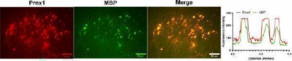
Double IF staining of Prox1 (red) and MBP (green) with IF (×200) and their intensity profile obtained using ImageJ software, along an ideal straight line (white).
Index in PubMed under a CC BY license. PMID: 41078399

Click image to see more details
(A) IHC images of Prox1 proteins in sciatic nerve tissue of control sciatic nerves and injured sciatic nerves on Day 3, 4, 5, 6, 7 and 14 post-injuries (×200) and the quantification of Prox1 proteins in top right panel (P value was normalized to control group, **** P < 0.0001, n = 5, One-Way ANOVA analysis was used). (B) Western blot and quantitative analysis of Prox1 protein expression in sciatic nerve tissue from control nerves and injured nerves at days 3, 4, 5, 6, 7, and 14 post-injury (**** P < 0.0001, n = 3, One-Way ANOVA analysis was used).
Index in PubMed under a CC BY license. PMID: 41078399

Click image to see more details
MBP protein level was significantly increased by PROX1 overexpression. (A) qPCR quantification of PROX1 overexpression, (B) WB images of MBP and GAPDH protein from cells transfected with NC, pcDNA-vector or pcDNA-prox1, (C) the quantification of (B). (**** P < 0.0001, n = 3, One-Way ANOVA analysis was used).
Index in PubMed under a CC BY license. PMID: 41078399

Click image to see more details
Western blot analysis of PROX1 using anti-PROX1 antibody (A01985-1).
Electrophoresis was performed on a 10% SDS-PAGE gel at 80V (Stacking gel) / 120V (Resolving gel) for 2 hours. The sample well of each lane was loaded with 30 ug of sample under reducing conditions.
Lane 1: human HepG2 whole cell lysates,
Lane 2: human HUH7 whole cell lysates,
Lane 3: human SH-SY5Y whole cell lysates,
Lane 4: rat RH35 whole cell lysates.
After electrophoresis, proteins were transferred to a nitrocellulose membrane at 150 mA for 50-90 minutes. Blocked the membrane with 5% non-fat milk/TBS for 1.5 hour at RT. The membrane was incubated with rabbit anti-PROX1 antigen affinity purified polyclonal antibody (A01985-1) at 0.5 μg/mL overnight at 4°C, then washed with TBS-0.1%Tween 3 times with 5 minutes each and probed with a goat anti-rabbit IgG-HRP secondary antibody (Catalog # BA1054) at a dilution of 1:5000 for 1.5 hour at RT. The signal is developed using an ECL Plus Western Blotting Substrate (Catalog # AR1196-200) with Tanon 5200 system. A specific band was detected for PROX1 at approximately 90 kDa. The expected band size for PROX1 is at 83 kDa.

Click image to see more details
IF analysis of PROX1 using anti-SGT1/ECD antibody (A01985-1) and anti-Tubulin Alpha antibody (M03989-3).
PROX1 was detected in immunocytochemical section of SiHa cell. Enzyme antigen retrieval was performed using IHC enzyme antigen retrieval reagent (AR0022) for 15 mins. The cells were blocked with 10% goat serum. And then incubated with 5 μg/mL rabbit anti-PROX1 Antibody (A01985-1) and mouse anti-Tubulin Alpha antibody (M03989-3) overnight at 4°C. DyLight®488 Conjugated Goat Anti-Rabbit IgG (BA1127) and DyLight®594 Conjugated Goat Anti-Mouse IgG (BA1141) were used as secondary antibody at 1:500 dilution and incubated for 30 minutes at 37°C. The section was counterstained with DAPI. Visualize using a fluorescence microscope and filter sets appropriate for the label used.

Click image to see more details
Immunoprecipitating (IP) PROX1 in SH-SY5Y whole cell lysate.
Western blot analysis of PROX1 using anti-PROX1 antibody (A01985-1);
Lane 1: SH-SY5Y whole cell lysates (30ug);
Lane 2: Rabbit control IgG instead of anti-PROX1 antibody in SH-SY5Y whole cell lysate;
Lane 3: anti-PROX1 antibody (2μg) + SH-SY5Y whole cell lysate (500μg).
After electrophoresis, proteins were transferred to a membrane. Then the membrane was incubated with rabbit anti-PROX1 antigen affinity purified polyclonal antibody (A01985-1) at a dilution of 0.5 μg/mL and probed with a goat anti-rabbit IgG-HRP secondary antibody (Catalog # BA1054). The signal is developed using ECL Plus Western Blotting Substrate (Catalog # AR1196-200). A specific band was detected for PROX1 at approximately 90 kDa. The expected band size for PROX1 is at 83 kDa.

Click image to see more details
Flow Cytometry analysis of SH-SY5Y cells using anti-PROX1 antibody (A01985-1).
Overlay histogram showing SH-SY5Y cells stained with A01985-1 (Blue line). To facilitate intracellular staining, cells were fixed with 4% paraformaldehyde and permeabilized with permeabilization buffer. The cells were blocked with 10% normal goat serum. And then incubated with rabbit anti-PROX1 Antibody (A01985-1, 1 μg/1x106 cells) for 30 min at 20°C. DyLight®488 conjugated goat anti-rabbit IgG (BA1127, 5-10 μg/1x106 cells) was used as secondary antibody for 30 minutes at 20°C. Isotype control antibody (Green line) was rabbit IgG (1 μg/1x106) used under the same conditions. Unlabelled sample without incubation with primary antibody and secondary antibody (Red line) was used as a blank control.
Specific Publications For Anti-PROX1 Antibody Picoband® (A01985-1)
Loading publications
Recommended Resources
Here are featured tools and databases that you might find useful.
- Boster's Pathways Library
- Protein Databases
- Bioscience Research Protocol Resources
- Data Processing & Analysis Software
- Photo Editing Software
- Scientific Literature Resources
- Research Paper Management Tools
- Molecular Biology Software
- Primer Design Tools
- Bioinformatics Tools
- Phylogenetic Tree Analysis
Customer Reviews
Have you used Anti-PROX1 Antibody Picoband®?
Share your experimental results or join a short interview to earn up to $1,000 in product credits or other rewards.
0 Reviews For Anti-PROX1 Antibody Picoband®
Customer Q&As
Have a question?
Find answers in Q&As, reviews.
Can't find your answer?
Submit your question
