Click image to see more details
-

-
-
-
-
+1
Product Info Summary
| SKU: | PB10089 |
|---|---|
| Size: | 100 μg/vial |
| Reactive Species: | Human, Mouse, Rat |
| Host: | Rabbit |
| Application: | Flow Cytometry, IHC, IHC-F, ICC, WB |
Customers Who Bought This Also Bought

Product info
Product Name
Anti-Proteasome 20S alpha 3/PSMA3 Antibody Picoband®
SKU/Catalog Number
PB10089
Size
100 μg/vial
Form
Lyophilized
Description
Boster Bio Anti-Proteasome 20S alpha 3/PSMA3 Antibody Picoband® catalog # PB10089. Tested in Flow Cytometry, IHC, IHC-F, ICC, WB applications. This antibody reacts with Human, Mouse, Rat. The brand Picoband indicates this is a premium antibody that guarantees superior quality, high affinity, and strong signals with minimal background in Western blot applications. Only our best-performing antibodies are designated as Picoband, ensuring unmatched performance.
Storage & Handling
Store at -20˚C for one year from date of receipt. After reconstitution, at 4˚C for one month. It can also be aliquotted and stored frozen at -20˚C for six months. Avoid repeated freeze-thaw cycles.
Cite This Product
Anti-Proteasome 20S alpha 3/PSMA3 Antibody Picoband® (Boster Biological Technology, Pleasanton CA, USA, Catalog # PB10089)
Host
Rabbit
Contents
Each vial contains antibody formulated with stabilizing components, 0.9 mg NaCl, 0.2 mg Na2HPO4, and 0.05 mg NaN3.
*This antibody is supplied in a stabilized formulation.
Compatibility with conjugation reactions depends on the chemistry of the conjugation method used.
For conjugation methods that are not compatible with the stabilizing components present in this formulation, a carrier-free antibody format is required.
Clonality
Polyclonal
Isotype
Rabbit IgG
Immunogen
A synthetic peptide corresponding to a sequence in the middle region of human PSMA3, identical to the related mouse and rat sequences.
Cross-reactivity
No cross-reactivity with other proteins
Reactive Species
PB10089 is reactive to PSMA3 in Human, Mouse, Rat
Observed Molecular Weight
28 kDa
Calculated molecular weight
28.4 kDa
Background of PSMA3
Proteasome subunit alpha type-3, also known as macropain subunit C8 and proteasome component C8, is a protein that in humans is encoded by the PSMA3 gene. The proteasome is a multicatalytic proteinase complex with a highly ordered ring-shaped 20S core structure. The core structure is composed of 4 rings of 28 non-identical subunits; 2 rings are composed of 7 alpha subunits and 2 rings are composed of 7 beta subunits. Proteasomes are distributed throughout eukaryotic cells at a high concentration and cleave peptides in an ATP/ubiquitin-dependent process in a non-lysosomal pathway. An essential function of a modified proteasome, the immunoproteasome, is the processing of class I MHC peptides. This gene encodes a member of the peptidase T1A family, that is a 20S core alpha subunit. Two alternative transcripts encoding different isoforms have been identified.
Antibody Validation
Boster validates all antibodies on WB, IHC, ICC, Immunofluorescence, and ELISA with known positive control and negative samples to ensure specificity and high affinity, including thorough antibody incubations.
Application & Images
Applications
PB10089 is guaranteed for Flow Cytometry, IHC, IHC-F, ICC, WB Boster Guarantee
Recommend Dilution
| Application | Dilution | Species |
|---|---|---|
| Immunohistochemistry (Paraffin-embedded Section) | 0.5-1μg/ml | Human, Mouse, Rat |
| Western blot | 0.1-0.5μg/ml | Human, Mouse, Rat |
| Immunohistochemistry (Frozen Section) | 0.5-1μg/ml | Human |
| Immunocytochemistry | 0.5-1μg/ml | Human |
| Flow Cytometry (Fixed) | 1-3μg/1x106 cells Human |
Tested application
Suggested blocking solution with 5% non-fat milk or BSA; (*)Recommended protein loading: 20-40 µg per lane
Use TE buffer pH 9.0 for antigen retrieval; (*) citrate buffer pH 6.0 is an alternative.
Validation Images & Assay Conditions
Click image to see more details
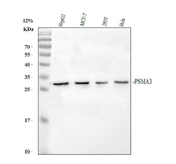
Western blot analysis of PSMA3 using anti-PSMA3 antibody (PB10089).
Electrophoresis was performed on a 12% SDS-PAGE gel at 80V (Stacking gel) / 120V (Resolving gel) for 2 hours. The sample well of each lane was loaded with 30 ug of sample under reducing conditions.
Lane 1: human HepG2 whole cell lysates,
Lane 2: human MCF-7 whole cell lysates,
Lane 3: human 293T whole cell lysates,
Lane 4: human Hela whole cell lysates.
After electrophoresis, proteins were transferred to a nitrocellulose membrane at 150 mA for 50-90 minutes. Blocked the membrane with 5% non-fat milk/TBS for 1.5 hour at RT. The membrane was incubated with rabbit anti-PSMA3 antigen affinity purified polyclonal antibody (PB10089) at 0.5 μg/mL overnight at 4°C, then washed with TBS-0.1%Tween 3 times with 5 minutes each and probed with a goat anti-rabbit IgG-HRP secondary antibody (Catalog # BA1054) at a dilution of 1:5000 for 1.5 hour at RT. The signal is developed using an ECL Plus Western Blotting Substrate (Catalog # AR1196-200) with Tanon 5200 system. A specific band was detected for PSMA3 at approximately 28 kDa. The expected band size for PSMA3 is at 28 kDa.

Click image to see more details
IHC analysis of PSMA3 using anti-PSMA3 antibody (PB10089).
PSMA3 was detected in paraffin-embedded section of mouse brain tissues. Heat mediated antigen retrieval was performed in citrate buffer (pH6, epitope retrieval solution) for 20 mins. The tissue section was blocked with 10% goat serum. The tissue section was then incubated with 1μg/ml rabbit anti-PSMA3 Antibody (PB10089) overnight at 4°C. Biotinylated goat anti-rabbit IgG was used as secondary antibody and incubated for 30 minutes at 37°C. The tissue section was developed using Strepavidin-Biotin-Complex (SABC)(Catalog # SA1022) with DAB as the chromogen.

Click image to see more details
IHC analysis of PSMA3 using anti-PSMA3 antibody (PB10089).
PSMA3 was detected in paraffin-embedded section of rat brain tissues. Heat mediated antigen retrieval was performed in citrate buffer (pH6, epitope retrieval solution) for 20 mins. The tissue section was blocked with 10% goat serum. The tissue section was then incubated with 1μg/ml rabbit anti-PSMA3 Antibody (PB10089) overnight at 4°C. Biotinylated goat anti-rabbit IgG was used as secondary antibody and incubated for 30 minutes at 37°C. The tissue section was developed using Strepavidin-Biotin-Complex (SABC)(Catalog # SA1022) with DAB as the chromogen.

Click image to see more details
IHC analysis of PSMA3 using anti-PSMA3 antibody (PB10089).
PSMA3 was detected in paraffin-embedded section of human intestinal cancer tissues. Heat mediated antigen retrieval was performed in citrate buffer (pH6, epitope retrieval solution) for 20 mins. The tissue section was blocked with 10% goat serum. The tissue section was then incubated with 1μg/ml rabbit anti-PSMA3 Antibody (PB10089) overnight at 4°C. Biotinylated goat anti-rabbit IgG was used as secondary antibody and incubated for 30 minutes at 37°C. The tissue section was developed using Strepavidin-Biotin-Complex (SABC)(Catalog # SA1022) with DAB as the chromogen.

Click image to see more details
Flow Cytometry analysis of HeLa cells using anti-PSMA3 antibody (PB10089).
Overlay histogram showing HeLa cells stained with PB10089 (Blue line). To facilitate intracellular staining, cells were fixed with 4% paraformaldehyde and permeabilized with permeabilization buffer. The cells were blocked with 10% normal goat serum. And then incubated with rabbit anti-PSMA3 Antibody (PB10089,1μg/1x106 cells) for 30 min at 20°C. DyLight®488 conjugated goat anti-rabbit IgG (BA1127, 5-10μg/1x106 cells) was used as secondary antibody for 30 minutes at 20°C. Isotype control antibody (Green line) was rabbit IgG (1μg/1x106) used under the same conditions. Unlabelled sample without incubation with primary antibody and secondary antibody (Red line) was used as a blank control.
Specific Publications For Anti-Proteasome 20S alpha 3/PSMA3 Antibody Picoband® (PB10089)
Loading publications
Recommended Resources
Here are featured tools and databases that you might find useful.
- Boster's Pathways Library
- Protein Databases
- Bioscience Research Protocol Resources
- Data Processing & Analysis Software
- Photo Editing Software
- Scientific Literature Resources
- Research Paper Management Tools
- Molecular Biology Software
- Primer Design Tools
- Bioinformatics Tools
- Phylogenetic Tree Analysis
Customer Reviews
Have you used Anti-Proteasome 20S alpha 3/PSMA3 Antibody Picoband®?
Share your experimental results or join a short interview to earn up to $1,000 in product credits or other rewards.
0 Reviews For Anti-Proteasome 20S alpha 3/PSMA3 Antibody Picoband®
Customer Q&As
Have a question?
Find answers in Q&As, reviews.
Can't find your answer?
Submit your question
4 Customer Q&As for Anti-Proteasome 20S alpha 3/PSMA3 Antibody Picoband®
Question
We appreciate helping with my inquiry over the phone. Here are the WB image, lot number and protocol we used for bone marrow using anti-Proteasome 20S alpha 3/PSMA3 antibody PB10089. Let me know if you need anything else.
Verified Customer
Verified customer
Asked: 2019-10-23
Answer
Thanks for the data. You have provided everything we needed. Our lab team are working to resolve your inquiry as quickly as possible, and we appreciate your patience and understanding! Please let me know if there is anything you need in the meantime.
Boster Scientific Support
Answered: 2019-10-23
Question
We are currently using anti-Proteasome 20S alpha 3/PSMA3 antibody PB10089 for mouse tissue, and we are content with the IHC-F results. The species of reactivity given in the datasheet says human, mouse, rat. Is it true that the antibody can work on feline tissues as well?
Verified Customer
Verified customer
Asked: 2018-01-19
Answer
The anti-Proteasome 20S alpha 3/PSMA3 antibody (PB10089) has not been validated for cross reactivity specifically with feline tissues, though there is a good chance of cross reactivity. We have an innovator award program that if you test this antibody and show it works in feline you can get your next antibody for free. Please contact me if I can help you with anything.
Boster Scientific Support
Answered: 2018-01-19
Question
Is there a BSA free version of anti-Proteasome 20S alpha 3/PSMA3 antibody PB10089 available?
Verified Customer
Verified customer
Asked: 2017-10-20
Answer
We appreciate your recent telephone inquiry. I can confirm that some lots of this anti-Proteasome 20S alpha 3/PSMA3 antibody PB10089 are BSA free. For now, these lots are available and we can make a BSA free formula for you free of charge. It will take 3 extra days to prepare. If you require this antibody BSA free again in future, please do not hesitate to contact me and I will be pleased to check which lots we have in stock that are BSA free.
Boster Scientific Support
Answered: 2017-10-20
Question
I have attached the WB image, lot number and protocol we used for bone marrow using anti-Proteasome 20S alpha 3/PSMA3 antibody PB10089. Please let me know if you require anything else.
R. Williams
Verified customer
Asked: 2014-08-28
Answer
Thank you very much for the data. Our lab team are working to resolve this as quickly as possible, and we appreciate your patience and understanding! You have provided everything we needed. Please let me know if there is anything you need in the meantime.
Boster Scientific Support
Answered: 2014-08-28
