
Click image to see more details
-

-
-
-
-
+2
Product Info Summary
| SKU: | M00207-1 |
|---|---|
| Size: | 100 μl |
| Reactive Species: | Human, Mouse, Rat |
| Host: | Rabbit |
| Application: | Flow Cytometry, IF, IHC, ICC, WB |
Customers Who Bought This Also Bought

Product info
Product Name
Anti-Rho A + B + C Rabbit Monoclonal Antibody
SKU/Catalog Number
M00207-1
BM4336 is an alternative SKU for this antibody, used in previous lots.
Size
100 μl
Form
Liquid
Description
Boster Bio Anti-Rho A + B + C Rabbit Monoclonal Antibody catalog # M00207-1. Tested in WB, IHC, ICC/IF, Flow Cytometry applications. This antibody reacts with Human, Mouse, Rat.
Storage & Handling
Store at -20°C for one year. For short term storage and frequent use, store at 4°C for up to one month. Avoid repeated freeze-thaw cycles.
Cite This Product
Anti-Rho A + B + C Rabbit Monoclonal Antibody (Boster Biological Technology, Pleasanton CA, USA, Catalog # M00207-1)
Host
Rabbit
Contents
Rabbit IgG in stabilizing components, phosphate buffered saline, pH 7.4, 150mM NaCl, 0.02% sodium azide and 50% glycerol.
*This antibody is supplied in a stabilized formulation.
Compatibility with conjugation reactions depends on the chemistry of the conjugation method used.
For conjugation methods that are not compatible with the stabilizing components present in this formulation, a carrier-free antibody format is required.
Clonality
Monoclonal
Clone Number
DGF-18
Isotype
Rabbit IgG
Immunogen
A synthesized peptide derived from human Rho A + B + C
Reactive Species
M00207-1 is reactive to RHOA in Human, Mouse, Rat
Observed Molecular Weight
22 kDa
Antibody Validation
Boster validates all antibodies on WB, IHC, ICC, Immunofluorescence, and ELISA with known positive control and negative samples to ensure specificity and high affinity, including thorough antibody incubations.
Application & Images
Applications
M00207-1 is guaranteed for Flow Cytometry, IF, IHC, ICC, WB Boster Guarantee
Assay Dilutions Recommendation
The recommendations below provide a starting point for assay optimization. The actual working concentration varies and should be decided by the user.
WB 1:500-2000
IHC 1:50-200
ICC/IF 1:50-200
FC 1:50
Positive Control
WB: human HUVEC whole cell, human THP-1 whole cell, human Hela whole cell, human U251 whole cell, rat brain tissue, rat lung tissue, mouse brain tissue, mouse lung tissue, human 293T whole cell, human SH-SY5Y whole cell, human C6 whole cell, human neuro-2α whole cell
IHC: human lung carcinoma tissue
ICC/IF: Hela cell
Validation Images & Assay Conditions
Click image to see more details
Western blot analysis of Rho A+B+C using anti-Rho A+B+C antibody (M00207-1).
Electrophoresis was performed on a 12% SDS-PAGE gel at 70V (Stacking gel) / 90V (Resolving gel) for 2-3 hours. The sample well of each lane was loaded with 30 ug of sample under reducing conditions.
Lane 1: human HUVEC whole cell lysates,
Lane 2: human THP-1 whole cell lysates,
Lane 3: human Hela whole cell lysates,
Lane 4: human U251 whole cell lysates,
Lane 5: rat brain tissue lysates,
Lane 6: rat lung tissue lysates,
Lane 7: mouse brain tissue lysates,
Lane 8: mouse lung tissue lysates.
After electrophoresis, proteins were transferred to a nitrocellulose membrane at 150 mA for 50-90 minutes. Blocked the membrane with 5% non-fat milk/TBS for 1.5 hour at RT. The membrane was incubated with rabbit anti-Rho A+B+C antigen affinity purified monoclonal antibody (M00207-1) at 1:500 overnight at 4°C, then washed with TBS-0.1%Tween 3 times with 5 minutes each and probed with a goat anti-rabbit IgG-HRP secondary antibody at a dilution of 1:500 for 1.5 hour at RT. The signal is developed using an Enhanced Chemiluminescent detection (ECL) kit (Catalog # AR1196-200) with Tanon 5200 system. A specific band was detected for Rho A+B+C at approximately 22 kDa. The expected band size for Rho A+B+C is at 22 kDa.

Click image to see more details
Western blot analysis of Rho A+B+C using anti-Rho A+B+C antibody (M00207-1).
Electrophoresis was performed on a 12% SDS-PAGE gel at 70V (Stacking gel) / 90V (Resolving gel) for 2-3 hours. The sample well of each lane was loaded with 30 ug of sample under reducing conditions.
Lane 1: human 293T whole cell lysates,
Lane 2: human Hela whole cell lysates,
Lane 3: human SH-SY5Y whole cell lysates,
Lane 4: human U251 whole cell lysates,
Lane 5: rat brain tissue lysates,
Lane 6: rat C6 whole cell,
Lane 7: mouse brain tissue lysates,
Lane 8: mouse neuro-2a whole cell.
After electrophoresis, proteins were transferred to a nitrocellulose membrane at 150 mA for 50-90 minutes. Blocked the membrane with 5% non-fat milk/TBS for 1.5 hour at RT. The membrane was incubated with rabbit anti-Rho A+B+C antigen affinity purified monoclonal antibody (Catalog # M00207-1) at 1:500 overnight at 4°C, then washed with TBS-0.1%Tween 3 times with 5 minutes each and probed with a goat anti-rabbit IgG-HRP secondary antibody at a dilution of 1:500 for 1.5 hour at RT. The signal is developed using an Enhanced Chemiluminescent detection (ECL) kit (Catalog # AR1196-200) with Tanon 5200 system. A specific band was detected for Rho A+B+C at approximately 22 kDa. The expected band size for Rho A+B+C is at 22 kDa.

Click image to see more details
Immunofluorescent analysis of Hela cells, using Rho A + B + C Antibody .

Click image to see more details
Western blot analysis of Rho A+B+C using anti-Rho A+B+C antibody (M00207-1).
Electrophoresis was performed on a 12% SDS-PAGE gel at 70V (Stacking gel) / 90V (Resolving gel) for 2-3 hours. The sample well of each lane was loaded with 30 ug of sample under reducing conditions.
Lane 1: human 293T whole cell lysates,
Lane 2: human 293T cells transfected with RHOP whole cell lysayes,
Lane 3: human 293T cells transfected with RHOP whole cell lysayes.
After electrophoresis, proteins were transferred to a nitrocellulose membrane at 150 mA for 50-90 minutes. Blocked the membrane with 5% non-fat milk/TBS for 1.5 hour at RT. The membrane was incubated with rabbit anti-Rho A+B+C antigen affinity purified monoclonal antibody (M00207-1) at 1:2500 overnight at 4°C, then washed with TBS-0.1%Tween 3 times with 5 minutes each and probed with a goat anti-rabbit IgG-HRP secondary antibody at a dilution of 1:10000 for 1 hour at RT. The signal is developed using an Enhanced Chemiluminescent detection (ECL) kit (Catalog # AR1196-200) with ChemiDoc MP system. A specific band was detected for Rho A+B+C at approximately 22 kDa. The expected band size for Rho A+B+C is at 22 kDa.

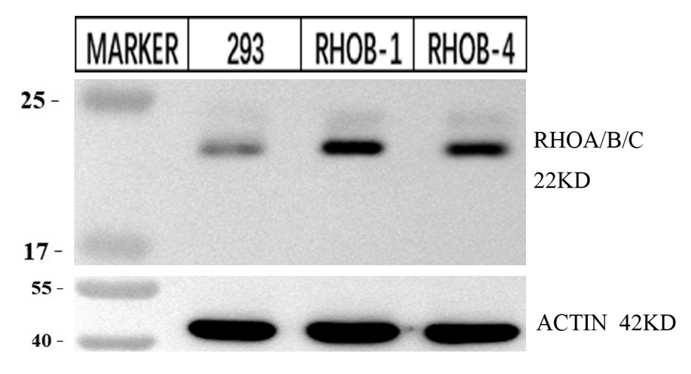
Click image to see more details
Western blot analysis of Rho A+B+C using anti-Rho A+B+C antibody (M00207-1).
Electrophoresis was performed on a 12% SDS-PAGE gel at 70V (Stacking gel) / 90V (Resolving gel) for 2-3 hours. The sample well of each lane was loaded with 30 ug of sample under reducing conditions.
Lane 1: Normal group-rat colon tissue lysates,
Lane 2: Model group-rat colon tissue lysates,
Lane 3: Triditional Chinese medicine treatment (low dose)-rat colon tissue lysates,
Lane 4: Triditional Chinese medicine treatment (medium dose)-rat colon tissue lysates,
Lane 5: Triditional Chinese medicine treatment(high dose)-rat colon tissue lysates,
Lane 6: Western medicine treatment-rat colon tissue lysates.
After electrophoresis, proteins were transferred to a nitrocellulose membrane at 150 mA for 50-90 minutes. Blocked the membrane with 5% non-fat milk/TBS for 1.5 hour at RT. The membrane was incubated with rabbit anti-Rho A+B+C antigen affinity purified monoclonal antibody (Catalog # M00207-1) at 1:1000 overnight at 4°C, then washed with TBS-0.1%Tween 3 times with 5 minutes each and probed with a HRP Conjugated AffiniPure Goat Anti-rabbit IgG (H+L) antibody at a dilution of 1:5000 for 1 hour at RT. The signal is developed using an Enhanced Chemiluminescent detection (ECL) kit (Catalog # AR1196-200) with ChemiDoc MP system. A specific band was detected for Rho A+B+C at approximately 22 kDa. The expected band size for Rho A+B+C is at 22 kDa.

Click image to see more details
Immunohistochemical analysis of paraffin-embedded human lung carcinoma, using Rho A + B + C Antibody .
Specific Publications For Anti-Rho A + B + C Rabbit Monoclonal Antibody (M00207-1)
Loading publications
Recommended Resources
Here are featured tools and databases that you might find useful.
- Boster's Pathways Library
- Protein Databases
- Bioscience Research Protocol Resources
- Data Processing & Analysis Software
- Photo Editing Software
- Scientific Literature Resources
- Research Paper Management Tools
- Molecular Biology Software
- Primer Design Tools
- Bioinformatics Tools
- Phylogenetic Tree Analysis
Customer Reviews
Have you used Anti-Rho A + B + C Rabbit Monoclonal Antibody?
Share your experimental results or join a short interview to earn up to $1,000 in product credits or other rewards.
2 Reviews For Anti-Rho A + B + C Rabbit Monoclonal Antibody
This antibody is highly specific and efficient, suitable for detecting RHOA/B/C protein in rat colon by Western blot, with only minimal non-specific bands.
Excellent

| SKU | M00207-1 |
|---|---|
| Application | Western Blot |
| Sample | rat colon tissue |
| Sample Processing Description | RIPA lysis buffer with protease inhibitor PMSF (100:1) was used to lyse the sample for 10 minutes, followed by centrifugation at 12,000 rpm for 15 minutes. The supernatant was mixed with 5× loading buffer, denatured at 100°C for 10 minutes, and then loaded onto SDS-PAGE. |
| Other Reagents | Blocking buffer |
| Primary Antibody | Rho A + B + C Rabbit Monoclonal Antibody |
| Primary Incubation | 1:1000, overnight at 4 ℃ |
| Secondary Antibody | HRP Conjugated AffiniPure Goat Anti-Rabbit IgG (H+L) |
| Secondary Incubation | 1:5000, 1 hour in room temperature |
| Detection | Substrate: ECL, Imaging system:ChemiDoc MP |
| Results Summary | The figure shows the Western blot results of the target protein RHOA/B/C and the internal control Actin in rat colon tissue across the following groups: normal, disease model, low/middle/high dose traditional Chinese medicine treatment, and Western medicine treatment. The target bands are clear and distinct, and the experimental results are satisfactory. |
Shiyu Zhang, LUTCM
Verified customer
Submitted 2026-01-06
The target band of this antibody is clear and at the correct position, with minimal nonspecific bands, and the results are good.
Excellent
| SKU | M00207-1 |
|---|---|
| Application | Western Blot |
| Sample | huaman 293 cells |
| Sample Processing Description | RHOB detection was performed using total protein extracted from normal 293 cells and 293 cells with RHOB overexpression. |
| Other Reagents | RIPA lysis buffer, Protease inhibitor, Electrophoresis buffer, Transfer buffer, Blocking buffer |
| Primary Antibody | ATF4 Rabbit Monoclonal Antibody |
| Primary Incubation | 1:2500, overnight at 4 ℃ |
| Secondary Antibody | HRP Goat Anti-Rabbit IgG |
| Secondary Incubation | 1:10000, 1 hour in room temperature |
| Detection | Substrate: ECL, Imaging system:ChemiDoc MP |
| Results Summary | We generated a stable RHOB overexpression cell line using 293 cells. The results show that the RHOB expression level in the transfected samples is significantly higher than in the normal 293 cells, indicating successful transfection. |
Jie Zhang, Zhejiang A&F University
Verified customer
Submitted 2025-12-04
Customer Q&As
Have a question?
Find answers in Q&As, reviews.
Can't find your answer?
Submit your question
