Click image to see more details
Product Info Summary
| SKU: | A06793-1 |
|---|---|
| Size: | 100 μg/vial |
| Reactive Species: | Human, Rat |
| Host: | Rabbit |
| Application: | ELISA, IHC, WB |
Customers Who Bought This Also Bought

Product info
Product Name
Anti-RPL8 Antibody Picoband®
SKU/Catalog Number
A06793-1
Size
100 μg/vial
Form
Lyophilized
Description
Boster Bio Anti-RPL8 Antibody Picoband® catalog # A06793-1. Tested in ELISA, IHC, WB applications. This antibody reacts with Human, Rat. The brand Picoband indicates this is a premium antibody that guarantees superior quality, high affinity, and strong signals with minimal background in Western blot applications. Only our best-performing antibodies are designated as Picoband, ensuring unmatched performance.
Storage & Handling
At -20°C for one year from date of receipt. After reconstitution, at 4°C for one month. It can also be aliquotted and stored frozen at -20°C for six months. Avoid repeated freezing and thawing.
Cite This Product
Anti-RPL8 Antibody Picoband® (Boster Biological Technology, Pleasanton CA, USA, Catalog # A06793-1)
Host
Rabbit
Contents
Each vial contains 4 mg Trehalose, 0.9 mg NaCl, 0.2 mg Na2HPO4.
Clonality
Polyclonal
Isotype
Rabbit IgG
Immunogen
E.coli-derived human RPL8 recombinant protein (Position: R3-N257).
Cross-reactivity
No cross-reactivity with other proteins.
Reactive Species
A06793-1 is reactive to RPL8 in Human, Rat
Observed Molecular Weight
34 kDa
Calculated molecular weight
28.0 kDa
Background of RPL8
60S ribosomal protein L8 is a protein that in humans is encoded by the RPL8 gene. Ribosomes, the organelles that catalyze protein synthesis, consist of a small 40S subunit and a large 60S subunit. Together these subunits are composed of 4 RNA species and approximately 80 structurally distinct proteins. This gene encodes a ribosomal protein that is a component of the 60S subunit. The protein belongs to the L2P family of ribosomal proteins. It is located in the cytoplasm. In rat, the protein associates with the 5.8S rRNA, very likely participates in the binding of aminoacyl-tRNA, and is a constituent of the elongation factor 2-binding site at the ribosomal subunit interface. Alternatively spliced transcript variants encoding the same protein exist. As is typical for genes encoding ribosomal proteins, there are multiple processed pseudogenes of this gene dispersed through the genome.
Antibody Validation
Boster validates all antibodies on WB, IHC, ICC, Immunofluorescence, and ELISA with known positive control and negative samples to ensure specificity and high affinity, including thorough antibody incubations.
Application & Images
Applications
A06793-1 is guaranteed for ELISA, IHC, WB Boster Guarantee
Assay Dilutions Recommendation
The recommendations below provide a starting point for assay optimization. The actual working concentration varies and should be decided by the user.
Western blot, 0.25-0.5 μg/ml, Human, Rat
Immunohistochemistry(Paraffin-embedded Section), 2-5 μg/ml, Human
ELISA, 0.1-0.5 μg/ml, -
Positive Control
WB: human HepG2 whole cell, human Jurkat whole cell, human MCF-7 whole cell, human PC-3 whole cell, rat brain tissue, rat testis tissue
IHC: human laryngeal squamous cell carcinoma tissue, human liver cancer tissue, human pancreatic cancer tissue
Validation Images & Assay Conditions
Click image to see more details
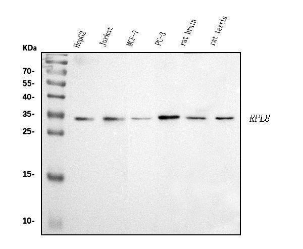
Western blot analysis of RPL8 using anti-RPL8 antibody (A06793-1).
Electrophoresis was performed on a 5-20% SDS-PAGE gel at 70V (Stacking gel) / 90V (Resolving gel) for 2-3 hours. The sample well of each lane was loaded with 30 ug of sample under reducing conditions.
Lane 1: human HepG2 whole cell lysates,
Lane 2: human Jurkat whole cell lysates,
Lane 3: human MCF-7 whole cell lysates,
Lane 4: human PC-3 whole cell lysates,
Lane 5: rat brain tissue lysates,
Lane 6: rat testis tissue lysates.
After electrophoresis, proteins were transferred to a nitrocellulose membrane at 150 mA for 50-90 minutes. Blocked the membrane with 5% non-fat milk/TBS for 1.5 hour at RT. The membrane was incubated with rabbit anti-RPL8 antigen affinity purified polyclonal antibody (Catalog # A06793-1) at 0.5 μg/mL overnight at 4°C, then washed with TBS-0.1%Tween 3 times with 5 minutes each and probed with a goat anti-rabbit IgG-HRP secondary antibody at a dilution of 1:5000 for 1.5 hour at RT. The signal is developed using an Enhanced Chemiluminescent detection (ECL) kit (Catalog # EK1002) with Tanon 5200 system. A specific band was detected for RPL8 at approximately 34 kDa. The expected band size for RPL8 is at 34 kDa.

Click image to see more details
IHC analysis of RPL8 using anti-RPL8 antibody (A06793-1).
RPL8 was detected in a paraffin-embedded section of human laryngeal squamous cell carcinoma tissue. Heat mediated antigen retrieval was performed in EDTA buffer (pH 8.0, epitope retrieval solution). The tissue section was blocked with 10% goat serum. The tissue section was then incubated with 2 μg/ml rabbit anti-RPL8 Antibody (A06793-1) overnight at 4°C. Biotinylated goat anti-rabbit IgG was used as secondary antibody and incubated for 30 minutes at 37°C. The tissue section was developed using Strepavidin-Biotin-Complex (SABC) (Catalog # SA1022) with DAB as the chromogen.

Click image to see more details
IHC analysis of RPL8 using anti-RPL8 antibody (A06793-1).
RPL8 was detected in a paraffin-embedded section of human liver cancer tissue. Heat mediated antigen retrieval was performed in EDTA buffer (pH 8.0, epitope retrieval solution). The tissue section was blocked with 10% goat serum. The tissue section was then incubated with 2 μg/ml rabbit anti-RPL8 Antibody (A06793-1) overnight at 4°C. Biotinylated goat anti-rabbit IgG was used as secondary antibody and incubated for 30 minutes at 37°C. The tissue section was developed using Strepavidin-Biotin-Complex (SABC) (Catalog # SA1022) with DAB as the chromogen.

Click image to see more details
IHC analysis of RPL8 using anti-RPL8 antibody (A06793-1).
RPL8 was detected in a paraffin-embedded section of human pancreatic cancer tissue. Heat mediated antigen retrieval was performed in EDTA buffer (pH 8.0, epitope retrieval solution). The tissue section was blocked with 10% goat serum. The tissue section was then incubated with 2 μg/ml rabbit anti-RPL8 Antibody (A06793-1) overnight at 4°C. Biotinylated goat anti-rabbit IgG was used as secondary antibody and incubated for 30 minutes at 37°C. The tissue section was developed using Strepavidin-Biotin-Complex (SABC) (Catalog # SA1022) with DAB as the chromogen.
Specific Publications For Anti-RPL8 Antibody Picoband® (A06793-1)
Loading publications
Recommended Resources
Here are featured tools and databases that you might find useful.
- Boster's Pathways Library
- Protein Databases
- Bioscience Research Protocol Resources
- Data Processing & Analysis Software
- Photo Editing Software
- Scientific Literature Resources
- Research Paper Management Tools
- Molecular Biology Software
- Primer Design Tools
- Bioinformatics Tools
- Phylogenetic Tree Analysis
Customer Reviews
Have you used Anti-RPL8 Antibody Picoband®?
Share your experimental results or join a short interview to earn up to $1,000 in product credits or other rewards.
0 Reviews For Anti-RPL8 Antibody Picoband®
Customer Q&As
Have a question?
Find answers in Q&As, reviews.
Can't find your answer?
Submit your question
