Click image to see more details
Product Info Summary
| SKU: | M01567-2 |
|---|---|
| Size: | 100 μl/vial |
| Reactive Species: | Human, Mouse, Rat |
| Host: | Rabbit |
| Application: | IF, IHC, ICC, WB |
Customers Who Bought This Also Bought

Product info
Product Name
Anti-RPS6 (Phospho-S235 + S236) Antibody (Monoclonal, 32R03)
SKU/Catalog Number
M01567-2
Size
100 μl/vial
Form
Liquid
Description
Boster Bio Anti-RPS6 (Phospho-S235 + S236) Antibody (Monoclonal, 32R03) catalog # M01567-2. Tested in WB, IHC, ICC/IF applications. This antibody reacts with Human, Mouse, Rat.
Storage & Handling
Store at -20°C for one year. For short term storage and frequent use, store at 4°C for up to one month. Avoid repeated freeze-thaw cycles.
Cite This Product
Anti-RPS6 (Phospho-S235 + S236) Antibody (Monoclonal, 32R03) (Boster Biological Technology, Pleasanton CA, USA, Catalog # M01567-2)
Host
Rabbit
Contents
Rabbit IgG in stabilizing components, phosphate buffered saline, pH 7.4, 150mM NaCl, 0.02% sodium azide and 50% glycerol.
This antibody is supplied in a stabilized formulation.
Compatibility with conjugation reactions depends on the chemistry of the conjugation method used.
For conjugation methods that are not compatible with the stabilizing components present in this formulation, a carrier-free antibody format is required.
Clonality
Monoclonal
Clone Number
32R03
Immunogen
Synthetic peptide within Human RPS6 aa 200 to the C-terminus (phospho S235 + S236).
Reactive Species
M01567-2 is reactive to RPS6 in Human, Mouse, Rat
Observed Molecular Weight
34 kDa
Calculated molecular weight
28.7 kDa
Background of RPS6
Ribosomal protein S6 (rpS6) is a component of the 40S ribosomal subunit and is therefore thought to be involved in regulating translation. Ribosomes, the organelles that catalyze protein synthesis, consist of a small 40S subunit and a large 60S subunit. Together these subunits are composed of 4 RNA species and approximately 80 structurally distinct proteins. This gene encodes a cytoplasmic ribosomal protein that is a component of the 40S subunit. The protein belongs to the S6E family of ribosomal proteins. It is the major substrate of protein kinases in the ribosome, with subsets of five C-terminal serine residues phosphorylated by different protein kinases. Phosphorylation is induced by a wide range of stimuli, including growth factors, tumor-promoting agents, and mitogens. Dephosphorylation occurs at growth arrest. The protein may contribute to the control of cell growth and proliferation through the selective translation of particular classes of mRNA. As is typical for genes encoding ribosomal proteins, there are multiple processed pseudogenes of this gene dispersed through the genome. While the true function of rpS6 is currently under investigation, studies have shown that it is involved in the regulation of cell size, cell proliferation, and glucose homeostasis.
Antibody Validation
Boster validates all antibodies on WB, IHC, ICC, Immunofluorescence, and ELISA with known positive control and negative samples to ensure specificity and high affinity, including thorough antibody incubations.
Application & Images
Applications
M01567-2 is guaranteed for IF, IHC, ICC, WB Boster Guarantee
Recommend Dilution
| Application | Dilution | Species |
|---|---|---|
| Western blot | 1:500-2000 | |
| Immunohistochemistry | 1:50-200 | |
| Immunocytochemistry/Immunofluorescence | 1:50-200 |
Tested application
Suggested blocking solution with 5% non-fat milk or BSA; (*)Recommended protein loading: 20-40 µg per lane
Use TE buffer pH 9.0 for antigen retrieval; (*) citrate buffer pH 6.0 is an alternative.
Validation Images & Assay Conditions
Click image to see more details
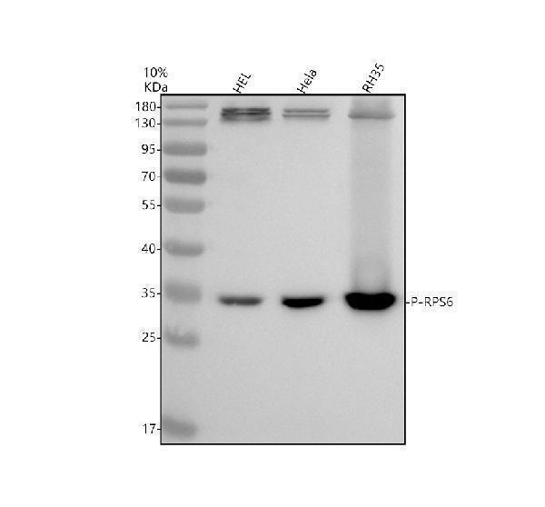
Western blot analysis of P-RPS6 using anti-P-RPS6 antibody (M01567-2).
Electrophoresis was performed on a 10% SDS-PAGE gel at 80V (Stacking gel) / 120V (Resolving gel) for 2 hours. The sample well of each lane was loaded with 30 ug of sample under reducing conditions.
Lane 1: human HEL whole cell lysates,
Lane 2: human Hela whole cell lysates,
Lane 3: rat RH35 whole cell lysates.
After electrophoresis, proteins were transferred to a nitrocellulose membrane at 150 mA for 50-90 minutes. Blocked the membrane with 5% non-fat milk/TBS for 1.5 hour at RT. The membrane was incubated with rabbit anti-P-RPS6 antigen affinity purified monoclonal antibody (M01567-2) at 1:500 overnight at 4°C, then washed with TBS-0.1%Tween 3 times with 5 minutes each and probed with a goat anti-rabbit IgG-HRP secondary antibody at a dilution of 1:5000 for 1.5 hour at RT. The signal is developed using an ECL Plus Western Blotting Substrate (Catalog # AR1196-200) with Tanon 5200 system. A specific band was detected for P-RPS6 at approximately 34 kDa. The expected band size for P-RPS6 is at 29 kDa.

Click image to see more details
IHC analysis of P-RPS6 using anti-P-RPS6 antibody (M01567-2).
P-RPS6 was detected in a paraffin-embedded section of mouse bladder tissue. Heat mediated antigen retrieval was performed in EDTA buffer (pH 8.0, epitope retrieval solution). The tissue section was blocked with 10% goat serum. The tissue section was then incubated with 1:50 rabbit anti-P-RPS6 Antibody (M01567-2) overnight at 4°C. Peroxidase Conjugated Goat Anti-rabbit IgG was used as secondary antibody and incubated for 30 minutes at 37°C. The tissue section was developed using HRP Conjugated Rabbit IgG Super Vision Assay Kit (Catalog # SV0002) with DAB as the chromogen.

Click image to see more details
IHC analysis of P-RPS6 using anti-P-RPS6 antibody (M01567-2).
P-RPS6 was detected in a paraffin-embedded section of rat bladder tissue. Heat mediated antigen retrieval was performed in EDTA buffer (pH 8.0, epitope retrieval solution). The tissue section was blocked with 10% goat serum. The tissue section was then incubated with 1:50 rabbit anti-P-RPS6 Antibody (M01567-2) overnight at 4°C. Peroxidase Conjugated Goat Anti-rabbit IgG was used as secondary antibody and incubated for 30 minutes at 37°C. The tissue section was developed using HRP Conjugated Rabbit IgG Super Vision Assay Kit (Catalog # SV0002) with DAB as the chromogen.
Specific Publications For Anti-RPS6 (Phospho-S235 + S236) Antibody (Monoclonal, 32R03) (M01567-2)
Loading publications
Recommended Resources
Here are featured tools and databases that you might find useful.
- Boster's Pathways Library
- Protein Databases
- Bioscience Research Protocol Resources
- Data Processing & Analysis Software
- Photo Editing Software
- Scientific Literature Resources
- Research Paper Management Tools
- Molecular Biology Software
- Primer Design Tools
- Bioinformatics Tools
- Phylogenetic Tree Analysis
Customer Reviews
Have you used Anti-RPS6 (Phospho-S235 + S236) Antibody (Monoclonal, 32R03)?
Share your experimental results or join a short interview to earn up to $1,000 in product credits or other rewards.
0 Reviews For Anti-RPS6 (Phospho-S235 + S236) Antibody (Monoclonal, 32R03)
Customer Q&As
Have a question?
Find answers in Q&As, reviews.
Can't find your answer?
Submit your question
