Click image to see more details
Product Info Summary
| SKU: | A01093-1 |
|---|---|
| Size: | 100 μg/vial |
| Reactive Species: | Human, Mouse |
| Host: | Rabbit |
| Application: | ELISA, Flow Cytometry, IF, ICC, WB |
Customers Who Bought This Also Bought

Product info
Product Name
Anti-Scavenging Receptor SR-BI/SCARB1 Antibody Picoband®
SKU/Catalog Number
A01093-1
Size
100 μg/vial
Form
Lyophilized
Description
Boster Bio Anti-Scavenging Receptor SR-BI/SCARB1 Antibody Picoband® catalog # A01093-1. Tested in ELISA, Flow Cytometry, ICC/IF, WB applications. This antibody reacts with Human, Mouse. The brand Picoband indicates this is a premium antibody that guarantees superior quality, high affinity, and strong signals with minimal background in Western blot applications. Only our best-performing antibodies are designated as Picoband, ensuring unmatched performance.
Storage & Handling
At -20°C for one year from date of receipt. After reconstitution, at 4°C for one month. It can also be aliquotted and stored frozen at -20°C for six months. Avoid repeated freezing and thawing.
Cite This Product
Anti-Scavenging Receptor SR-BI/SCARB1 Antibody Picoband® (Boster Biological Technology, Pleasanton CA, USA, Catalog # A01093-1)
Host
Rabbit
Contents
Each vial contains 4 mg Trehalose, 0.9 mg NaCl, 0.2 mg Na2HPO4.
Clonality
Polyclonal
Isotype
Rabbit IgG
Immunogen
E.coli-derived human Scavenging Receptor SR-BI/SCARB1 recombinant protein (Position: F70-R492).
Cross-reactivity
No cross-reactivity with other proteins
Reactive Species
A01093-1 is reactive to SCARB1 in Human, Mouse
Observed Molecular Weight
85 kDa
Calculated molecular weight
60.9 kDa
Background of SCARB1
Scavenger receptor class B member 1 (SRB1), also known as SR-BI, is a protein that in humans is encoded by the SCARB1 gene. SR-BI functions as a receptor for high-density lipoprotein. Scavenger receptor class B, type I (SR-BI) is an integral membrane protein found in numerous cell types/tissues, including the liver and adrenal. It is best known for its role in facilitating the uptake of cholesteryl esters from high-density lipoproteins in the liver. This process drives the movement of cholesterol from peripheral tissues towards the liver for excretion. This movement of cholesterol is known as reverse cholesterol transport and is a protective mechanism against the development of atherosclerosis, which is the principal cause of heart disease and stroke. SR-BI has also been identified in the livers of non-mammalian species (turtle, goldfish, shark, chicken, frog, and skate), suggesting it emerged early in vertebrate evolutionary history. The turtle also seems to upregulate SB-RI during egg development, indicating that cholesterol efflux may be at peak levels during developmental stages.
Antibody Validation
Boster validates all antibodies on WB, IHC, ICC, Immunofluorescence, and ELISA with known positive control and negative samples to ensure specificity and high affinity, including thorough antibody incubations.
Application & Images
Applications
A01093-1 is guaranteed for ELISA, Flow Cytometry, IF, ICC, WB Boster Guarantee
Recommend Dilution
| Application | Dilution | Species |
|---|---|---|
| Western blot | 0.25-0.5 μg/ml | Human, Mouse |
| Immunocytochemistry/Immunofluorescence | 5 μg/ml | Human |
| Flow Cytometry (Fixed) | 1-3 μg/1x106 cells | Human |
| ELISA | 0.1-0.5 μg/ml | - |
Tested application
Suggested blocking solution with 5% non-fat milk or BSA; (*)Recommended protein loading: 20-40 µg per lane
Validation Images & Assay Conditions
Click image to see more details
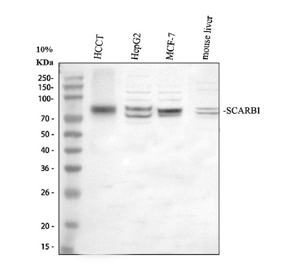
Western blot analysis of SCARB1 using anti-SCARB1 antibody (A01093-1).
Electrophoresis was performed on a 10% SDS-PAGE gel at 80V (Stacking gel) / 120V (Resolving gel) for 2 hours. The sample well of each lane was loaded with 30 ug of sample under reducing conditions.
Lane 1: human hepatocellular carcinoma tumor tissue (HCCT) lysates,
Lane 2: human HepG2 whole cell lysates,
Lane 3: human MCF-7 whole cell lysates,
Lane 4: mouse liver tissue lysates.
After electrophoresis, proteins were transferred to a nitrocellulose membrane at 150 mA for 50-90 minutes. Blocked the membrane with 5% non-fat milk/TBS for 1.5 hour at RT. The membrane was incubated with rabbit anti-SCARB1 antigen affinity purified polyclonal antibody (A01093-1) at 0.5 μg/mL overnight at 4°C, then washed with TBS-0.1%Tween 3 times with 5 minutes each and probed with a goat anti-rabbit IgG-HRP secondary antibody (Catalog # BA1054) at a dilution of 1:5000 for 1.5 hour at RT. The signal is developed using an ECL Plus Western Blotting Substrate (Catalog # AR1196-200) with Tanon 5200 system. A specific band was detected for SCARB1 at approximately 85 kDa. The expected band size for SCARB1 is at 61 kDa.

Click image to see more details
IF analysis of SCARB1 using anti-SCARB1 antibody (A01093-1).
SCARB1 was detected in an immunocytochemical section of Hela cells. Enzyme antigen retrieval was performed using IHC enzyme antigen retrieval reagent (AR0022) for 15 mins. The cells were blocked with 10% goat serum. And then incubated with 5 μg/mL rabbit anti-SCARB1 Antibody (A01093-1) overnight at 4°C. Cy3 Conjugated Goat Anti-Rabbit IgG (BA1032) was used as secondary antibody at 1:500 dilution and incubated for 30 minutes at 37°C. The section was counterstained with DAPI. Visualize using a fluorescence microscope and filter sets appropriate for the label used.

Click image to see more details
Flow Cytometry analysis of HepG2 cells using anti-SCARB1 antibody (A01093-1).
Overlay histogram showing HepG2 cells stained with A01093-1 (Blue line). The cells were fixed with 4% paraformaldehyde and blocked with 10% normal goat serum. And then incubated with rabbit anti-SCARB1 Antibody (A01093-1, 1 μg/1x106 cells) for 30 min at 20°C. Fluoro488 conjugated goat anti-rabbit IgG (BA1127, 5-10 μg/1x106 cells) was used as secondary antibody for 30 minutes at 20°C. Isotype control antibody (Green line) was rabbit IgG (1 μg/1x106) used under the same conditions. Unlabelled sample without incubation with primary antibody and secondary antibody (Red line) was used as a blank control.
Specific Publications For Anti-Scavenging Receptor SR-BI/SCARB1 Antibody Picoband® (A01093-1)
Loading publications
Recommended Resources
Here are featured tools and databases that you might find useful.
- Boster's Pathways Library
- Protein Databases
- Bioscience Research Protocol Resources
- Data Processing & Analysis Software
- Photo Editing Software
- Scientific Literature Resources
- Research Paper Management Tools
- Molecular Biology Software
- Primer Design Tools
- Bioinformatics Tools
- Phylogenetic Tree Analysis
Customer Reviews
Have you used Anti-Scavenging Receptor SR-BI/SCARB1 Antibody Picoband®?
Share your experimental results or join a short interview to earn up to $1,000 in product credits or other rewards.
0 Reviews For Anti-Scavenging Receptor SR-BI/SCARB1 Antibody Picoband®
Customer Q&As
Have a question?
Find answers in Q&As, reviews.
Can't find your answer?
Submit your question
