Click image to see more details
-

-
-
-
-
+3
Product Info Summary
| SKU: | A01090 |
|---|---|
| Size: | 100 μg/vial |
| Reactive Species: | Human, Mouse, Rat |
| Host: | Rabbit |
| Application: | ELISA, Flow Cytometry, IHC, WB |
Customers Who Bought This Also Bought

Product info
Product Name
Anti-SDHB Antibody Picoband®
SKU/Catalog Number
A01090
Size
100 μg/vial
Form
Lyophilized
Description
Boster Bio Anti-SDHB Antibody Picoband® catalog # A01090. Tested in ELISA, Flow Cytometry, IHC, WB applications. This antibody reacts with Human, Mouse, Rat. The brand Picoband indicates this is a premium antibody that guarantees superior quality, high affinity, and strong signals with minimal background in Western blot applications. Only our best-performing antibodies are designated as Picoband, ensuring unmatched performance.
Storage & Handling
Store at -20˚C for one year from date of receipt. After reconstitution, at 4˚C for one month. It can also be aliquotted and stored frozen at -20˚C for six months. Avoid repeated freeze-thaw cycles.
Cite This Product
Anti-SDHB Antibody Picoband® (Boster Biological Technology, Pleasanton CA, USA, Catalog # A01090)
Host
Rabbit
Contents
Each vial contains 4mg Trehalose, 0.9mg NaCl, 0.2mg Na2HPO4, 0.05mg NaN3.
Clonality
Polyclonal
Isotype
Rabbit IgG
Immunogen
E. coli-derived human SDHB recombinant protein (Position: A29-V280).
Cross-reactivity
No cross-reactivity with other proteins.
Reactive Species
A01090 is reactive to SDHB in Human, Mouse, Rat
Observed Molecular Weight
29 kDa
Calculated molecular weight
31.6 kDa
Background of SDHB
SDHB (Succinate Dehydrogenase Complex, Subunit B, iron sulfur protein), also known as iron-sulfur subunit of complex II (Ip) or SDH2, HOMOLOG OF, is a protein that in humans is encoded by the SDHB gene. SDHB is one of four protein subunits forming succinate dehydrogenase, the other three being SDHA, SDHC and SDHD. The SDHB subunit is connected to the SDHA subunit on the hydrophilic, catalytic end of the SDH complex. The SDHB gene is mapped on 1p36.13. It is stated that the nuclear-encoded Krebs cycle enzymes fumarate hydratase and succinate dehydrogenases like SDHB act as tumor suppressors, and germline mutations in these genes predispose individuals to leiomyomas and renal cancer and to paragangliomas, respectively. In affected members of families with paragangliomas-4, mutations were identified in the SDHB gene.
Antibody Validation
Boster validates all antibodies on WB, IHC, ICC, Immunofluorescence, and ELISA with known positive control and negative samples to ensure specificity and high affinity, including thorough antibody incubations.
Application & Images
Applications
A01090 is guaranteed for ELISA, Flow Cytometry, IHC, WB Boster Guarantee
Recommend Dilution
| Application | Dilution | Species |
|---|---|---|
| Western blot | 0.1-0.5μg/ml | Human, Mouse, Rat |
| Immunohistochemistry (Paraffin-embedded Section) | 2-5μg/ml | Human, Mouse, Rat |
| Flow Cytometry(Fixed) | 1-3 μg/1x106 cells | Human |
| ELISA | 0.1-0.5μg/ml |
Tested application
Suggested blocking solution with 5% non-fat milk or BSA; (*)Recommended protein loading: 20-40 µg per lane
Use TE buffer pH 9.0 for antigen retrieval; (*) citrate buffer pH 6.0 is an alternative.
Validation Images & Assay Conditions
Click image to see more details
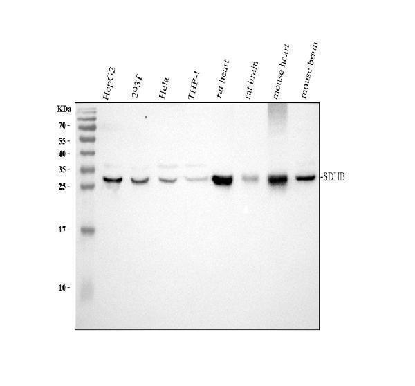
Western blot analysis of SDHB using anti-SDHB antibody (A01090).
Electrophoresis was performed on a 13% SDS-PAGE gel at 80V (Stacking gel) / 120V (Resolving gel) for 2 hours. The sample well of each lane was loaded with 30 ug of sample under reducing conditions.
Lane 1: human HepG2 whole cell lysates,
Lane 2: human 293T whole cell lysates,
Lane 3: human Hela whole cell lysates,
Lane 4: human THP-1 whole cell lysates,
Lane 5: rat heart tissue lysates,
Lane 6: rat brain tissue lysates,
Lane 7: mouse heart tissue lysates,
Lane 8: mouse brain tissue lysates.
After electrophoresis, proteins were transferred to a nitrocellulose membrane at 150 mA for 50-90 minutes. Blocked the membrane with 5% non-fat milk/TBS for 1.5 hour at RT. The membrane was incubated with rabbit anti-SDHB antigen affinity purified polyclonal antibody (A01090) at 0.5 μg/mL overnight at 4°C, then washed with TBS-0.1%Tween 3 times with 5 minutes each and probed with a goat anti-rabbit IgG-HRP secondary antibody (Catalog # BA1054) at a dilution of 1:5000 for 1.5 hour at RT. The signal is developed using an ECL Plus Western Blotting Substrate (Catalog # AR1196-200) with Tanon 5200 system. A specific band was detected for SDHB at approximately 29 kDa. The expected band size for SDHB is at 32 kDa.

Click image to see more details
IHC analysis of SDHB using anti-SDHB antibody (A01090).
SDHB was detected in a paraffin-embedded section of human colon cancer tissue. Heat mediated antigen retrieval was performed in EDTA buffer (pH 8.0, epitope retrieval solution). The tissue section was blocked with 10% goat serum. The tissue section was then incubated with 2 μg/ml rabbit anti-SDHB Antibody (A01090) overnight at 4°C. Peroxidase Conjugated Goat Anti-rabbit IgG was used as secondary antibody and incubated for 30 minutes at 37°C. The tissue section was developed using HRP Conjugated Rabbit IgG Super Vision Assay Kit (Catalog # SV0002) with DAB as the chromogen.

Click image to see more details
IHC analysis of SDHB using anti-SDHB antibody (A01090).
SDHB was detected in a paraffin-embedded section of human lung cancer tissue. Heat mediated antigen retrieval was performed in EDTA buffer (pH 8.0, epitope retrieval solution). The tissue section was blocked with 10% goat serum. The tissue section was then incubated with 2 μg/ml rabbit anti-SDHB Antibody (A01090) overnight at 4°C. Peroxidase Conjugated Goat Anti-rabbit IgG was used as secondary antibody and incubated for 30 minutes at 37°C. The tissue section was developed using HRP Conjugated Rabbit IgG Super Vision Assay Kit (Catalog # SV0002) with DAB as the chromogen.

Click image to see more details
IHC analysis of SDHB using anti-SDHB antibody (A01090).
SDHB was detected in a paraffin-embedded section of human mammary cancer tissue. Heat mediated antigen retrieval was performed in EDTA buffer (pH 8.0, epitope retrieval solution). The tissue section was blocked with 10% goat serum. The tissue section was then incubated with 2 μg/ml rabbit anti-SDHB Antibody (A01090) overnight at 4°C. Peroxidase Conjugated Goat Anti-rabbit IgG was used as secondary antibody and incubated for 30 minutes at 37°C. The tissue section was developed using HRP Conjugated Rabbit IgG Super Vision Assay Kit (Catalog # SV0002) with DAB as the chromogen.

Click image to see more details
IHC analysis of SDHB using anti-SDHB antibody (A01090).
SDHB was detected in a paraffin-embedded section of mouse small intestine tissue. Heat mediated antigen retrieval was performed in EDTA buffer (pH 8.0, epitope retrieval solution). The tissue section was blocked with 10% goat serum. The tissue section was then incubated with 2 μg/ml rabbit anti-SDHB Antibody (A01090) overnight at 4°C. Peroxidase Conjugated Goat Anti-rabbit IgG was used as secondary antibody and incubated for 30 minutes at 37°C. The tissue section was developed using HRP Conjugated Rabbit IgG Super Vision Assay Kit (Catalog # SV0002) with DAB as the chromogen.

Click image to see more details
IHC analysis of SDHB using anti-SDHB antibody (A01090).
SDHB was detected in a paraffin-embedded section of rat small intestine tissue. Heat mediated antigen retrieval was performed in EDTA buffer (pH 8.0, epitope retrieval solution). The tissue section was blocked with 10% goat serum. The tissue section was then incubated with 2 μg/ml rabbit anti-SDHB Antibody (A01090) overnight at 4°C. Peroxidase Conjugated Goat Anti-rabbit IgG was used as secondary antibody and incubated for 30 minutes at 37°C. The tissue section was developed using HRP Conjugated Rabbit IgG Super Vision Assay Kit (Catalog # SV0002) with DAB as the chromogen.

Click image to see more details
Flow Cytometry analysis of A431 cells using anti-SDHB antibody (A01090).
Overlay histogram showing A431 cells stained with A01090 (Blue line). To facilitate intracellular staining, cells were fixed with 4% paraformaldehyde and permeabilized with permeabilization buffer. The cells were blocked with 10% normal goat serum. And then incubated with rabbit anti-SDHB Antibody (A01090, 1 μg/1x106 cells) for 30 min at 20°C. Fluoro488 conjugated goat anti-rabbit IgG (BA1127, 5-10 μg/1x106 cells) was used as secondary antibody for 30 minutes at 20°C. Isotype control antibody (Green line) was rabbit IgG (1 μg/1x106) used under the same conditions. Unlabelled sample without incubation with primary antibody and secondary antibody (Red line) was used as a blank control.
Specific Publications For Anti-SDHB Antibody Picoband® (A01090)
Loading publications
Recommended Resources
Here are featured tools and databases that you might find useful.
- Boster's Pathways Library
- Protein Databases
- Bioscience Research Protocol Resources
- Data Processing & Analysis Software
- Photo Editing Software
- Scientific Literature Resources
- Research Paper Management Tools
- Molecular Biology Software
- Primer Design Tools
- Bioinformatics Tools
- Phylogenetic Tree Analysis
Customer Reviews
Have you used Anti-SDHB Antibody Picoband®?
Share your experimental results or join a short interview to earn up to $1,000 in product credits or other rewards.
0 Reviews For Anti-SDHB Antibody Picoband®
Customer Q&As
Have a question?
Find answers in Q&As, reviews.
Can't find your answer?
Submit your question
5 Customer Q&As for Anti-SDHB Antibody Picoband®
Question
We want using your anti-SDHB antibody for respiratory electron transport studies. Has this antibody been tested with western blotting on small intestine tissue? We would like to see some validation images before ordering.
Verified Customer
Verified customer
Asked: 2020-01-16
Answer
Thanks for your inquiry. This A01090 anti-SDHB antibody is validated on human a549, a549 whole cell lysates, rat heart tissue, brain tissue, mouse heart tissue, small intestine tissue, mammary cancer tissue. It is guaranteed to work for ELISA, IF, IHC-P, ICC, WB in human, mouse, rat. Our Boster guarantee will cover your intended experiment even if the sample type has not been be directly tested.
Boster Scientific Support
Answered: 2020-01-16
Question
We have observed staining in mouse fibroblast. Are there any suggestions? Is anti-SDHB antibody supposed to stain fibroblast positively?
Verified Customer
Verified customer
Asked: 2019-11-19
Answer
From what I have seen in literature fibroblast does express SDHB. From what I have seen in Uniprot.org, SDHB is expressed in muscle of leg, liver, uterus, brain, fibroblast, among other tissues. Regarding which tissues have SDHB expression, here are a few articles citing expression in various tissues:
Brain, Pubmed ID: 15489334
Fibroblast, Pubmed ID: 2494655
Liver, Pubmed ID: 2302193, 7622059, 24275569
Uterus, Pubmed ID: 14702039
Boster Scientific Support
Answered: 2019-11-19
Question
We ordered your anti-SDHB antibody for WB on uterus in the past. I am using human, and We are going to use the antibody for ICC next. My lab would like examining uterus as well as brain in our next experiment. Could give a recommendation on which antibody would work the best for ICC?
Verified Customer
Verified customer
Asked: 2018-12-26
Answer
I viewed the website and datasheets of our anti-SDHB antibody and it appears that A01090 has been tested on human in both WB and ICC. Thus A01090 should work for your application. Our Boster satisfaction guarantee will cover this product for ICC in human even if the specific tissue type has not been validated. We do have a comprehensive range of products for ICC detection and you can check out our website bosterbio.com to find out more information about them.
Boster Scientific Support
Answered: 2018-12-26
Question
Our lab were happy with the WB result of your anti-SDHB antibody. However we have seen positive staining in brain mitochondrion inner membrane using this antibody. Is that expected? Could you tell me where is SDHB supposed to be expressed?
Verified Customer
Verified customer
Asked: 2018-10-30
Answer
According to literature, brain does express SDHB. Generally SDHB expresses in mitochondrion inner membrane. Regarding which tissues have SDHB expression, here are a few articles citing expression in various tissues:
Brain, Pubmed ID: 15489334
Fibroblast, Pubmed ID: 2494655
Liver, Pubmed ID: 2302193, 7622059, 24275569
Uterus, Pubmed ID: 14702039
Boster Scientific Support
Answered: 2018-10-30
Question
We are currently using anti-SDHB antibody A01090 for rat tissue, and we are happy with the ICC results. The species of reactivity given in the datasheet says human, mouse, rat. Is it possible that the antibody can work on canine tissues as well?
Verified Customer
Verified customer
Asked: 2017-12-27
Answer
The anti-SDHB antibody (A01090) has not been validated for cross reactivity specifically with canine tissues, but there is a good chance of cross reactivity. We have an innovator award program that if you test this antibody and show it works in canine you can get your next antibody for free. Please contact me if I can help you with anything.
Boster Scientific Support
Answered: 2017-12-27
